Introduction
Pennellid copepods are parasites of various fishes and marine mammals (Boxshall and Halsey, Reference Boxshall and Halsey2004). Almost all species of this family are characterized by anterior anchoring and feeding parts that embed into the host tissue (Kabata, Reference Kabata1979). The genus Phrixocephalus, which is a member of the family Pennellidae, comprises 13 species (Walter and Boxshall, Reference Walter and Boxshall2026). Eleven of them are known to occur in Japan (Nagasawa and Uyeno, Reference Nagasawa and Uyeno2014). Copepods of this genus are found exclusively on the eyes of fishes, with a branching holdfast embedded in the host’s eye (Shiino, Reference Shiino1956). Molecular data were used to estimate evolutionary relationships within Pennellidae, including a species of this genus (Yumura et al., Reference Yumura, Adachi, Nitta, Kondo, Komeda, Wakabayashi, Fukuchi, Boxshall and Ohtsuka2022).
Phrixocephalus alatus Shiino, 1956 was first described based on a specimen obtained from a young individual of red seabream Pagrus major (Temminck and Schlegel, 1844) collected from Suruga Bay, northwestern Pacific Ocean (Shiino, Reference Shiino1956). This has been the only known host record and locality for the past 70 years. This species is the only known Phrixocephalus species that parasitizes sparid fishes. Here, we report a specimen of Ph. alatus from a yellowback seabream Dentex hypselosomus Bleeker, 1854, collected from the Sea of Japan. We also provide partial mitochondrial COI gene data for this species to contribute to further DNA barcoding.
Materials and methods
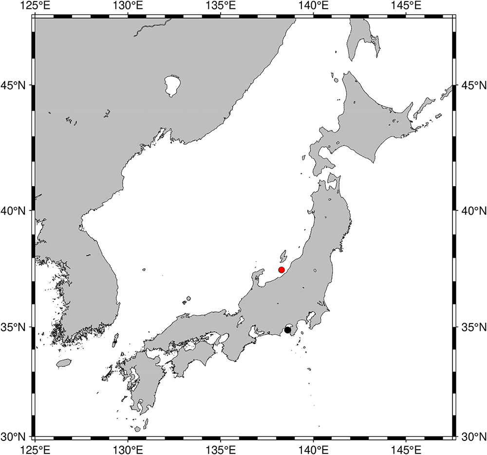
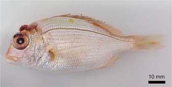
An individual of D. hypselosomus parasitized by a copepod was collected off Niigata Prefecture in the northeastern Sea of Japan during the July 2025 expedition of T/V Tenyo-Maru of Japan Fisheries Research and Education Agency, using a bottom trawl (Figures 1 and 2). The fish was frozen on board and subsequently thawed at the Atmosphere and Ocean Research Institute, the University of Tokyo, in July 2025. The copepod was carefully removed from the host and preserved in 99% ethanol. The copepod was photographed using a digital microscope (VHX-8000; Keyence). Some eggs were removed from the copepod and stored for use in other studies.
Total DNA was extracted from the trunk of the copepod using a DNeasy Blood and Tissue Kit (Qiagen, Venlo, Netherlands). A fragment of the mitochondrial COI gene was amplified by PCR with the primer pair LCO1490 (5′-GGTCAACAAATCATAAAGATATTGG-3′) and HCO2198 (5′-TAAACTTCAGGGTGACCAAAAAATCA-3′) (Folmer et al., Reference Folmer, Black, Hoeh, Lutz and Vrijenhoek1994). The PCR amplification was conducted in a total volume of 25 μL containing 9.4 μL of double-distilled water (DDW), 2.5 μL of DNA solution, 12.5 μL of Ex Premier DNA Polymerase Dye plus (2×) (TaKaRa, Shiga, Japan), and 0.3 μL of each forward and reverse primer (20 μM) under the following conditions: initial denaturation at 94°C for 1 min, followed by 30 cycles of denaturation at 98°C for 10 s, annealing at 42°C for 15 s, and extension at 68°C for 45 s. To degrade the remaining primers and nucleotides, 0.15 µL of each amplicon was mixed with 2.95 µL DDW and 0.6 µL ExoSAP-IT Express PCR Product Cleanup Reagent (Applied Biosystems, Waltham, USA) and incubated first at 37°C (165 s) and then at 80°C (1 min). Sequencing was performed using the Big Dye Terminator Cycle Sequence Kit, ver. 3.1 (Applied Biosystems) with the amplification primers. The sequenced product was purified using the Big Dye X Terminator Purification Kit (Applied Biosystems) and analysed using an ABI PRISM 3500xl sequencer (Applied Biosystems). The obtained sequence was trimmed to exclude the amplification primers in AliView (Larsson, Reference Larsson2014). The specimen was deposited in the crustacean collection (NSMT-Cr 33394) at the National Museum of Nature and Science, Tsukuba, Japan, after DNA extraction and removal of several eggs.
Localities of Phrixocephalus alatus. Red and black dots represent the sites of this study and Shiino (Reference Shiino1956), respectively.

Dentex hypselosomus collected from the Sea of Japan, parasitized by Phrixocephalus alatus. Scale bar: 10 mm.

Results
Family Pennellidae Burmeister, 1835
Genus Phrixocephalus Wilson, 1908
Phrixocephalus alatus Shiino, 1956
Phrixocephalus alatus: (A) Whole body; (B) Thoracic horns. Arowwheads indicate quadripartite thoracic horns. Scale bars: A, 1 mm; B, 0.5 mm.

Material examined
One female. NSMT-Cr 33394, obtained from the left eyeball of D. hypselosomus (total length: 87 mm, fork length: 71 mm) collected off Niigata Prefecture, the northeastern Sea of Japan (37°16′ N, 138°14′ E, 80 m), in July 2025, during the stock assessment ground fish survey of the T/V Tenyo-Maru.
Description and diagnosis
The specimen was identified as Ph. alatus based on the presence of the following morphological traits: body pale yellow in ethanol; two pairs of lateral processes on cephalothorax, each divided into several branches with additional secondary branches; short neck joined with trunk at a right angle; stubby, branched thoracic horns positioned behind middle of neck; sausage-shaped trunk, gradually widening posteriorly; prominent abdomen, higher than round bulge bearing egg strings; egg strings loosely coiled.
Remarks
Some morphological differences were observed from the original description of the species. In the new specimen examined, the lateral cephalothoracic processes overlapped both dorsally and ventrally (Figure 3A), unlike the original description. Additionally, the thoracic horns of the original description branched into at most three processes, whereas those of the present specimen branched into at most four processes (Figure 3B). Because the processes and horns comprising the holdfast are considered to function as anchors, differences in their shape may arise from host tissue condition, eyeball size, and subtle difference in attachment sites, and therefore represent intraspecific variation.
The present specimen differs from Ph. cincinnatus Wilson, 1908 in having thoracic horns situated in the middle of the neck (vs. close to the head and midway between the first thoracic horns and the trunk). Similarly, this specimen differs from Ph. arboreus Shiino, 1956 and Ph. longicollum Shiino, 1956 in having a short neck (vs. a long neck much longer than the trunk). In addition, this specimen can be distinguished from Ph. carcellesi Brian, 1944; Ph. crassus Shiino, 1956; Ph. diversus Wilson, 1917; Ph. reniformis Shiino, 1956; Ph. triangulus Wilson, 1917; and Ph. umbellatus Shiino, 1956 in having a sausage-shaped trunk (vs. a triangular trunk or a trunk with an anterior swelling). It also differs from Ph. curvatus Shiino, 1956, in having a prominent abdomen (vs. an abdomen as small as the weak oviducal bulges). The remaining two species, Ph. cepolae Yamaguti, 1939 and Ph. vipereus Shiino, 1956, are clearly distinguishable from the specimen in having highly complex and irregularly branched lateral cephalothoracic processes with multiple major trunks (vs. two pairs of lateral cephalothoracic processes, each divided into several branches that branch secondarily, in Ph. alatus). Therefore, among the currently recognized Phrixocephalus species, the present specimen is most consistent with Ph. alatus.
The partial mitochondrial COI sequence (658 bp) of Ph. alatus was obtained and has been deposited in the DDBJ/EMBL/GenBank databases (accession number: LC927251).
Discussion
Phrixocephalus alatus has been recorded only once, based on a single specimen parasitizing Pa. major collected from Suruga Bay in the Pacific Ocean (Shiino, Reference Shiino1956). The present finding is the second record of the species and demonstrates that this copepod also utilizes D. hypselosomus as a host.
The specimen of D. hypselosomus collected in the present study had a fork length of 71 mm and is estimated to be 0.5–1 year of age based on the relationship between age and body size (Oki and Tabeta, Reference Oki and Tabeta1998). The specimen described in the original description was obtained from the small individual of Pa. major, which had a fork length of 60 mm (Shiino, Reference Shiino1956). This may indicate that Ph. alatus preferentially infects young sparid fishes, as it has been reported that individuals of Pa. major with a fork length of approximately 60 mm are less than 100 days old after hatching (Arai et al., Reference Arai, Sakamoto and Maeda1995). Although sparid species are commercially important species at Japanese markets (Yamada et al., Reference Yamada, Tagawa, Kishida and Honjo1986; Irie and Yamada, Reference Irie and Yamada1991), Ph. alatus has not been reported for 70 years. This may be due to this copepod infecting small fish that are not commonly sold at the markets. In addition, limited research on parasitic copepods in Japanese waters may also be a reason for it not being reported in 70 years. Except for areas around Sado Island (e.g., Shiino, Reference Shiino1965; Ho and Do, Reference Ho and Do1985; Honma and Kitami, Reference Honma and Kitami1995), parasitic copepods have been rarely studied off the Japanese coast in the northern Sea of Japan.
Although the effects of this copepod on its host are unclear, it was reported that Ph. cincinnatus causes extensive damage to the host’s eye tissues, evident from the residual pus, tissue debris, and blood (Kabata, Reference Kabata and Kinne1984). Such damage may potentially reduce the growth and survival of the infected fish as reported by Ohtsuka et al. (Reference Ohtsuka, Harada, Miyahara, Nagahama, Ogawa and Ohta2007), who found that bastard halibut Paralichthys olivaceus (Temminck and Schlegel, 1846) parasitized by Ph. umbellatus had lower body weights than non-parasitized individuals. As many sparid species inhabiting Japanese waters are commercially important (Yamada et al., Reference Yamada, Tagawa, Kishida and Honjo1986; Irie and Yamada, Reference Irie and Yamada1991), it is necessary to investigate the effects of Ph. alatus on its host to clarify its potential impact on fisheries and sparid aquaculture.
Acknowledgements
We thank Drs. Masaya Iida and Kay Sakuma (Fisheries Resources Institute, Japan Fisheries Research and Education Agency), Mr. Kosei Yamazaki (Fukui Prefectural University), crew of T/V Tenyo-Maru, and students who participated in the expedition of Tenyo-Maru in 2025 for helping to collect the fish sample. We also thank Dr. Hironori Komatsu (National Museum of Nature and Science) and Mr. Hiroki Sato (The University of Tokyo) for helping to deposit the specimen and making the map of sampling sites, respectively. We greatly appreciate the constructive suggestions of the editor and the two reviewers, which substantially improved this manuscript. The sample used in this study was collected during a stock assessment ground fish survey, which was financially supported by the Ministry of Agriculture, Forestry and Fisheries, Japan.
Author contributions
Y.M. collected the sample and wrote the manuscript. N.Y. identified the specimen. S.K. supervised this study.
Declaration of interest
None.
Data availability statement
The authors confirm that the data supporting the findings of this study are available within the article.
Ethics and approach
This article did not require a research permit.
Funding statement
This research did not receive funding from any agency, commercial or not-for-profit sectors.