Introduction
It is common practice in dairy herd management to aim for one calf per year. As a consequence, dairy cows have concurrent pregnancy and lactation during a large part of the lactation period. This is a unique physiological condition where the lactating mammary gland fulfils its biological role by producing milk for the newborn calf, while at the same time it must prepare for the subsequent lactation to fulfil its role for the unborn calf. This ‘conflict of interest’ between the present and the subsequent lactation is evident from the shape of the lactation curves of pregnant and non-pregnant cows. The decrease in milk yield is more rapid in pregnant than in non-pregnant cows from approximately the sixth month of pregnancy at comparable stages of lactation (van Amburgh et al., Reference van Amburgh, Galton, Bauman and Everett1997; Bertilsson et al., Reference Bertilsson, Berglund, Ratnayake, SvennerstenSjaunja and Wiktorsson1997).
The decrease in milk yield seems to occur at the same stage of pregnancy independent of the stage of lactation. Therefore, the accelerated decline in yield of pregnant cows is likely regulated by endocrine signals originating from the conceptus. Progesterone, 17β-estradiol (hereafter oestrogen) and prolactin (Prl) are major hormones important for mammary gland function during the pregnancy–lactation cycle (Lamote et al., Reference Lamote, Meyer, Massart-Leen and Burvenich2004). Changes in plasma concentrations of these hormones are well described, but it is unclear as to how the bovine mammary gland responds to pregnancy at the cellular level.
The normal decline in milk yield with advancing lactation seems to be due mainly to a loss of secretory cells rather than to reduced synthetic activity of the individual cells (Knight and Wilde, Reference Knight and Wilde1993), but it is unknown whether the pregnancy-related decline in milk yield also is caused by a decline in the number of secretory cells.
On this basis, the main objective was to investigate whether the pregnancy-related decrease in milk yield is caused by alterations in mammary cell turnover. Secondly, we wanted to investigate whether pregnancy affects the mammary transcription of oestrogen, progesterone and prolactin receptors. Furthermore, we wanted to investigate the effect of feeding level on mammary cells, because results from a previous study have indicated that feeding a low-energy-density diet has an inhibitory effect on mammary cell proliferation during early lactation (Nørgaard et al., Reference Nørgaard, Sorensen, Sørensen, Andersen and Sejrsen2005). These objectives were addressed by taking mammary biopsies and measuring cell proliferation and apoptosis, and transcription of genes encoding hormone receptors and proteins indicative of secretory activity and turnover of mammary cells.
Materials and methods
Animals and experimental design
The experiment was carried out as a 2 × 2 factorial design; the main effects being pregnant or non-pregnant and low v. normal feeding level. Sixteen Danish Holstein Friesian cows were used. Thus, the main effects were compared with eight cows per treatment. The cows were part of a larger experiment investigating the effect of extended lactation and feeding level (Sehested and Danfær, Reference Sehested and Danfær2003).
Day of conception for the group of pregnant cows was day 88 ± 9 (mean ± SE) of lactation. The cows were fed a grass and maize silage-based mixed ration ad libitum during winter. In summer the cows were on pasture. In case of poor grass availability, the silage mix was offered as supplementation. Half of the cows were offered a concentrate mix (normal feeding level) while the other half were not (low feeding level). The concentrate mix provided the cows on normal feeding level an additional 48 MJ NEL (net energy for lactation) compared with cows on low feeding level. The mean parity for the treatment groups was 2.5 for low-pregnant, 1.5 for normal-pregnant, 1.5 for low-non-pregnant and 2.0 for normal-non-pregnant cows.
Recordings and tissue sampling
Milk yield was recorded weekly and individual feed intake of silage and concentrate was recorded three times a week. Clinical diseases and veterinary treatments were also recorded.
Mammary biopsies were obtained from the central part of the front quarters of all cows as described by Sørensen et al. (Reference Sørensen, Nørgaard, Theil, Vestergaard and Sejrsen2006). All samples were characterized as parenchyma by visual analysis of the fresh biopsy. Part of the tissue was immediately snap frozen in liquid nitrogen and kept at −80°C until further processing. Tissue for immunohistochemical detection of apoptosis and cell proliferation was fixed overnight in 4% neutral buffered formalin, dehydrated and embedded in paraffin. The biopsies were taken from the pregnant cows at day 189 ± 3 of pregnancy, corresponding to approximately 3 months before expected calving. The tissue was collected from the non-pregnant cows at day 224 ± 3 of lactation. As a consequence of re-breeding of cows in the pregnant group, they were on average 276 ± 10 days into lactation at the time of biopsy sampling. This has no bearing on the results, since we observed that mammary cell turnover and gene expression is relatively stable at this stage of lactation (Sørensen et al., Reference Sørensen, Nørgaard, Theil, Vestergaard and Sejrsen2006).
Analysis
Quantification of mammary cell proliferation and apoptosis was done using slides with 5 μm tissue sections from the paraffin-embedded tissue. Slides were deparaffinized in xylene and rehydrated in a graded series of ethanol. For both assays, slides were handled on Shandon Coverplates and Shandon Sequenza Slide Racks (Thermo Electron Corporation, Waltham, MA, USA).
To measure cell proliferation, the nuclear proliferation antigen Ki-67 was detected: epitopes were demasked by boiling in 10 mM citrate buffer (pH 6.0) in a microwave oven for a total period of 21 min. The specimens were incubated for 15 min with the primary antibody monoclonal mouse anti-human Ki-67 antigen clone MIB-1 (DakoCytomation Denmark A/S, Glostrup, Denmark). The identification of nuclei with antigens was made with the EnVision+ System, Peroxidase-HRP kit (DakoCytomation Denmark A/S) according to manufacturer’s instructions. Mammary cell proliferation data from one pregnant cow fed low feeding level were not included in the data analysis because the percentage of Ki-67 staining was inexplicably high (309 positive of 1691 alveolar cells), which could not be explained either in the health of the cow or in the biopsy sections, which appeared normal.
Apoptotic cells were detected using the TUNEL (terminal deoxynucleotidyl transferase dUTP nick end labeling) ApopTag Plus Peroxidase In Situ Apoptosis Detection Kit (Chemicon International Inc., Temecula, CA, USA) according to manufacturer’s instructions, with the following modifications: the slides were incubated with proteinase K (Chemicon International Inc.) solution for 15 min and with equilibration buffer for 10 min at 4°C, and apoptotic cells were stained with diaminobenzidine (DAB) for 10 min.
In both Ki-67 and TUNEL assays, cells were counterstained with Mayers haematoxylin for 30 s and slides were dehydrated in a graded series of ethanol ending in xylene and subsequently coverslipped with DePeX mounting medium (VWR International Ltd, Lutterworth, UK). In both assays, positive staining was quantified in five to nine randomly chosen fields for each animal by light microscopy at 400× magnification.
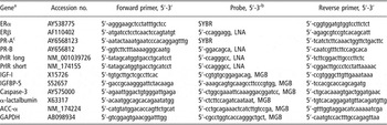
For real-time PCR, total mRNA from approximately 10 mg tissue was isolated using columns and reagents of the RNeasy Mini Kit (Qiagen Ltd, West Sussex, UK), and RNA concentration was determined spectrophotometrically at 260 nm. Synthesis of cDNA was performed using SuperScript II Reverse Transcriptase and Oligo(dT)12–18 Primer (Invitrogen, Taastrup, Denmark). The cDNA was quantified by real-time PCR on an ABI Prism 7900 SDS (Applied Biosystems, Naerum, Denmark). Melting curves based on SYBR green detection or negative controls of genomic DNA were used to ensure that only cDNA and not genomic DNA was amplified. Primers and MGB (minor groove binding) probes were designed using Primer Express software (Applied Biosystems), see Table 1. Primer sequences for oestrogen receptor (ER) α were identical to those described by Pfaffl et al. (Reference Pfaffl, Lange, Daxenberger and Meyer2001). Locked nucleic acid (LNA) probes (Roche Diagnostics, Hvidovre, Denmark) were used for the detection of long and short prolactin receptor (PrlR), ERβ and progesterone receptor (PR) B, see Table 1. Transcription of the reference gene glyceraldehyde 3-phosphate dehydrogenase (GAPDH) was not affected by treatments and therefore was used to normalize the transcription of target genes.
Gene names, accession numbers and primer and probe sequences used in real-time PCR

aER = oestrogen receptor α or β isoforms; PR = progesterone receptor isoform A (short) or B (long); PrlR = prolactin receptor short or long isoforms; IGFBP = insulin-like growth factor binding protein; ACC-α = acetyl CoA carboxylase isoform α; GAPDH = glyceraldehyde 3-phosphate dehydrogenase.
bSYBR = SYBR Green non-specific detection; LNA = locked nucleic acid; MGB = minor grove binding.
cAlternative splice site information based on human PR short isoform accession no. AB085843.
Statistical analysis
Statistical evaluation of milk yield, transcription, cell proliferation and apoptosis was performed using the MIXED procedure of SAS (SAS Institute, 1999). The statistical models used to evaluate gene expression included fixed effects of pregnancy status, feeding level, pregnancy × feeding level interaction, parity (parity 1 or parity >1) and a random cow effect. Milk yield was evaluated by a similar model except that treatment × week interaction was included as fixed effects and week of lactation was included as a random effect to account for repeated measurements. The type of covariance structure was assumed to be compound symmetry.
The statistical evaluation of cell proliferation and apoptosis was carried out using the model as described for gene expression, but also including the five to nine fields as a random component. Cell turnover data were arcsin-transformed and assumed to be normally distributed.
Lactation persistency was determined as the regression slope for milk yield for the first 100 days after insemination (or the equivalent stage of lactation for non-pregnant cows) with the preceding 30-day milk yield as the covariate.
Statistical evaluation of gene expression was based on reference gene-corrected threshold cycle (Ct) values, i.e. Ct of target gene minus Ct of reference gene (Theil et al., Reference Theil, Sorensen, Therkildsen and Oksbjerg2006). The mRNA quantities relative to the treatment group of pregnant normal fed cows (ΔΔCt) was calculated as E −ΔΔCt, where E is 1 + PCR efficiency of target gene as determined by 10−1/slope of standard curve −1 (Rasmussen, Reference Rasmussen2001). In cases with 100% PCR efficiency, the calculation was simplified to 2−ΔΔCt (Pfaffl, Reference Pfaffl2001).
Values are presented as least squares means and 95% confidence limits. Pearson correlation coefficients were calculated on mean milk yield, cell proliferation and apoptosis and quantitative mRNA abundance.
Results
After termination of the experiment, all cows in the pregnant group calved at the expected time. No clinical diseases were recorded or treated except for mastitis in two cows within 7 days following biopsy sampling.
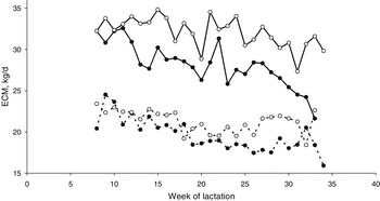
All treatment groups had a reduction (P < 0.001) in milk yield with progress of lactation. Milk yield between weeks 8 and 39 of calving was compared between groups (Figure 1). On average, pregnant cows had 2.6 kg/day lower (P = 0.20) milk yield than non-pregnant cows, and cows on low feeding level had 9.8 kg/day lower (P < 0.001) milk yield than cows fed normal feeding level. There was no interaction (P = 0.59) between main effects. Pregnant cows had an accelerated (P < 0.001) decline in milk yield from the time of insemination compared with non-pregnant cows at the same stage of lactation, i.e. lactation persistency was reduced by pregnancy.
Average daily energy-corrected milk yield (ECM) of non-pregnant cows (open circle), and pregnant cows (closed circle) inseminated week 12 (day 88 ± 9 (mean ± SEM)) after parturition on either normal (full line) or low (dotted line) feeding level. Biopsies were taken at week 27 (day 189 ± 3) of pregnancy, i.e. week 39 (day 276 ± 10) of lactation for pregnant cows and week 32 (day 224 ± 3) of lactation for non-pregnant cows. N = 16. Values are least squares means.

Average daily feed intake was 135 (130–140) MJ NEL (net energy for lactation) for cows on normal feeding level and 110 (105–114) MJ NEL for cows on low feeding level.
The percentage of mammary alveolar cells staining positive for apoptosis in the TUNEL assay varied between 0.1% and 0.2% and was affected neither by pregnancy (P = 0.52) nor by diet (P = 0.80) (Table 2). The percentage of proliferative cells as determined by Ki-67 antigen staining was also unaffected by feeding level (P = 0.36), but pregnant cows showed a lower (P = 0.06) rate of cell proliferation than non-pregnant cows.
Percentages of mammary alveolar cells undergoing apoptosis (TUNEL) or proliferation (Ki-67) in pregnant or non-pregnant cows at either normal (N) or low (L) feeding level

an = 4, values are least squares means and 95% confidence limits.
bP values are for differences between pregnancy status, feeding level, and pregnancy and feeding level interaction (P × F), i.e. the difference between treatments.
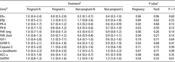
The abundance of mRNA for genes encoding the pregnancy-related hormone receptors was affected by pregnancy but not by feeding level (Table 3). ERα (P = 0.06), but not ERβ (P = 0.84) mRNA, and both PR-A (P = 0.02) and PR-B (P = 0.03) mRNA as well as both the long (P = 0.06) and short (P = 0.04) PrlR mRNA were more abundant in pregnant compared with non-pregnant cows.
Abundance of mRNA quantified by real-time PCR in mammary tissue of pregnant or non-pregnant cows at either normal (N) or low (L) feeding level

aResults are relative to the treatment group of pregnant normal feeding level cows, values in parenthesis are 95% confidence limits.
bER = oestrogen receptor α or β isoforms; PR = progesterone receptor isoform A (short) or B (long); PrlR = prolactin receptor short or long isoforms; IGFBP = insulin-like growth factor binding protein; ACC-α = acetyl CoA carboxylase isoform α; GAPDH = glyceraldehyde 3-phosphate dehydrogenase.
cP values are for differences between pregnancy status, feeding level, and pregnancy and feeding level interaction (P × F), i.e. the difference between treatments.
Neither pregnancy status nor feeding level affected the abundance of IGF-I, IGF binding protein (BP)-5 and caspase-3 mRNAs. The same was the case for abundance of mRNA of α-lactalbumin, while acetyl CoA carboxylase (ACC)-α tended to be more (P = 0.08) abundant in pregnant cows.
Discussion
Pregnancy status
In agreement with data by Bertilsson et al. (Reference Bertilsson, Berglund, Ratnayake, SvennerstenSjaunja and Wiktorsson1997) and Sorensen and Knight (Reference Sorensen and Knight2002), we observed reduced milk yield in pregnant cows compared with non-pregnant cows. In our study the difference appeared almost immediately after conception, while it did not appear until week 16 to week 20 of pregnancy in the studies by Bertilsson et al. and Sorensen and Knight. The reason for this difference between studies in the effect of pregnancy on milk yield in early pregnancy is not obvious: it is not a random effect due to the low number of animals, since the same was observed in the large experiment with 125 cows (Sehested and Danfær, Reference Sehested and Danfær2003). We did not anticipate this difference. Therefore, we did not collect mammary tissue in early pregnancy and thus it is not possible to address the exact biological basis for the effect of pregnancy on lactation at this stage. However, the slightly inhibiting effect of pregnancy on milk yield in early pregnancy is probably due to reduced secretory activity in the mammary gland and not to a reduction in the number of secretory cells because subtle changes in cell number would have to accumulate over time in order to significantly affect milk yield.
In contrast, our data suggest that the reduced milk yield of pregnant cows in late lactation is related to reduced mammary cell number, since we observed reduced (P = 0.06) mammary cell proliferation and unchanged apoptosis in the pregnant cows at 25 to 26 weeks of pregnancy. The data on mRNA abundance of acetyl CoA carboxylase (ACC) suggest that the secretory capacity is also decreased (P = 0.08). However, the abundance of α-lactalbumin mRNA was not different between pregnant and non-pregnant cows. We are not aware of other studies comparing mammary cell turnover in pregnant and non-pregnant cows at a comparable stage of lactation.
We observed increased ERα gene expression in the pregnant cows. Combined with anticipated increased plasma oestrogen, this should be in favour of increased mammary cell proliferation, since it has been shown in many classical studies that oestrogen stimulates mammary epithelial cell proliferation. These studies, however, were conducted with developing mammary tissue and not with lactating mammary cells as in this study, where we observed reduced and not increased mammary cell proliferation. In fact, it has been well documented that oestrogen inhibits milk synthesis in lactating cows (Akers, Reference Akers2002), so the increased ERα may be involved in mediating the negative effect of oestrogen on milk yield. This is supported by the observation that the mRNA abundance of IGF-I and IGFBP-5 was unaffected by pregnancy, since the IGF system is believed to be involved in mediating the effect of oestrogen on cell proliferation (Hanstein et al., Reference Hanstein, Djahansouzi, Dall, Beckmann and Bender2004; Connor et al., Reference Connor, Meyer, Li, van Amburgh, Boisclair and Capuco2007). Thus, in animals with concurrent pregnancy and lactation, the effects of oestrogen may be different from those in non-lactating animals with developing mammary glands such as prepubertal and pubertal animals. This suggestion complies well with a pregnancy-mediated increase in both oestrogen and ERα and a subsequent inhibition of secretory activity of mammary cells.
In agreement with the data of Schams et al. (Reference Schams, Kohlenberg, Amselgruber, Berisha, Pfaffl and Sinowatz2003), the expression of ERβ mRNA was much lower than ERα expression (data not shown). Similarly, oestrogen receptor distribution studies, using western blotting and immunohistochemistry, have shown that ERβ concentration is either low (Schams et al., Reference Schams, Kohlenberg, Amselgruber, Berisha, Pfaffl and Sinowatz2003) or undetectable (Connor et al., Reference Connor, Wood, Sonstegard, da Mota, Bennett, Williams and Capuco2005) in the bovine mammary gland. This suggests that ERβ has little importance for mammary development and function in cows, which is in contrast to other species and to malignant mammary tissue (Warner et al., Reference Warner, Saji and Gustafsson2000; Palmieri et al., Reference Palmieri, Cheng, Saji, Zelada-Hedman, Warri, Weihua, Van Noorden, Wahlstrom, Coombes, Warner and Gustafsson2002). Thus the effect of oestrogen on the lactating mammary gland seems to be mainly mediated by ERα.
Like oestrogen, progesterone is a potential candidate for mediating effects of pregnancy on mammary cell turnover. Progesterone has been shown to be a pregnancy-associated stimulant of lobulo-alveolar development (Clarke, Reference Clarke2000; Mulac-Jericevic and Conneely, Reference Mulac-Jericevic and Conneely2004). There are two common progesterone receptors, PR-A and PR-B (Anderson and Clarke, Reference Anderson and Clarke2004). The PR-A and PR-B gene expressions were positively correlated (r = 0.86, P < 0.001). Both were increased by pregnancy but correlated neither to cell proliferation nor to apoptosis. This concurs with the observations by Smith et al. (Reference Smith, Capuco and Akers1987), who, in multiparous ewes, found that mammary progesterone binding is increased during the period of pregnancy characterized by rapid lobulo-alveolar development, i.e. in late pregnancy (Sørensen et al., Reference Sørensen, Nørgaard, Theil, Vestergaard and Sejrsen2006).
Prolactin is the third major hormone that is affected by pregnancy. Prolactin stimulates cell proliferation in bovine mammary epithelial cells (Olazabal et al., Reference Olazabal, Munoz, Ogueta, Obregon and Garcia-Ruiz2000). Two PrlR, the short and long isoform, have been identified in several bovine tissues (Schuler et al., Reference Schuler, Nagel, Gao, Horseman and Kessler1997). In the present study, PrlR long and short isoforms were regulated similarly (r = 0.96, P < 0.001) according to treatments, but long-isoform mRNA was significantly more abundant (data not shown) than short-isoform PrlR mRNA. The long-isoform to short-isoform PrlR ratio in the sheep mammary gland increases during the second half of pregnancy and decreases during lactation (Cassy et al., Reference Cassy, Charlier, Bélair, Guillomot, Charron, Bloch and Djiane1998). In contrast to Cassy et al. (Reference Cassy, Charlier, Bélair, Guillomot, Charron, Bloch and Djiane1998), we did not find that pregnancy mediated a change in ratio, and there was no correlation between the PrlR isoforms and cell turnover. This might be due to the fact that cows were already lactating.
Placental lactogen (PL) is also a potential mediator of pregnancy-related changes in the mammary gland acting through the long but not the short PrlR isoform. The effects of PL are described in heifers, where PL can support mammogenesis (Schams et al., Reference Schams, Russe, Schallenberger, Prokopp and Chan1984) and stimulate mammary cell proliferation and differentiation (Byatt et al., Reference Byatt, Eppard, Veenhuizen, Curran, Curran, McGrath and Collier1994). Placental lactogen is detected in the maternal circulation from day 60 of pregnancy and peak at day 220 of pregnancy (Takahashi, Reference Takahashi2006). Thus, although increased plasma concentrations of PL are expected (biopsies were taken on day 189 of pregnancy), there were no indications of major impact on mammary cell turnover – at least at this stage of pregnancy in multiparous cows.
Feeding level
The level of apoptosis in mammary alveoli was unaffected by feeding level, which is in accordance with our previous study. Also cell proliferation was unaffected by feeding level and this concurs with our previous results (Nørgaard et al., Reference Nørgaard, Sorensen, Sørensen, Andersen and Sejrsen2005) from mid-lactation, but not from early lactation, where a decrease in nutrient availability led to lower cell proliferation. The findings regarding cell proliferation and apoptosis were supported by the observed lack of effect of feeding level on the gene expression of IGF-I, IGFBP-5 and caspase-3.
Gene expression of α-lactalbumin and ACC-α mRNA was not affected by the treatments even though milk yield was significantly affected by treatments. We have also previously found that gene expression and activity of enzymes involved in milk synthesis were not closely linked to the actual level of milk yield (Sørensen et al., Reference Sørensen, Nørgaard, Theil, Vestergaard and Sejrsen2006). It is intriguing that α-lactalbumin gene expression is not well correlated (r = 0.08, P = 0.79) to milk yield, because α-lactalbumin is part of lactose synthase, the first rate-limiting enzyme regulating lactose synthesis, which is the main regulator of milk volume.
When considering the potential effects of Prl and PrlR it is important to keep in mind that the biopsies were taken throughout the year. Thus, plasma Prl concentrations most likely have varied significantly (Sorensen and Knight, Reference Sorensen and Knight2002). These variations can be interpreted as plasma Prl concentrations have little impact (Akers, Reference Akers2002). However, regulation of PrlR expression and its affinity for Prl may counteract variations in plasma Prl concentrations, and the quantitative expression of PrlR may be a major determinant in Prl bioactivity. In dry dairy cows, a short-day photoperiod results in reduced plasma Prl, but increased PrlR gene expression and PrlR affinity for Prl (Auchtung et al., Reference Auchtung, Rius, Kendall, McFadden and Dahl2005). Low feeding level results in lower plasma Prl (Lacasse et al., Reference Lacasse, Block and Petitclerc1994), and a greater gene expression of Prl receptors in cows on low feeding level may counterbalance a low Prl plasma concentration.
Conclusions
Pregnancy was accompanied by a reduction in milk yield that was seen almost immediately after insemination. The reason for the almost immediate reduction was not investigated in the present study, but it is suggested that pregnancy-driven increases in plasma oestrogen and mammary ERα may mediate a reduced secretory activity of mammary cells. The gradual reduction in milk yield with advancing pregnancy may then be explained by a gradual decrease in cell number in pregnant cows compared with non-pregnant cows as a result of lower mammary cell proliferation concurrent with unchanged apoptosis. Oestrogen, progesterone and Prl receptor gene expression were regulated by pregnancy, but their stimuli may be less important for mammary redevelopment during lactation than at other developmental stages.
Mammary cell turnover was unaffected by feeding level at this stage of lactation. The changes in mRNA abundance of genes encoding hormone receptors and proteins indicative of secretory activity and turnover of mammary cells were not affected by feeding level.
Acknowledgements
The authors thank Hans Christian Christiansen, Lars Bilde Gildbjerg, Helle Handll and Ole Hartvig Olsen for data collection, preparation and analysis. The study was funded partly by the Danish Directorate for Food, Fisheries and Agri Business (DFFE) and by the Danish Research Center for Organic Farming (DARCOF) as part of the project Organic Dairy Production Systems. Support was also obtained from Danish Institute for Agricultural Sciences and The Royal Agricultural and Veterinary University.