Introduction
The presence of cervical lymph node metastases is one of the most important prognostic factors in patients with laryngeal carcinoma.Reference Kramer, Marcial, Pajak, MacLean and Davis 1 , Reference Ferlito, Rinaldo, Silver, Robbins, Medina and Rodrigo 2 Although the morbidity associated with laryngeal cancer has significantly reduced in recent times, the oncological outcome still remains unimpressive.Reference Ferlito, Rinaldo, Silver, Robbins, Medina and Rodrigo 2 A few researchers advocate a policy of ‘watchful waiting’ in the treatment of laryngeal cancer patients with no clinically apparent neck nodes (i.e. N0).Reference Clayman and Frank 3 Studies have shown that 20–30 per cent of laryngeal cancer patients with a clinical N0 neck harbour occult cervical metastasis.Reference Clayman and Frank 3 – Reference Waldfahrer, Hauptmann and Iro 5 The available radiological investigative tools (e.g. ultrasonography, computed tomography (CT) and magnetic resonance imaging (MRI)) are helpful in identifying lymph node metastasis but have a low sensitivity.Reference Ferlito, Rinaldo, Devaney, Nakashiro and Hamakawa 4
Although neck dissection is often included in the treatment protocol of these patients, the value of elective treatment of the clinically N0 neck with selective neck dissection or modified radical neck dissection requires investigation.
In patients with clinically apparent neck nodes (i.e. N+), a modified radical neck dissection is often favoured, which includes dissection of levels I to V.Reference Clayman and Frank 3
The appropriate extent of neck dissection for different indications has been widely discussed in the literature.Reference Ferlito, Rinaldo, Silver, Robbins, Medina and Rodrigo 2 – Reference Ferlito, Silver, Suarez and Rinaldo 6 When healthcare resources become scarce, especially with regards to staff, materials and time, operative procedures need to be economised. In such circumstances, the efficiency of time-consuming surgical interventions must be re-evaluated.
The dissection of certain neck levels is considered to be time-consuming and associated with particular complications. Neck level I contains important structures such as lymphatics, the submandibular gland, the marginal branch of the facial nerve, the lingual and facial arteries, the lingual and hypoglossal nerves, and the facial vein. All of these important structures are vulnerable to injury during neck dissection, with very significant consequences. Neck level IIB contains the accessory nerve as its cardinal structure, as well as multiple vascular structures in the area of the submuscular recess. Special care must be taken during the dissection of this level, prolonging the operation time.
Level I is not included as part of the main lymphatic drainage of tumours of the larynx.Reference Ferlito, Silver, Suarez and Rinaldo 6 However, in laryngeal tumour patients surgical dissection of the neck frequently reveals metastatic lymph nodes in level IIA, while levels I and IIB are considered to be at low metastatic risk.
Therefore, this study aimed to verify whether inclusion of levels I and IIB during neck dissection is justified in laryngeal cancer patients who are clinically N0 or N+.
Materials and methods
This was a retrospective review of patients who had undergone surgical treatment for laryngeal tumour, including neck dissection, at the Department of Otorhinolaryngology, Head and Neck Surgery of Philipps University Marburg, Germany, over a 10-year period (April 1998 to August 2008).
Patients who had received primary chemoradiotherapy or who had undergone previous neck treatment were excluded from the study.
The following clinical data were obtained from patients' medical records: sex, site of primary lesion, tumour-node-metastasis (TNM) staging status,Reference Wittekind, Meyer and Bootz 7 operative procedure (especially type of neck dissection), and number and level of cervical lymph node metastases. Patients' necks were examined with ultrasonography and thereafter appropriately staged. Lymph nodes larger than 10 mm were considered suspicious for metastasis. In addition, and depending on the location of the primary tumour, CT or (in selected cases) MRI were also performed.
After neck dissection, the neck node specimens were separated into different containers according to their neck level and sent to the Institute of Pathology for processing and examination. Pathological examination included assessment of the number of metastatic nodes in levels I, IIA, IIB, III and IV. The T and N stages were determined from the pathologist's report, while the M stage was determined by radiological investigation.
Data were collated and analysed using the Statistical Package for the Social Sciences version 10.0.7 software program. Ninety-five per cent confidence intervals were calculated according to the method of Clopper and Pearson.Reference Clopper and Pearson 8
Results
A total of 73 patients with squamous cell carcinoma of the larynx were included in the study. There were 67 (91.8 per cent) men and six (8.2 per cent) women, giving a sex ratio (male:female) of 11:1. Patients' ages ranged from 41.7 to 79.2 years, with a mean age of 60.1 years. Forty-one (56.2 per cent) patients suffered from supraglottic carcinoma, 17 (23.3 per cent) from glottic carcinoma, three (4.1 per cent) from subglottic carcinoma and 12 (16.4 per cent) from transglottic laryngeal carcinoma. In 27 (37.0 per cent) patients, the tumour had crossed the midline to the contralateral side.
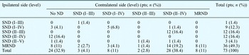
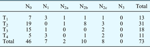
The types of neck dissection performed in these patients are shown in Table I. Ipsilateral neck dissection was performed in all the patients, but 49 (67.1 per cent) patients also had associated contralateral neck dissection. In the ipsilateral neck, clinical nodal staging was N0 in 48 patients and N+ in 25 patients. In those who also underwent contralateral neck dissection, clinical nodal staging was N0 in 43 patients and N+ in six patients. The indications for contralateral neck dissection included transglottic laryngeal carcinoma, tumours crossing the midline to the contralateral side, and suspicious contralateral lymph nodes. There were no patients with distant metastases. The distribution of pathological staging is shown in Table II.
Types of neck dissection

Pts = patients; ND = neck dissection; SND = selective neck dissection; MRND = modified radical neck dissection
Pathological staging
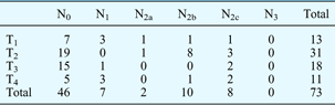
Data represent patient numbers. N = node stage; T = tumour stage
Neck dissection involving level I was performed in 46 (63.1 per cent) patients, of whom 19 (41.3 per cent) had a bilateral neck dissection. The decision to undertake a contralateral neck dissection was based on the localisation and extent of the primary tumour.
However, histological analysis of neck node specimens identified metastasis in only two (4.3 per cent) patients undergoing ipsilateral level I neck dissection. These patients also had metastases in their ipsilateral level IIA nodes. One (5.3 per cent) patient undergoing a bilateral neck dissection had bilateral nodal metastases in levels I and IIA, as well as ipsilateral metastases in level IIB; the ipsilateral side had been clinically N+ and the contralateral side clinically N0. Of these three patients with histopathological neck node metastasis, two had supraglottic carcinoma and one had subglottic carcinoma. These tumours were staged as T1 N2b, T1 N2c and T2 N2b, based on histopathological analysis.
All the patients had level II included in their neck dissection; 49 (67.1 per cent) patients had bilateral neck dissection while 24 (33 per cent) had ipsilateral neck dissection. The neck node specimens of 22 (30 per cent) patients were pathologically N+ in level IIA. These nodal metastases were ipsilateral in 16 (73 per cent) patients, bilateral in five (23 per cent) and contralateral in one (4 per cent). The clinical nodal staging was N0 in one patient and N+ in 21 patients. Only four patients were pathologically N+ in ipsilateral level IIB; their clinical staging was N0 in one patient and N+ in the remaining three patients. Only one of these patients had associated, synchronous, bilateral neck node metastases in level I. These four primary tumours were staged pathologically as T1 N2c, T2 N2b, T2 N2c and T4 N1.
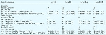
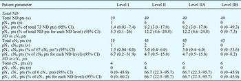
Patients with more than one metastasis underwent post-operative adjuvant chemoradiotherapy. The distribution of cervical lymph node metastases in levels I, IIA and IIB is shown in Tables III and IV.
Distribution of lymph node metastases in ipsilateral levels I, II, IIA and IIB

ND = neck dissection; pts = patients; pN+ = pathologically node-positive; CI = confidence interval; cN0 = clinally node-negative; cN+ = clinically node-positive
Distribution of lymph node metastases in contralateral levels I, II, IIA and IIB

*43 patients + 24 patients who did not undergo contralateral surgery. ND = neck dissection; pts = patients; pN+ = pathologically node-positive; CI = confidence interval; cN0 = clinally node-negative; cN+ = clinically node-positive
Discussion
The dissection of neck levels I and IIB is time-consuming because of their anatomical location. There is an associated risk of injury to adjacent structures, including blood vesselsReference Wenig and Applebaum 9 and the accessory nerve (leading to shoulder dysfunction).Reference Cheng, Hao, Lin and Yeh 10 , Reference Leipzig, Suen, English, Barnes and Hooper 11 The present study findings agree with previous reports that not all clinically node-negative necks harbour occult metastasis, and that performing neck dissection thus increases operation time and morbidity risk unnecessarily in truly node-negative patients. Thus, the issue is the identification of cases in which neck dissection is justified, as opposed to those in which it can be omitted without impairing the oncological outcome.
Numerous studies have demonstrated that lymph node metastases from laryngeal squamous cell carcinoma to level I are infrequent.Reference Candela, Shah, Jaques and Shah 12 – Reference dos Santos, Goncalves, Magrin, Johnson, Ferlito and Kowalski 15 In patients with a clinically N0 neck, only a few cases of occult metastasis to this region have been described. Some studies have found that neck dissection specimens from patients with a clinically N0 neck contain no metastases.Reference Buckley and MacLennan 14 , Reference Byers, Wolf and Ballantyne 16 , Reference Mercante, Bacciu, Oretti and Ferri 17 The present study findings agree with the above-mentioned studies.
In the present study, ipsilateral metastasis to level I was found in only 4.1 per cent of patients, and contralateral metastasis to level I in only 1.4 per cent. Based on the fact that patients with a clinically N0 neck frequently have no level I metastases, several authors have recommended surgical therapy that preserves this region.Reference Wenig and Applebaum 9 , Reference Candela, Shah, Jaques and Shah 12 – Reference Buckley and MacLennan 14 , Reference Byers, Wolf and Ballantyne 16 , Reference Mercante, Bacciu, Oretti and Ferri 17 The findings from the present study support this recommendation.
Some authors have stated that routine performance of neck dissection including level I in laryngeal cancer patients with a clinically N+ neck is unjustified.Reference Li, Wei, Guo, Yuen and Lam 13 , Reference dos Santos, Goncalves, Magrin, Johnson, Ferlito and Kowalski 15 , Reference Mercante, Bacciu, Oretti and Ferri 17 , Reference Ferlito and Rinaldo 18 In the present study, all patients with pathologically proven nodal metastases in level I also had metastases in level IIA. Therefore, dissection of level I is recommended in cases in which ultrasound or CT reveal suspicious lymph nodes in levels I or IIA.
-
• Lymph node metastases greatly affect laryngeal carcinoma prognosis
-
• Level I and IIB dissection is time-consuming and increases costs
-
• Preservation of these levels in clinically node-negative necks is cost-effective and lowers comorbidity, without oncological risk
-
• Patients with clinically apparent nodes in levels I, IIA and IIB should have these levels dissected
The physiological lymphatic drainage of the supraglottic and glottic larynx is mainly to the lymph nodes in levels II and III.Reference Werner, Dünne and Myers 19 The introduction of radiological investigative tools such as ultrasound and CT has helped to improve identification of neck node metastasis. According to the current understanding of the lymphatic drainage pathway, laryngeal carcinomas metastasise firstly to levels II and III before reaching other lymph node regions. However, numerous evaluations of patients with laryngeal carcinoma have shown that metastases are rarely found in level IIB.Reference Chone, Crespo, Rezende, Carvalho and Altemani 20 – Reference Villaret, Piazza, Peretti, Calabrese, Ansarin and Chiesa 27 Metastases in this region are often associated with metastases in sublevel IIA.Reference Santoro, Franchi, Gallo, Burali and de'Campora 28
In the present study, ipsilateral level II nodes were dissected in all 73 patients, and associated contralateral level I nodes in 49 patients. Level IIB metastases were found in 5.5 per cent of ipsilateral dissections and 0 per cent of contralateral dissections, a rather low incidence. In only one patient (2 per cent), with an advanced tumour stage (pathologically T4) but a clinically N0 neck, was a single metastasis found in level IIB. These results support the recommendation to preserve level IIB in patients with a clinically N0 neck.Reference Chone, Crespo, Rezende, Carvalho and Altemani 20 – Reference Lim, Lee, Koo and Choi 26 , Reference Santoro, Franchi, Gallo, Burali and de'Campora 28 This approach seems to be oncologically safe and may prevent patients from developing the frequent complication of shoulder dysfunction, thus reducing post-operative morbidity. In our patients with a clinically N+ neck, all those with metastases in level IIB had additional metastases in level IIA. Therefore, it is debatable whether it is necessary to dissect level IIB in all patients with a clinically N+ neck, or whether it would be preferable to preserve level IIB in those patients with a clinically N+ neck in whom pre-operative staging investigations show no suspicious lymph nodes in levels IIA or IIB.
However, we recognise that not all patients in our study underwent bilateral neck surgery, and a broad spectrum of different types of neck dissection were performed. This may have biased our study findings. As not all patients underwent contralateral neck dissection, and level I dissection was not performed in all patients, the prevalence of metastasis may have been underestimated. Moreover, the 95 per cent confidence intervals calculated for all metastatic rates were often rather wide, because of the low number of cases.
Conclusion
In this study, laryngeal cancer patients with a clinically N+ neck had a higher prevalence of lymph node metastases in level IIA compared with levels I and IIB. Therefore dissection of levels I, IIA and IIB is recommended in patients with clinically apparent lymph nodes in these levels. However, patients with a clinically N0 neck rarely had metastases in levels I and IIB. These findings support previous recommendations for preservation of levels I and IIB in patients with a clinically N0 neck. This approach has the advantages of better post-operative quality of life and greater cost-effectiveness (due to reduced surgical time), without any impairment of oncological outcomes.