Cardiac allograft vasculopathy is a leading cause of mortality among paediatric heart transplant recipients surviving 1 year after transplantation, accounting for 25% of deaths within 10 years after transplant. Reference Dipchand, Edwards and Kucheryavaya1
Coronary angiography is the gold standard diagnostic method for cardiac allograft vasculopathy. Reference Mehra, Crespo-Leiro and Dipchand2 However, coronary angiography, as a luminography that measures lumen diameter, often leads to false-negative diagnoses of cardiac allograft vasculopathy. This is primarily because the lumen diameter is not compromised in early cardiac allograft vasculopathy, and its diffuse pathologic nature makes distinguishing the normal and the suspected diseased segments difficult. Reference Rickenbacher, Pinto and Chenzbraun3–Reference Cai, Rangasetty, Barbageelata, Fujise and Koerner5 Intravascular ultrasonography can be conducted simultaneously with coronary angiography. It is already widely used given its high sensitivity for cardiac allograft vasculopathy. Reference Kuhn, Jutzy and Deming6,Reference Mendiz, Gamboa, Renedo, Lev, Favaloro and Bertolotti7 For diagnosing cardiac allograft vasculopathy using Intravascular ultrasonography 1 year after heart transplantation, a cut-off maximal intimal thickness of >0.3 mm or ≥0.5 mm or a difference between baseline maximal intimal thickness and at 1 year of ≥0.5 mm has been used in adult patients. Reference Cai, Rangasetty, Barbageelata, Fujise and Koerner5,Reference Kobashigawa, Tobis and Starling8,Reference Nelson, Rossing, Ihlemann, Boesgaard, Engstrøm and Gustafsson9 However, the appropriate cut-off level at 1 year after transplant in children remains inconclusive. Recent paediatric studies have reported using a cut-off of intimal thickness >0.3 mm to define cardiac allograft vasculopathy. Reference Zoeller, Miyamoto, Younoszai and Landeck10,Reference Boruta, Miyamoto, Younoszai, Patel and Landeck11
In our practice, several paediatric heart transplant recipients had an maximal intimal thickness of ≥0.5 mm at 1 month after transplantation that did not significantly decrease on follow-up. Several studies have reported the progression of intimal hyperplasia in the first year after heart transplantation. Reference Klauss, Ackermann and Spes12–Reference Li, Tanaka, Oeser, Kobashigawa and Tobis14 Results of studies on whether donor-transmitted atherosclerosis, defined as maximal intimal thickness ≥0.5 mm at baseline Intravascular ultrasonography, is a significant risk factor for the later development of cardiac allograft vasculopathy in adult heart recipients are inconsistent. Reference Wong and Yeung13,Reference Botas, Pinto and Chenzbraun15–Reference Moayedi, Fan and Tremblay-Gravel18
We aimed to investigate the effect of donor-transmitted atherosclerosis on late aggravation of cardiac allograft vasculopathy in paediatric heart recipients aged ≥7 years.
Methods
Patients
All consecutive patients who underwent heart transplantation at the Department of Pediatrics at Asan Medical Center between May 2007 and May 2019 were considered for inclusion in the study. Before heart transplantation, the urgency of the transplant for each patient was determined using the status codes for medical urgency per the Korean Network for Organ Sharing (KONOS). 19 The KONOS grades range from 0 to 3, with 0 implying the highest urgency. We excluded patients without intravascular ultrasonography results at 1 month (baseline) and 1 year after transplantation. Medical records were reviewed for collecting demographic and clinical data.
Patients were managed per the post-heart transplantation management protocol that included immunosuppression with a calcineurin inhibitor, mycophenolate mofetil, and prednisolone. During follow-up, the immunosuppressant was substituted with a mammalian target of rapamycin inhibitor in case of serious adverse drug reactions or if needed to control refractory rejection. Allograft rejection requiring intervention was defined based on the histopathologic evidence of an acute rejection of ≥grade 2 or an unexplainable compromised cardiac systolic function with compatible symptoms. A statin was administered to every patient from the early post-transplant period, with the patient under surveillance for serious adverse effects.
According to the hospital standard management protocol, all patients underwent coronary angiographic examination (for all three major coronary arteries) at 1 month and 1 year after transplantation. For patients aged ≥7 years, an intravascular ultrasonography examination was additionally conducted. Patients with a baseline maximal intimal thickness of ≥0.5 mm were included into the donor-transmitted atherosclerosis (+) group and those with <0.5 mm into the donor-transmitted atherosclerosis (−) group. After 1 year of transplantation, coronary angiography and intravascular ultrasonography examinations were conducted every 0.5–3 years based on the latest results. The angiographic diagnosis of cardiac allograft vasculopathy was made according to the guidelines of the International Society for Heart and Lung Transplantation (ISHLT). Reference Mehra, Crespo-Leiro and Dipchand2 Late aggravation of cardiac allograft vasculopathy was defined as new angiographic diagnosis of cardiac allograft vasculopathy or an aggravation of luminal stenosis from the baseline on angiographic examination performed at 1 year or later.
This retrospective study was approved by the Institutional Review Board of Asan Medical Center (2020–1922), and the requirement for informed patient consent was waived.
Intravascular ultrasonography procedure and analysis
After selection of the left main coronary artery, 200 µg of nitroglycerine was administered. Intravascular ultrasonography catheters were advanced over a guidewire to the distal segment of the left anterior descending coronary artery. Grayscale intravascular ultrasonography imaging was performed using a commercial scanner (Boston Scientific Scimed, Inc., Minneapolis, Minnesota, United States) using motorised transducer pullback at 0.5 mm/s. Intravascular ultrasonography images were stored on a computer disc for offline analysis later.
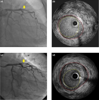
Using computerised planimetry (EchoPlaque 3.0; Indec Systems, Mountain View, California), offline qualitative and quantitative intravascular ultrasonography analyses were performed according to the standard methods in the core lab of the CardioVascular Research Foundation (Seoul, Korea). Reference Mintz, Nissen and Anderson20 Maximal intimal thickness, the lumen area, and the external elastic membrane (EEM) area at the minimal lumen area site were measured (Fig 1). The percentage intimal index was calculated as (EEM area − lumen area)/EEM area × 100.
Angiographic and intravascular ultrasonography (IVUS) examinations of a 17.6-year-old male recipient. ( a ) Baseline angiographic examination of the left coronary arteries showed no definite stenosis. The yellow-colored arrow indicates the examination site for IVUS. ( b ) The external elastic membrane (red circle) and lumen area (yellow circle) were measured. Maximal intimal thickness (MIT) was 1.01 mm; hence, this patient was classified into the group with donor-transmitted atherosclerosis. ( c ) At 1 year after transplantation, angiographic examination showed a definite stenosis of the mid-segment of the left anterior descending coronary artery. ( d ) MIT was measured at 1.78 mm at the same site as in the baseline study.

Statistical analysis
Categorical data are presented as frequencies with percentages, and continuous data are presented as means ± standard deviations. The chi-square test and unpaired t-test were used to compare data between the groups. The paired t-test was used to compare data between the two time points. Univariate and multivariate logistic regression analyses were performed to identify risk factors for donor-transmitted atherosclerosis. Rates of survival free from late aggravation of cardiac allograft vasculopathy in each group were estimated using the Kaplan–Meier method and compared using the log-rank test.
The effect of the results of intravascular ultrasonography at 1 year after transplantation on the late aggravation of cardiac allograft vasculopathy, correcting for possible covariates including donor-transmitted atherosclerosis, was examined using the Cox proportional hazards model. Of the results of intravascular ultrasonography at 1 year after transplantation, maximal intimal thickness and the difference between the baseline maximal intimal thickness and that after 1 year were selected, and two multivariate models including one each were constructed. Adjusted covariates were selected by backward elimination using variables with p < 0.05 on the univariate analysis.
Statistical analyses were conducted using SPSS version 21 (IBM Co., Armonk, NY). Statistical significance was defined as a p < 0.05.
Results
Demographic and peri-transplant variables
During the study period, 77 heart transplants were performed. Seven patients died before 1 year from the transplantation. Forty-eight patients with intravascular ultrasonography results at the two time points were included in the study. Among them, 23 (47.9%) had donor-transmitted atherosclerosis at the baseline. Most of the other 22 excluded patients were aged <7 years at transplantation. In three patients who aged >7 years at transplantation, part of stored images could not be analysed due to a failure of reading the recorded disc. Mean follow-up duration of the 22 excluded patients was 6.16 ± 4.40 years. After 1 year following transplantation, three of them died from the graft rejection (two) or the late aggravation of cardiac allograft vasculopathy (one). In three patients including one died patient, the late aggravation of cardiac allograft vasculopathy was observed.
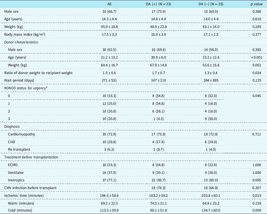
Demographic and patient characteristics from the peri-transplant period are summarised in Table 1. Patients with donor-transmitted atherosclerosis had a greater mean donor age and body weight (39.9 years versus 23.2 years (p < 0.001) and 67.9 kg versus 53.6 kg (p = 0.002), respectively). Patients who ended up with donor-transmitted atherosclerosis had higher average KONOS grades (p = 0.045) and higher rates of inotropic use than those who received heart transplant with clean coronary arteries (95.7% versus 60.0%, p = 0.005) before transplantation. The total (163.2 minutes versus 203.8 minutes, p = 0.015) and cold ischaemic (90.1 minutes versus 134.7 minutes, p = 0.008) times were shorter among patients with donor-transmitted atherosclerosis.
Demographic and clinical characteristics of 48 patients during the peri-transplant period

CMV, = cytomegalovirus, DA = donor-transmitted atherosclerosis, ECMO = extracorporeal membrane oxygenator, KONOS = Korea Network for Organ Sharing.
Data are means ± standard deviation or frequency (percentages).
* A smaller KONOS grade indicated higher transplantation urgency.
Angiographic and intravascular ultrasonography data and prediction of donor-transmitted atherosclerosis
Coronary angiography showed luminal irregularity in 3/48 (6.3%) patients at baseline; all three were also diagnosed with donor-transmitted atherosclerosis by concurrent intravascular ultrasonography. Nine (5 in grade 1, 3 in grade 2, and 1 in grade 3 per the ISHLT guidelines Reference Mehra, Crespo-Leiro and Dipchand2 ) out of 23 patients with donor-transmitted atherosclerosis and only 1 (grade 1) out of 25 patients without donor-transmitted atherosclerosis, progressed to a diagnosis of cardiac allograft vasculopathy established by angiography 1 year after transplantation.
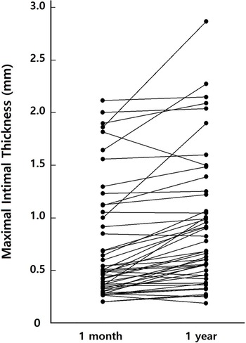
Intravascular ultrasonography at 1 year post-transplant revealed an increase in maximal intimal thickness compared to baseline for all recipients (p < 0.001), as well as in subgroups with donor-transmitted atherosclerosis (p = 0.003) and without (p = 0.005) (Table 2; Fig 2). And a similar pattern was seen for the intimal index in all patients (p < 0.001), in patients with donor-transmitted atherosclerosis (p < 0.001) and without (p = 0.008). Both maximal intimal thickness and intimal index were greater in patients with donor-transmitted atherosclerosis, compared to those without donor-transmitted atherosclerosis, at baseline (p < 0.001 both) and 1 year after transplantation (p < 0.001 both). In those with donor-transmitted atherosclerosis, the luminal area was decreased 1 year after transplantation compared to baseline (p = 0.003), whereas the luminal area did not significantly change in patients without donor-transmitted atherosclerosis (p = 0.725). The EEM area was greater in patients with donor-transmitted atherosclerosis than in those without at both time points (baseline, p < 0.001; 1 year after transplant, p = 0.048).
Changes in maximal intimal thickness (MIT) from baseline to 1 year after transplantation. MIT had increased in 39 patients. In nine patients with reduced MIT at 1 year, the reduction was relatively small (0.03–0.06 mm).

Intravascular ultrasound examination results at baseline and at 1 year after transplantation

DA = donor-transmitted atherosclerosis, EEM = external elastic membrane, MIT = maximal intimal thickness.
Data are means ± standard deviation.
* P < 0.05 on comparing the two examination periods.
** P < 0.05 on comparing the two groups.
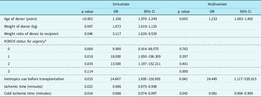
Univariate logistic regression revealed donor age (p < 0.001), donor weight (p = 0.007), the donor weight to recipient weight ratio (p = 0.046), KONOS grades 1 (p = 0.018) and 2 (p = 0.035), inotropic use before transplantation (p = 0.015), total ischaemic time (p = 0.022), and cold ischaemic time (p = 0.014) as significant predictors for donor-transmitted atherosclerosis (Table 3). Donor age (odds ratio, 1.232; 95% confidence interval, 1.083–1.402; p = 0.002), use of inotropics before transplantation (odds ratio, 24.445; 95% confidence interval, 1.117–535.015; p = 0.042), and cold ischaemic time (odds ratio, 0.981; 95% confidence interval, 0.964–0.999; p = 0.042) were also significant predictors for donor-transmitted atherosclerosis per the multivariate analysis.
Results of logistic regression analysis for prediction of donor-transmitted coronary atherosclerosis with demographic and peri-operative characteristics

CI = confidence interval, KONOS = Korean Network for Organ Sharing.
* A smaller KONOS grade indicates a greater urgency for transplantation.
Analyses of survival free from late aggravation of cardiac allograft vasculopathy
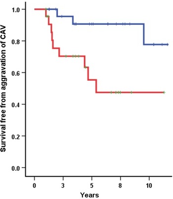
The mean follow-up duration after transplantation was 5.97 ± 3.58 years (with donor-transmitted atherosclerosis, 5.44 ± 3.39 years; without donor-transmitted atherosclerosis, 6.47 ± 3.75 years; p = 0.327). Thirteen subjects (11 [47.8%] with donor-transmitted atherosclerosis and 2 [8.0%] without donor-transmitted atherosclerosis) experienced late aggravation of cardiac allograft vasculopathy. Figure 3 presents the late cardiac allograft vasculopathy aggravation-free survival in each group. The log-rank test showed patients with donor-transmitted atherosclerosis had worse survival free from aggravation of cardiac allograft vasculopathy than those without (p = 0.008).
Kaplan–Meier survival free from the aggravation of cardiac allograft vasculopathy, conditional on survival beyond 1 year after transplant. The log-rank test showed that the survival of patients with donor-transmitted atherosclerosis (in red) was worse than that of patients without (in blue) (p = 0.008).

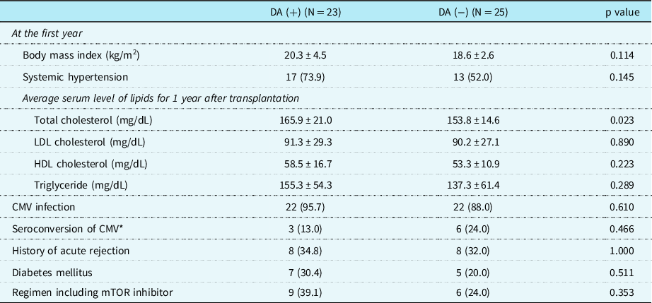
Late clinical characteristics of patients in each group are summarised in Table 4. The average serum total cholesterol level in the first year after transplantation was higher in patients with donor-transmitted atherosclerosis than in those without (165.9 versus 153.8 mg/dL; p = 0.023). Other characteristics did not differ between groups. The number of patients experienced acute rejection did not differ between groups (p = 1.000). The number of rejection episodes was 1–3 times, and allograft rejection based on clinical judgement or histologically proven acute cellular or antibody-mediated rejection occurred separately in several patients.
Late clinical characteristics of patients after the transplantation

CMV, cytomegalovirus; DA, donor-transmitted atherosclerosis; HDL, high density lipoprotein; LDL, low density lipoprotein; mTOR, mammalian target of rapamycin.
Data are means ± standard deviation or frequency (percentages).
* Change of CMV infection status from negative.
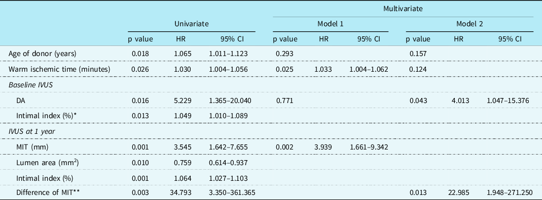
Per the univariate Cox proportional hazards model, donor age (p = 0.018), warm ischaemic time (p = 0.026), donor-transmitted atherosclerosis (p = 0.016), and the intimal index (p = 0.013) at the baseline and maximal intimal thickness (p = 0.001), lumen area (p = 0.010), and intimal index (p = 0.001) at 1 year after transplantation were significant variables predicting cardiac allograft vasculopathy along with the difference between baseline maximal intimal thickness and at 1 year after transplantation (p = 0.003; Table 5). The total cholesterol level was not a significant variable (p = 0.408). Per the first multivariate model including maximal intimal thickness at 1 year (hazard ratio, 3.939; 95% CI, 1.661–9.342; p = 0.002), warm ischaemic time (hazard ratio, 1.033; 95% CI, 1.004–1.062; p = 0.025) was a significant covariate, and donor-transmitted atherosclerosis was not significant (p = 0.771). However, per the second multivariate model including the difference in maximal intimal thickness (hazard ratio, 22.985; 95% CI, 1.948–271.250; p = 0.013), donor-transmitted atherosclerosis (HR, 4.013; 95% CI, 1.047–15.376; p = 0.043) was a significant covariate.
Results of the Cox proportional hazards model analysis for prediction of late aggravation of cardiac allograft vasculopathy after 1 year from heart transplantation

CI, confidence interval; DA, donor-transmitted atherosclerosis; HR, hazard ratio; IVUS, intravascular ultrasound; MIT, maximal intimal thickness.
* Intimal index = (total area – lumen area)/total area.
** Difference of MIT = (MIT at 1 year) – baseline MIT.
Discussion
To our knowledge, this is the first study on the effect of donor-transmitted atherosclerosis on the late aggravation of cardiac allograft vasculopathy in children. Our results showed that patients with donor-transmitted atherosclerosis had worse late cardiac allograft vasculopathy aggravation-free survival than patients without.
The mean maximal intimal thickness of 0.71 mm at baseline implies that it would be inappropriate to use a cut-off level of 0.3–0.5 mm for cardiac allograft vasculopathy diagnosis in children considering that intimal hyperplasia progresses during the first year after heart transplantation. Reference Wong and Yeung13,Reference Li, Tanaka, Oeser, Kobashigawa and Tobis14 Among the total 48 total patients, 47.9% had maximal intimal thickness ≥0.5 mm. This incidence of donor-transmitted atherosclerosis is similar to that reported in previous studies including adult recipients (48.5–51.9%), Reference Tuzcu, Kapadia and Tutar21,Reference Kim, Kang and Lee22 and the mean donor age was 31.2 years in this study, in line with that reported in those adult studies (31.4–33.4 years). Reference Tuzcu, Kapadia and Tutar21,Reference Kim, Kang and Lee22 Donor age was a significant predictor of donor-transmitted atherosclerosis. Similar to this study, previous studies including adult recipients have also reported donor age as a significant predictor of increased maximal intimal thickness in the early post-transplant period. Reference Tuzcu, Kapadia and Tutar21–Reference St Goar, Pinto and Alderman23 Per this study, other significant predictors of donor-transmitted atherosclerosis were the use of inotropics before transplantation and a shorter cold ischaemic time. Patients with donor-transmitted atherosclerosis likely required a heart transplant more urgently and hence the heart of an aged donor was used. Comparison of groups by donor-transmitted atherosclerosis status revealed that a greater number of patients with donor-transmitted atherosclerosis required the use inotropics and had a relatively urgent KONOS status, although the urgency status was not a significant predictor per the multivariate analysis. Shorter cold ischaemic time in patients with donor-transmitted atherosclerosis implies a shorter preparation process for transplantation because of the urgent status of the recipients.
Intravascular ultrasonography examination at 1 year after transplantation is frequently used for cardiac allograft vasculopathy diagnosis or in predicting late aggravation of cardiac allograft vasculopathy in the current clinical practice. Reference Cai, Rangasetty, Barbageelata, Fujise and Koerner5,Reference Kobashigawa, Tobis and Starling8,Reference Nelson, Rossing, Ihlemann, Boesgaard, Engstrøm and Gustafsson9 We hypothesised that the most significant predictor of the late aggravation of cardiac allograft vasculopathy would be a variable among the results of intravascular ultrasonography examination at 1 year after transplantation. Therefore, we constructed a multivariate Cox proportional hazards models to examine whether donor-transmitted atherosclerosis is a significant covariate. Several demographic and clinical variables have been reported to be risk factors for cardiac allograft vasculopathy Reference Moayedi, Fan and Tremblay-Gravel18,Reference Lázaro, Bonet and López24,Reference Jeewa, Dreyer, Kearney and Denfield25 – recipient age, acute rejection, cytomegalovirus infection, use of an mammalian target of rapamycin inhibitor, and serum lipid levels were hence assessed as covariates. In addition, the traditional risk factors for coronary atherosclerosis, body mass index, systemic hypertension, and diabetes mellitus, were also assessed. To avoid collinearity, we select a representative variable among the intravascular ultrasonography results at 1 year after transplant. Hence, two multivariate models were constructed, one including maximal intimal thickness at 1 year and the other, the difference in maximal intimal thickness at the two time points. Per the first Cox multivariate model including maximal intimal thickness at 1 year, donor-transmitted atherosclerosis was not a significant covariate. However, per the second Cox multivariate model including the difference in maximal intimal thickness, donor-transmitted atherosclerosis was a significant covariate. These results could be possibly explained by intimal hyperplasia in the baseline examination being significantly contained within maximal intimal thickness at 1 year and being independent of later aggravation of intimal thickening. Several recent studies including adult heart transplant recipients have shown that donor-transmitted atherosclerosis is a significant risk factor for the development of cardiac allograft vasculopathy in the first year after transplantation. Reference Wong and Yeung13,Reference Yamasaki, Sakurai and Hirohata17,Reference Moayedi, Fan and Tremblay-Gravel18 The findings of this study are in line with those observations. In addition, this study seems to be meaningful in that the observation period was extended by more than 1 year. The mechanism for the late aggravation of cardiac allograft vasculopathy by donor-transmitted atherosclerosis is uncertain. In a study of adult recipients, authors suggested that more endothelial dysfunction, altered shear stress, and local insult in certain sites with donor-transmitted atherosclerosis may accelerate plaque progression. Reference Yamasaki, Sakurai and Hirohata17 We think that their assumptive suggestion is useful for an explanation of results in this study. The prediction of the plaque progression at 1-year post-transplant by early endothelial dysfunction was reported by another authors. Reference Davis, Yeung and Meredith26 However, the mechanism for the late aggravation of cardiac allograft vasculopathy by donor-transmitted atherosclerosis in this study cannot be definitively explained. Just additive coronary arterial intimal growth without endothelial dysfunction could have been a causative factor for the later aggravation of cardiac allograft vasculopathy in patients with donor-transmitted atherosclerosis.
Limitations
This retrospective, single-institution study has several limitations. First, the sample size may have been insufficient to conclusively determine the predictive ability of diverse clinical variables including traditional risk factors for atherosclerosis in logistic regression analyses. Donor age which was a predictor of donor-transmitted atherosclerosis was not predictive of late aggravation of cardiac allograft vasculopathy in this study. However, a future study including more number of children could determine whether donor age is an independent predictor of late aggravation of cardiac allograft vasculopathy. Second, data on donors were limited. Only demographic data were available, and additional information such as smoking history was not available. Third, only the left main coronary artery and the left anterior descending coronary artery were evaluated with intravascular ultrasonography and the other major branches – the right coronary artery and the left circumflex coronary artery were not evaluated. However, left coronary arteries are often selected for screening of cardiac allograft vasculopathy on intravascular ultrasonography examination in current practice. According to a report by Floré V, et al, intravascular ultrasonography of the left anterior descending coronary artery is sufficient to detect early cardiac allograft vasculopathy. Reference Floré, Brown and Pettit27 Fourth, the difference in maximal intimal thickness was used to represent the change in intravascular ultrasonography results between the two time points, which might not have been the best variable representing the progression of coronary vasculopathy. Given that the pathologic mechanism of cardiac allograft vasculopathy includes diffuse exacerbation of coronary arteries, the use of three-dimensional intravascular ultrasonography may have been more appropriate. Fifth, this study included only paediatric recipients aged ≥7 years at transplantation owing to body size restrictions for intravascular ultrasonography examination. Therefore, the results may not be generalisable to children of all age ranges. Future studies including a larger number of children and considering the aforementioned limitations are required.
Conclusions
Paediatric recipients aged ≥7 years at heart transplantation with donor-transmitted atherosclerosis have worse survival free from the late aggravation of cardiac allograft vasculopathy. Donor-transmitted atherosclerosis is also a significant covariate when predicting late cardiac allograft vasculopathy aggravation along with the difference between baseline maximal intimal thickness and that at 1 year after transplantation.
Acknowledgements
None.
Financial support
This research received no specific grant from any funding agency, commercial, or not-for-profit sectors.
Conflicts of interest
The authors have no conflicts of interest to disclose.
Ethical standards
All study procedures complied with the ethical standards of the Helsinki Declaration.
Authorship
Mi Jin Kim involved in data collection, drafting, and approval of the manuscript. Jeong Jin Yu involved in conception and study design, data collection and analysis, and revision and final approval of the manuscript. Seulgi Cha involved in data interpretation and approval of the manuscript. Jae Suk Baek involved in data interpretation and critical revision and approval of the manuscript. Eun Seok Choi involved in critical revision and approval of the manuscript. Bo Sang Kwon involved in data interpretation and approval of the manuscript. Chun Soo Park involved in critical revision and approval of the manuscript. Tae-Jin Yun involved in data interpretation and approval of the manuscript. Young-Hwue Kim involved in conception and study design, critical revision, and approval of the final manuscript.