Introduction
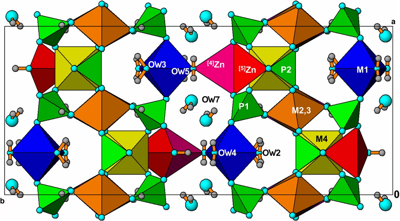
The schoonerite mineral group was established recently (Grey et al., Reference Grey, Keck, Kampf, MacRae and Cashion2017a, Reference Grey, Kampf, Keck, MacRae, Cashion and Gozukara2018), with the general formula [4](Zn,Fe)x[5](Zn,Fe)1–x[6](M1M2M31–x□xFe3+)(PO4)3(OH)y(H2O)9–y·2H2O, where □ = a vacancy and the schoonerite-group minerals (SGM) are distinguished by the dominant cations in the octahedral sites M1 to M3. The value of y is determined by the valence state of Fe and the vacancy concentration; y = 2 for schoonerite, which contains only divalent cations, and y increases above 2 with increasing Fe3+ in oxidised SGM. Schoonerite is reported to have M1 = Mn2+, M2 = M3 = Fe2+ (Kampf, Reference Kampf1977), and x = 0. The first oxidised SGM to be described was wilhelmgümbelite, from the Hagendorf-Süd pegmatite, Hagendorf, Bavaria (Grey et al., Reference Grey, Keck, Kampf, MacRae, Glenn and Price2017b), for which the dominant cations of dominant valency in the M1 to M3 sites are M1 = M2 = Fe3+ and M3 = Fe2+. The structure of wilhelmgümbelite differs from that of schoonerite by having partial disorder of the 5-coordinated Zn site, [5]Zn in schoonerite, so that the Zn is distributed between the [5]Zn site and an adjacent vacant tetrahedral site, [4]Zn, with the two sites separated by ~1.0 Å. The location of the split Zn sites are shown in Fig. 1. The value of x in wilhelmgümbelite is 0.3 (30% of Zn partitioned from [5]Zn to [4]Zn). Wilhelmgümbelite has a matching deficiency in the M3 site of 30%, in contrast to schoonerite that does not have split Zn sites or metal-atom vacancies.
Crystal structure of oxidised schoonerite-group minerals, viewed along [001].

In our ongoing studies of secondary phosphate minerals from Hagendorf-Süd, we have identified and characterised two more oxidised schoonerite-related minerals, schmidite (IMA2017-012, Grey et al., Reference Grey, Keck, Kampf, MacRae, Glenn, Cashion and Gozukara2017c) and wildenauerite (IMA2017-058, Grey et al., Reference Grey, Keck, Kampf, MacRae, Cashion, Glenn, Davidson and Gozukara2017d) that share the same structural features displayed by wilhelmgümbelite, of partitioning of the Zn between [5]Zn and [4]Zn sites, and partial occupancy of the M3 site. They differ, however, in the dominant cations in M1 to M3.
Schmidite is named for Dr. Hans Schmid, born in 1925 in Wackersdorf in the Oberpfalz region, Bavaria, died in 2013 in Greece, and is buried in Athens. He studied mining engineering and geology at the University of Clausthal, Germany. While working on his doctoral dissertation he discovered a very large lithium phosphate (triphylite) occurrence in the Hagendorf-Süd pegmatite. This was commercially mined during 1950 to 1955, producing ~1600 tons for production of Li products. In 1955 he published a seminal paper on the textures and structures of pegmatites of the Oberpfälzer, titled “Verbandsverhältnisse der Pegmatite des Oberpfälzer und Bayerischen Waldes (Hagendorf-Pleystein-Hühnerkobel)”, published in Neues Jahrbuch für Mineralogie – Abhandlungen (Schmid, Reference Schmid1955). Wildenauerite comes from the old name for the Hagendorf-Süd quarry = Wildenauer-Grube, at the mine where the mineral was found. The quarry name is in honour of the mining family, Wildenauer, and their long-time activities in feldspar mining at the Hagendorf Süd pegmatite. Two co-type specimens were used for all measurements on schmidite (IGC-1 and IGC-31). They are housed in the mineralogical collections at Museums Victoria, registration numbers M53810 and M53811. The holotype specimen of wildenauerite (IGC-14) is housed in the mineralogical collections at Museums Victoria, registration number M53979.
Occurrence, mineral assemblage and paragenesis
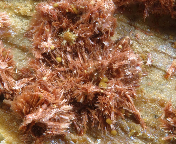
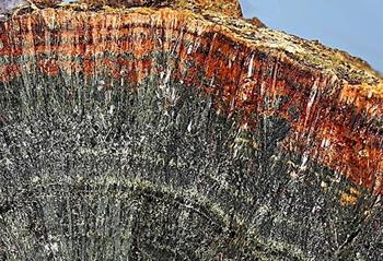
The two new secondary phosphates were found in specimens collected by one of the authors (EK) from the Hagendorf-Süd pegmatite mine, Hagendorf, a branch of Amberger Kaolinwerke (AKW), Hirschau, Oberpfalz, Bavaria, Germany (49°39′1″N, 12°27′35″E). Schmidite, in close association with altered phosphophyllite, was the most common secondary phosphate assemblage in a heavily corroded triphylite nodule, located at 67 m from the surface of the mine. The triphylite in the nodule had been replaced by phosphophyllite and vivianite, and these minerals, together with apatite, had been further altered to a rich spectrum of secondary phosphate minerals including laueite, whitmoreite, jahnsite-subgroup minerals, mitridatite, scholzite–parascholzite, stewartite, zincostrunzite, hopeite, parahopeite and schoonerite-group minerals. Schmidite from the 67 m nodule (specimen IGC-1), is shown in Fig. 2. Schmidite was also found in fissures in phosphophyllite, collected at depths between 60 and 67 m (specimen IGC-31). Other closely associated minerals in specimen IGC-31 are vivianite and decomposed, white chalky zwieselite. Wildenauerite has a quite different occurrence, mineral assemblage and likely paragenesis. It occurs as dense narrow bands of laminated crystals, forming terminations of radial growths of rockbridgeite, shown in Fig. 3. The only other mineral in contact with wildenauerite is jahnsite-(CaMnFe). Minerals found nearby include strunzite, whitmoreite/earlshannonite, laueite and recrystallised mats of acicular crystals of rockbridgeite.
Red schmidite crystals on a substrate of yellowish, microcrystalline, altered phosphophyllite. Specimen IGC-1, Museums Victoria, registration number M53810. Field of view = 2 mm (photo by Christian Rewitzer).

Red wildenauerite terminating radial green Zn-bearing rockbridgeite. Field of view = 3 cm (photo by Volker Betz).

Mücke (Reference Mücke1981) has published tables classifying the different parageneses of the phosphate minerals at Hagendorf. The association of schmidite with phosphophyllite places it in Mücke's Classification Table IC (rockbridgeite-free subparagenesis). Mücke made a distinction between two types of red oxidised schoonerite-group minerals; those that have formed from oxidation of original unoxidised green schoonerite (dull, matt surfaces of crystals, washed-out colour) and those freshly crystallised from solution (smooth surfaces of crystals and copper-red colour). The specimens IGC-1 and IGC-31 correspond to these two categories, respectively.
Wildenauerite's association with rockbridgeite locates it in Mücke's (Reference Mücke1981) classification Table II (rockbridgeite subparageneses), under paragenesis IID1. Commonly, schoonerite-group minerals associated with rockbridgeite at Hagendorf-Süd are of a green colour due to mixed Fe2+/Fe3+. Wildenauerite is an oxidised orange–red mineral with the iron predominantly as Fe3+. It is likely that wildenauerite originally formed as the green mixed-valence species and was subsequently oxidised.
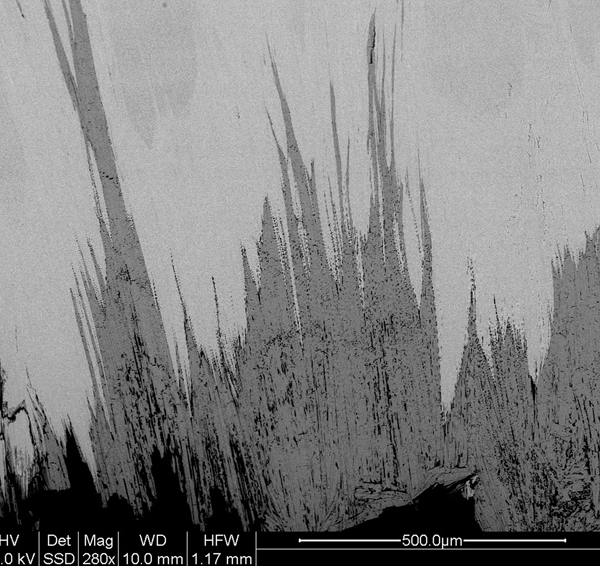
Sectioned wildenauerite/rockbridgeite occurrences show ‘fingers’ of porous wildenauerite penetrating into the rockbridgeite from the surface, shown in Fig. 4. The assemblage has the appearance of a fluid-mediated mineral replacement reaction (coupled dissolution and precipitation), which must occur at low temperature (≤100°C) in order for the highly hydrated wildenauerite to be stable. The solution-mobilised zinc required for the formation of wildenauerite at Hagendorf-Süd comes from altered sphalerite.
Back-scattered electron image of fingers of wildenauerite (dark grey) penetrating rockbridgeite (light grey).

Physical properties
Schmidite occurs as aggregates of orange–brown to red laths on the surface of altered phosphophyllite in the corroded triphylite nodule as shown in Fig. 2 for specimen IGC-1. The lath dimensions are typically 0.1 to 0.5 mm long but only a few μm thick. Schmidite also forms sprays of shiny copper-red, needle-like rectangular laths in fissures in altered phosphophyllite in specimen IGC31. The schmidite laths are elongated on [100] and flattened on {010}. They are brittle and have perfect cleavage parallel to {010}. The crystals remain suspended in bromoform (density = 2.89 g·cm–3) and have a calculated density of 2.82 g·cm–3, based on the empirical formula for co-type IGC-31.
Wildenauerite occurs as bands of compacted platelets which form terminations of radial Zn-bearing rockbridgeite as shown in Fig. 3. The bands are typically 1 to 5 mm wide. The crystals are oriented along the radial direction so that the 11 Å axis of wildenauerite (the long dimension of ruler-shaped laths) is parallel to the 5.2 Å axis of rockbridgeite. Separated platelets are orange to red brown in colour and have a pearly lustre. Cleavage is perfect parallel to {010}. The density measured by flotation in a mixture of methylene iodide and toluene is 2.79(1) g·cm–3. The calculated density based on the empirical formula and the room temperature unit cell is 2.76 g·cm–3.
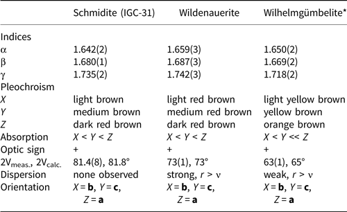
The optical properties of schmidite and wildernauerite, together with wilhelmgümbelite from Grey et al. (Reference Grey, Keck, Kampf, MacRae, Glenn and Price2017b), are given in Table 1. The indices of refraction were measured in white light. 2V was measured using extinction data analysed with EXCALIBR (Gunter et al., Reference Gunter, Bandli, Bloss, Evans, Su and Weaver2004). The Gladstone–Dale compatibility index (Mandarino, Reference Mandarino1981) is 0.016 (superior) for schmidite based on the optical data, empirical formula and density calculated from the powder X-ray diffraction room-temperature unit-cell parameters. For wildenauerite the compatibility index is 0.007 (superior) based on the empirical formula and the measured density.
Optical properties of schmidite, wildenauerite and wilhelmgümbelite.

*from Grey et al. (Reference Grey, Keck, Kampf, MacRae, Glenn and Price2017b)
Mössbauer spectroscopy
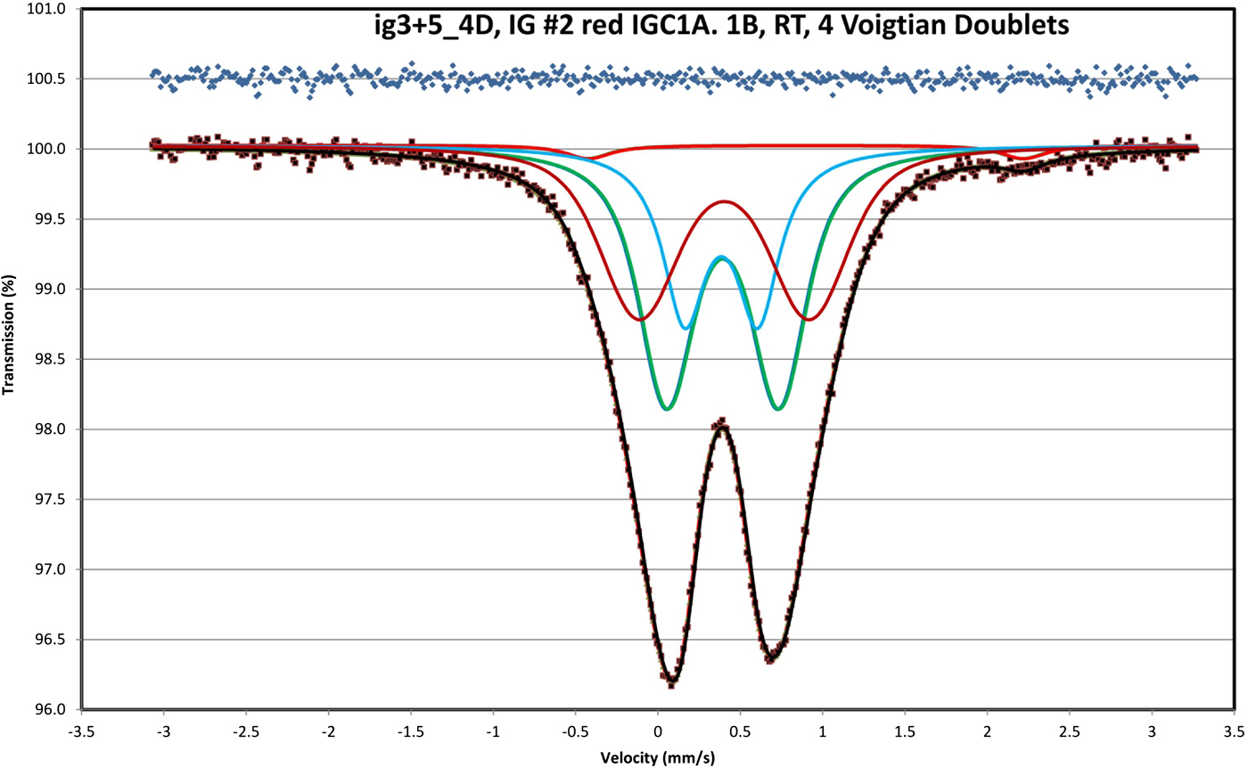
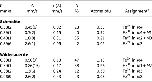
The Mössbauer spectra were taken using a conventional constant acceleration drive with a symmetrical sawtooth waveform (Wissel). The source of 57Co in rhodium was maintained at room temperature. The spectra were fitted using four Voigtian doublets (three for Fe3+ and one for Fe2+) comprising a Lorentzian half width at half maximum (HWHM) component of typically 0.14 mm/s and a variable Gaussian component to deal with line broadening. The results for isomer shifts δ, quadrupole splitting Δ, Gaussian component σ(Δ) and doublet relative areas are reported in Table 2. The isomer shifts are given relative to α-Fe at room temperature. The fitted spectrum for schmidite, specimen IGC-1, is shown in Fig. 5. Tentative site assignments are given in Table 1. The fitting of a number of nested doublets corresponding to different metal-atom sites, to the single broad ferric envelope as shown in Fig. 5, presents ambiguities as evidenced by the large standard deviations associated with the quadrupole splittings in Table 2. The resulting site assignments given in Table 2 are thus tentative. The most useful information obtained from the Mössbauer spectra, in relation to the crystal structure analysis, is the relative amounts of ferrous and ferric iron. This information can be used to constrain the atom valencies in the analysis of metal-atom site occupations using refined bond distances (bond-valency analysis). The ferric contents as a percentage of total Fe from the fitting of the Mössbauer spectra are 98% Fe3+ in schmidite and 97% Fe3+ in wildenauerite.
Fitted Mössbauer spectrum for schmidite (specimen IGC-1).

Mössbauer parameters (mm/s) for schmidite and wildenauerite: δ, isomer shift; Δ, quadrupole splitting; σ(Δ), Gaussian component; A, area of doublet.

*Assignments based on increasing octahedral distortion in order M4 < M1, M2 < M3.
Thermal analysis
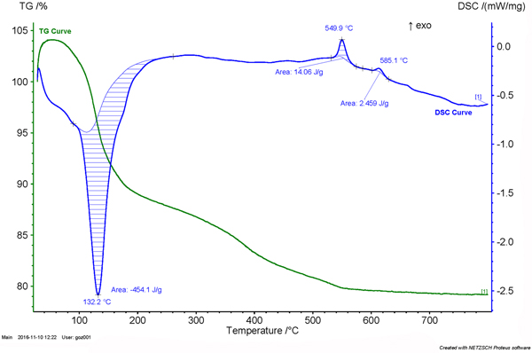
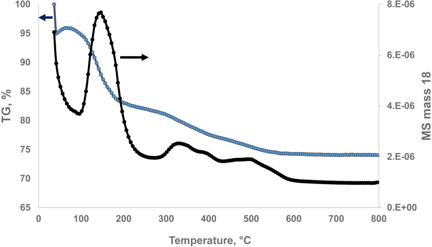
Thermogravimetric analysis was performed on schmidite (11 mg of specimen IGC-1) and wildenauerite (14 mg of specimen IGC-14) using a Netzsch STA 449 F1 Jupiter Simultaneous TGA/DSC thermal analyser. The analysis was performed in argon, with a ramp rate of 10°C/min, between 30 and 800°C. Evolved gas analysis was made using a coupled Thermostar Pfeiffer mass spectrometer (MS). The TGA and DSC curves for schmidite are shown in Fig. 6. The TGA curve and the MS curve for evolved H2O for wildenauerite are shown in Fig. 7. A strong endotherm centred at 132°C for schmidite (at 142°C for wildenauerite) corresponds to the main loss of water from the sample, as confirmed by the MS results. A shoulder at 60–90°C probably represents loss of the weakly bound interlayer water molecule, OW7. A very broad, shallow endotherm extending from 250 to 500°C corresponds to loss of hydroxyl ions and residual strongly held coordinated water molecules. The MS curve in Fig. 7 shows loss of H2O is complete by 600°C. An exotherm at 550°C indicates recrystallisation. The initial part of the TGA curve was affected by buoyancy for both samples, so the mass loss was determined from the initial and final (after cooling back to room temperature) microbalance readings, giving a total loss of 23.3 wt.% H2O for schmidite and 24.5 wt.% H2O for wildenauerite.
Thermogravimetric (green) and differential scanning calorimetry (blue) curves for schmidite (specimen IGC-1).

Thermogravimetric and mass spectrometry (H2O) curves for wildenauerite.

Chemical composition
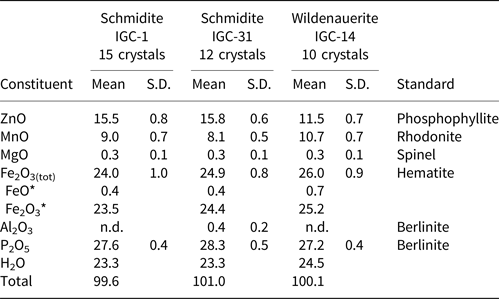
Electron microprobe (EMP) analyses were conducted using wavelength-dispersive spectrometry on a JEOL JXA 8500F Hyperprobe operated at an accelerating voltage of 15 kV and a beam current of 4 nA. The beam was defocused to 2 μm or 5 μm, depending on the size of the crystals. Water was determined by thermal analysis and the FeO/Fe2O3 ratio was obtained from the Mössbauer spectra for specimens IGC-1 and IGC-14. The schmidite specimen IGC-31 was assumed to have the same FeO content as for IGC-1. The results are given in Table 3.
Representative compositions (wt.%) for schmidite and wildenauerite.

* FeO from Mossbauer spectra for IGC-1 and IGC14. FeO for IGC31 assumed to be same as for IGC-1.
S.D. – standard deviation; n.d. - not detected.
The empirical formulae, scaled to 3 P with OH– adjusted for charge balance are: Schmidite (IGC-1): Zn1.47Mn2+0.98Mg0.05Fe2+0.04Fe3+2.27(PO4)3(OH)2.89(H2O)8.54, Schmidite (IGC-31): Zn1.46Mn2+0.86Mg0.05Fe2+0.04Fe3+2.30Al0.06(PO4)3(OH)2.95(H2O)8.26 and Wildenauerite (IGC-14): Zn1.11Mn2+1.18Mg0.05Fe2+0.08Fe3+2.47(PO4)3(OH)3.25(H2O)9.03. According to the crystal structure for schoonerite (Kampf, Reference Kampf1977), the number of OH + H2O per formula unit (pfu) is 11. The empirical formulae all have > 11 (OH + H2O), with the largest discrepancy for wildenauerite. This is most likely due to the presence of some non-structural, adsorbed water on the very thin crystals. The empirical formulae also consistently have less than the theoretical five metal atoms reported for schoonerite (Kampf, Reference Kampf1977), ranging from 4.77 M in specimen IGC-31 to 4.89 M for wildenauerite.
As SGM are considered to derive from the Li-containing primary phosphate mineral, triphylite (Moore and Kampf, Reference Moore and Kampf1977), the possibility exists that the apparent metal-atom deficiency may be explained by Li incorporation. To check this we conducted laser induced breakdown spectroscopy (LIBS) on the specimens. Results from LIBS analyses, however, gave only ~0.01 wt.% Li. This is an order-of-magnitude too low to explain the metal-atom deficiency. As will be discussed in the following section, the deficiency is confirmed from the structure analysis to be due to metal-atom vacancies at the M3 site.
Crystallography
Powder X-ray diffraction
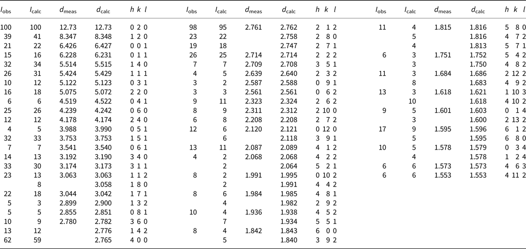
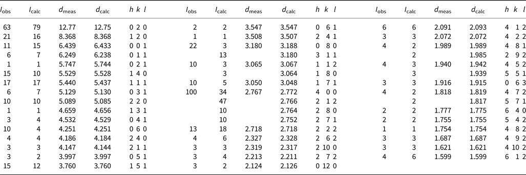
Powder X-ray diffraction data on ground samples were collected at room temperature in the 2θ range 5 to 70° using a Philips diffractometer employing CoKα radiation. The indexed powder patterns (space group: orthorhombic, Pmab) are given in Table 4 for schmidite (specimen IGC-1) and Table 5 for wildenauerite. The unit-cell parameters were refined using the Rietveld program FULLPROF (Rodríguez-Carvajal, Reference Rodríguez-Carvajal2001), giving: a = 11.059(1) Å, b = 25.452(1) Å, c = 6.427(1) Å and V = 1809.0 Å3 for schmidite; and a = 11.082(1) Å, b = 25.498(2) Å, c = 6.436(1) Å and V = 1818.6 Å3 for wildenauerite.
Powder diffraction data (d in Å) for schmidite (specimen IGC-1).

Powder diffraction data (d in Å) for wildenauerite.

Single-crystal studies
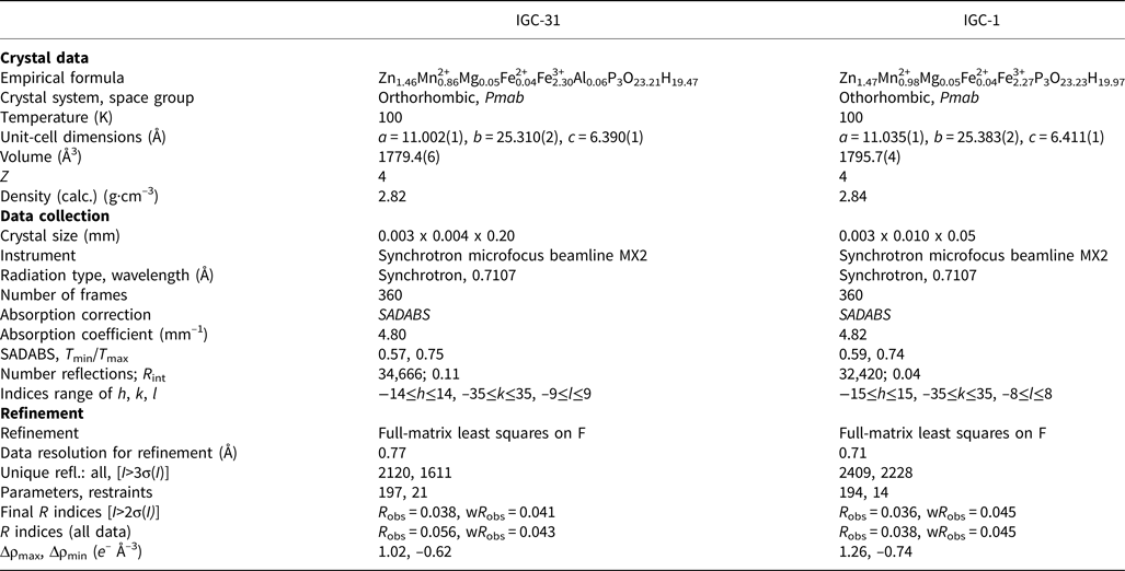
Single-crystal studies were carried out on the macromolecular beam line MX2 of the Australian Synchrotron. Data were collected using an ACRF detector and monochromatic radiation with a wavelength of 0.7107 Å. A phi scan was employed with a framewidth of 1° and a counting time per frame of 1 s. The crystals were maintained at 100 K in an open-flow nitrogen cryostream. The intensity data sets were processed using XDS software to produce data files which were analysed using WinGX (Farrugia, Reference Farrugia1999). The single-crystal structure analysis and refinement for wildenauerite (specimen IGC-14) has been published by Grey et al. (Reference Grey, Kampf, Keck, MacRae, Cashion and Gozukara2018). The data collection and refinement details for schmidite (specimens IGC-1 and IGC-31) are given in Table 6.
Data collection and refinement details for schmidite.

A structural model for schmidite, in space group Pbcm was obtained in WinGX using SHELXT (Sheldrick, Reference Sheldrick2008). A structural plot confirmed that the model conformed to that published for schoonerite (Kampf, Reference Kampf1977). A difference-Fourier map showed a large peak (13.4 e −Å–3 for specimen IGC-31) located at 1.0 Å from the [5]Zn site. The displaced site is coordinated tetrahedrally, at distances consistent with ZnO4. The same splitting of the [5]Zn site into [5]Zn/[4]Zn sites occurs in the oxidised SGM, wilhelmgümbelite (Grey et al., Reference Grey, Keck, Kampf, MacRae, Glenn and Price2017b) and wildenauerite (Grey et al., Reference Grey, Kampf, Keck, MacRae, Cashion and Gozukara2018). The unit cell was transformed to correspond to the setting reported for schoonerite, Pmab, and refinements were conducted using JANA2006 (Petříček et al., Reference Petříček, Dušek and Palatinus2014).
Structure refinement
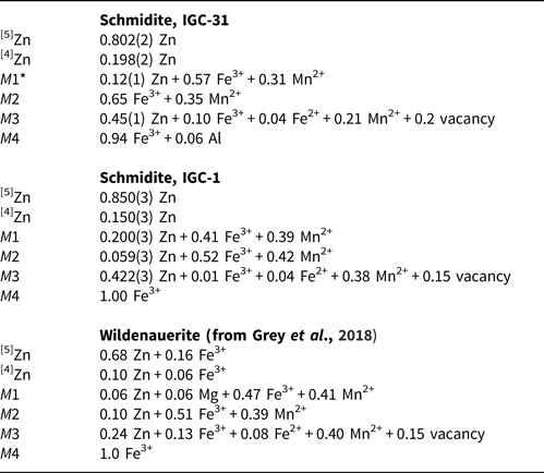
A preliminary refinement of the data for IGC-31, with Zn in the [5]Zn/[4]Zn sites and Fe in the octahedrally co-ordinated sites M1 to M4 gave a distribution of 0.8/0.2 for [5]Zn/[4]Zn, and showed a deficiency at the M3 site. A similar metal-atom deficiency occurs also in wilhelmgümbelite (site Fe2), and was found to equate to the amount of Zn displaced from the [5]Zn site (Grey et al., Reference Grey, Keck, Kampf, MacRae, Glenn and Price2017b). In IGC-31, the displaced Zn value of 0.2 is close to the metal-atom deficiency in the empirical formula (0.23), so the vacancy concentration at M3 was fixed at 0.2. Previous refinements of SGM (Kampf, Reference Kampf1977; Grey et al., Reference Grey, Keck, Kampf, MacRae, Glenn and Price2017b, Reference Grey, Kampf, Keck, MacRae, Cashion and Gozukara2018) have shown that Fe3+ is ordered in the smallest octahedral site, M4, and we also allocated the 0.06 atoms pfu of Al in specimen IGC-31 to this site. It was then necessary to establish the distribution of the three major elements, Zn, Fe and Mn, between the M1, M2 and M3 sites. The Mössbauer analysis showed that the Fe is present predominantly (98%) as Fe3+, while bond-valence calculations, combined with the EMP analyses confirm that the Mn is present as Mn2+ (details of this calculation for oxidised SGM have been reported by Grey et al., Reference Grey, Kampf, Keck, MacRae, Cashion and Gozukara2018), so we require the distribution of Zn, Fe3+ and Mn2+ between M1, M2 and M3. There is sufficient atomic number contrast between Zn and Fe/Mn to allow the Zn content in the three sites to be determined by site-occupancy refinement. There is a large difference in the ionic radii of Fe3+ and Mn2+, so once the Zn occupancies have been determined, the amounts of Fe3+ and Mn2+ could be determined from the calculated bond-valence sums (BVS, Brown and Altermatt, Reference Brown and Altermatt1985) for the sites. The validity of this approach was checked by comparing the summed metal-atom values with the results from the EMP analyses. The resulting site occupancies are given in Table 7. They give summed cations of Zn1.57Mn2+0.87Fe2+0.04Fe3+2.26Al0.06 for specimen IGC-31, very close to the empirical formula. The minor Mg component was not included in the site-occupancy refinement.
Site occupancies for metal atoms in schmidite and wildenauerite.

*Labelling M1 to M4, corresponds to labelling Fe to Fe3 for wilhelmgümbelite (Grey et al., Reference Grey, Keck, Kampf, MacRae, Glenn and Price2017b).
With the site occupancies established, the refinement proceeded smoothly and the H atoms associated with all water molecules were located in difference-Fourier maps and their positions were refined with soft restraints, O−H = 0.85(2) Å and H–O–H = 109.47(2)°. A common isotropic displacement parameter was refined for the H atoms, while all non-H atoms were refined with anisotropic displacement parameters. The final refinement for IGC-31 converged at wR obs = 0.041 for 1611 observed reflections with I > 3σ(I) and wR obs = 0.043 for all 2120 reflections, using 197 refined parameters.
The same refinement procedure was applied to the data for specimen IGC-1. In this case the site occupancies (Table 7) give summed cations of Zn1.68Mn2+1.19Fe2+0.04Fe3+1.94. The Zn and Mn values are both ~0.2 atoms pfu higher than that obtained from the mean EMP analyses, but they lie within the range of EMP analyses for IGC-1 crystals. In the refinement for IGC-1, the hydrogen atoms were located for all water molecules except for OW6.
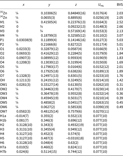
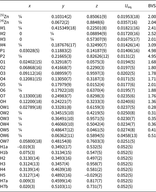
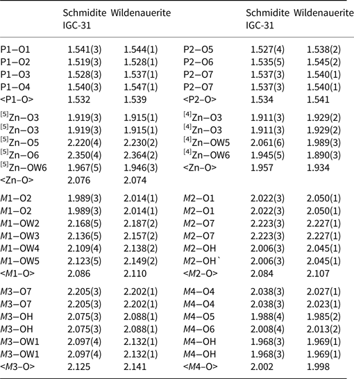
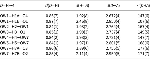
The refined coordinates, equivalent isotropic displacement parameters and bond-valence sums from the single-crystal refinements are reported in Table 8 for IGC-31 and Table 9 for IGC-1. Tables of anisotropic displacement parameters are in crystallographic information files, deposited with the Principal Editor of Mineralogical Magazine and available as Supplementary material (see below). Selected interatomic distances are reported in Table 10 for schmidite (IGC-31) and wildenauerite (from Grey et al., Reference Grey, Kampf, Keck, MacRae, Cashion and Gozukara2018), and the H bonding in schmidite specimen IGC-31 is reported in Table 11. The latter largely agrees with that reported for schoonerite (Kampf, Reference Kampf1977), although the H atoms were not located in the schoonerite refinement. Surprisingly, we could not locate the H atom associated with the OH site. As seen from Tables 8 and 9, the OH site has an elevated BVS of 1.4 for both IGC-31 and IGC-1, so at least partial replacement of OH– by O2– is most likely to occur at this site to provide charge balance for the increased Fe3+ content relative to schoonerite.
Refined coordinates, equivalent isotropic displacement parameters (Å2) and calculated bond-valence sums (BVS) for schmidite, specimen IGC-31.

Refined coordinates, equivalent isotropic displacement parameters (Å2) and calculated bond-valence sums (BVS) for schmidite, specimen IGC-1.

Selected interatomic distances (Å) in schmidite and wildenauerite.

Hydrogen bonds for schmidite, IGC-31 [Å and °].

Discussion
The crystal structure for schoonerite (Kampf, Reference Kampf1977) and for the oxidised SGM wilhelmgümbelite (Grey et al., Reference Grey, Keck, Kampf, MacRae, Glenn and Price2017b) have been described previously in some detail and the main features only will be summarised here. A projection of the schoonerite crystal structure along [001] is shown in Fig. 1. It comprises heteropolyhedral slabs parallel to (010) with H-bonded water molecules between the slabs. The slabs have a complex arrangement of octahedra, PO4 tetrahedra and ZnO5 polyhedra. The M2- and M3-centred octahedra share edges along the 6.4 Å c axis and are corner-connected to parallel 6.4 Å chains of alternating M4-centred octahedra and PO4 tetrahedra. The PO4 tetrahedra share edges with 5-coordinated ZnO5 polyhedra. Isolated M1-centred octahedra are coordinated to the slab via trans connections to PO4 tetrahedra. An important structural modification occurs in the oxidised SGM, wilhelmgümbelite, schmidite and wildenauerite, whereby there is partial partitioning of Zn from the 5-coordinated site, [5]Zn, to an adjacent vacant tetrahedral site, [4]Zn, shown in Fig. 1. The two sites share a common face and are separated by ~1.0 Å.
Based on the refinement results for wilhelmgumbelite (Grey et al., Reference Grey, Keck, Kampf, MacRae, Glenn and Price2017b) and schmidite, as well as for wildenauerite and a number of other SGM reported by Grey et al. (Reference Grey, Kampf, Keck, MacRae, Cashion and Gozukara2018), a general formula and nomenclature for SGM was approved by the International Mineralogical Association Commission on New Minerals, Nomenclature and Classification (Grey et al., Reference Grey, Keck, Kampf, MacRae and Cashion2017a). The structural formula is [4](Zn,Fe)x[5](Zn,Fe)1–x[6](M1M2M31–x□xFe3+)(PO4)3(OH)y(H2O)9–y·2H2O, where □ = vacancy, x ≤ 0.3 for the known SGM, and the cations in the octahedrally coordinated M1, M2 and M3 sites are Zn, Mn2+, Fe2+ and Fe3+, as well as minor Mg. The value of y is determined by the valence state of Fe and the vacancy concentration; y = 2 for schoonerite, which contains only divalent cations, and y increases above 2 with increasing Fe3+. In the structural formula, ‘2H2O’ signifies that two H2O molecules are in the interlayer region and link to other structural components only through hydrogen bonding. To avoid further complication of an already complex formula, the charge balance is achieved by varying only the OH–/H2O ratio, whereas the elevated BVS for the OH site (Tables 8 and 9) suggests that some minor substitution of O2– at this site is also involved in the charge balance.
The different SGM can be distinguished by the dominant cation occupancies of the M1, M2 and M3 sites and their dominant valencies (Hatert and Burke, Reference Hatert and Burke2008). Wilhelmgümbelite has M1 = Fe3+, M2 = Fe3+ and M3 = Fe2+, while schoonerite is reported to have M1 = Mn2+, M2 = Fe2+ and M3 = Fe2+ (Kampf, Reference Kampf1977) The cation occupancies of the M1, M2 and M3 sites for schmidite and wildenauerite are given in Table 7. Both minerals contain approximately equal amounts of divalent and trivalent cations in the M1 and M2 sites. The dominant divalent cation is Mn2+ while the dominant trivalent cation is Fe3+, so the simplified cation occupancies for M1 and M2 are represented by (Fe3+0.5Mn2+0.5). The minerals are then distinguished by the dominant cation in M3, which is Zn in schmidite and Mn2+ in wildenauerite. The resulting simplified formulae, ignoring the Zn partitioning and vacancies at M3, are: Zn(Fe3+0.5Mn2+0.5)2ZnFe3+(PO4)3(OH)3(H2O)8 for schmidite; and Zn(Fe3+0.5Mn2+0.5)2Mn2+Fe3+(PO4)3(OH)3(H2O)8 for wildenauerite.
The variation of the M 2+:M 3+ ratio about 0.5:0.5 in the M1 and M2 sites shown for the SGM from Hagendorf-Süd in Table 7 is also observed in SGM minerals from the Palermo #1 pegmatite, New Hampshire (Grey et al., Reference Grey, Kampf, Keck, MacRae, Cashion and Gozukara2018). This general phenomenon for SGM means that using a dominant cation rule for M1 and M2 would make it difficult to unambiguously allocate potentially new members to one end-member (Fe3+-dominant in M1 and/or M2) or the other (Mn2+ dominant) as the amounts of each element fluctuate considerably about the 50/50 composition in different specimens (compare IGC-1 and IGC-31 for schmidite in Table 7). By assigning Mn2+0.5Fe3+0.5 to M1 and M2 in the ideal formula, it is implicit that the compositions of these sites can vary between Mn2+0.25Fe3+0.75 and Mn2+0.75Fe3+0.25, (i.e. the site composition boundaries are halfway between the 50/50 compositions and the 100% Fe3+ or Mn2+ end-member compositions). This formulation, with wide compositional fields for M1 and M2 should ensure that it will be more straightforward for museum and other specimens of SGM to be characterised unambiguously.
Zinc (dominant in the [5]Zn/[4]Zn sites) and Fe3+ (dominant in the M4 site) are essential elements in all SGM. Manganese is not essential to the stability of SGMs, as evidenced by the low-Mn mineral, wilhelmgümbelite. Zinc that is in excess of that required in the [5]Zn/[4]Zn sites tends to order in the M3 site. The oxidised SGMs are similar in appearance but can be distinguished by their chemistry and associated minerals. Wilhelmgümbelite has MnO contents of only 2 to 3 wt.%, whereas schmidite has MnO levels of 7–10 wt.%, and wildenauerite has 10–11% MnO. Schmidite has significantly higher ZnO (15–16 wt.%) than wildenauerite (11 wt.%). It is worth noting that SGM-related minerals with higher ZnO or MnO than the above values are compositionally consistent with flurlite (23–27 wt.% ZnO; Grey et al., Reference Grey, Keck, Mumme, Pring and MacRae2015) and manganflurlite (13–15 wt.% MnO; Kampf et al., Reference Kampf, Grey, MacRae and Keck2017), which have a structure related to that of schoonerite by topological isomorphism. Schmidite and wilhelmgümbelite are found in close association with phosphophyllite while wildenauerite is associated closely with Zn-bearing rockbridgeite, from which it is derived by a mineral replacement reaction. Wildenauerite occurs as dense compacts of red laths whereas schmidite and wilhelmgümbelite form radiating sprays of laths that vary in colour from light yellow–brown (wilhelmgümbelite) to red (schmidite)
Acknowledgements
This research was undertaken in part using the MX2 beamline at the Australian Synchrotron, part of ANSTO, and made use of the Australian Cancer Research Foundation (ACRF) detector. Cameron Davidson is thanked for preparing sample mounts for EMP analyses and for SEM. We thank Volker Betz and Christian Rewitzer for the colour photos of the minerals and Steve Tassios for the LIBS experiment.
Supplementary material
To view supplementary material for this article, please visit https://doi.org/10.1180/mgm.2018.123