Introduction
The prevention and treatment of ototoxic inner ear damage is currently a popular research field.
Ototoxicity develops mostly after oncological treatments. The ototoxic effects of cisplatin and carboplatin are well known. There are also many published studies on various chemoprotective agents used to prevent cisplatin-based ototoxicity.
In recent years, the use of intratympanic steroid applications for the treatment of cochlear disorders has gained popularity. The main advantage of intratympanic injection lies in achieving high local concentrations of active substance within the ear while avoiding systemic side effects.
In this study, we aimed to reduce cochlear ototoxicity using various compounds. Ototoxic inner ear damage was created in rats, followed by intratympanic injection of dexamethasone, memantine or piracetam. The animals were sacrificed and levels of apoptosis were determined histopathologically at the stria vascularis, the spiral ganglion and the organ of Corti. Data were used to compare the protective effects of the three drugs.
Materials and methods
Animals and ototoxicity model
The study was approved by the Kocaeli University Ethical Committee for Experimental Animals.
The study was performed on 36 healthy, adult, female, Wistar albino rats. The rats weighed 240–330 g and were housed under standard conditions (i.e. a cycle of 12 hours of light then 12 hours of darkness, at 21–22°C, with free access to water and food).
In the course of the study, any animals which died or developed otitis media were excluded. Rats with a normal otoscopic examination were included.
Animals were divided into 3 groups of 12 rats each. The left ear was used for intratympanic treatment while the right ear was injected with saline and served as a control.
In all groups, under ether anaesthesia, rats were injected intraperitoneally with 16 mg/kg cisplatin. Forty-eight hours after this injection, under general anaesthesia induced by intraperitoneal injection of 45 mg/kg ketamine plus 5 mg/kg xylazine, either dexamethasone (4 mg/ml), memantine hydrochloride (10 mg/ml) or piracetam (200 mg/ml) was administered via a single intratympanic injection of 0.2 ml of drug solution. Injections were repeated 4 times with 48 hour intervals in between.
On the 10th day after cisplatin administration, animals were sacrificed by decapitation under anaesthesia inducted by intraperitoneal injection of 100 mg/kg ketamine.
Histological examination
Inner ears were dissected from all animals under ketamine anaesthesia, and fixed in 4 per cent neutral formalin. Tissues were then decalcified with Decal solution (Decal Corporation, Tallman, New York, USA) for 2 hours. After histological procedures, tissues were embedded in paraffin and serial, 3 µm thick sections were cut. Serial cochlear sections were stained with mouse anti single stranded DNA monoclonal antibody (MAB 3299; LV1505484, 2002–2007; EMD Millipore, Billerica, Massachusetts, USA) and counter-stained with Mayer's haematoxylin and eosin (H&E). Adjacent sections were stained with H&E only, for routine examination.
Detection of cell apoptosis
Apoptosis is a genetically and biochemically regulated mechanism of programmed cell death that plays an important role in a variety of human diseases including cancer, immune disorders, and neurological, cardiovascular and infectious diseases. Decreased stability of apoptotic DNA toward thermal denaturation is induced by the proteolysis of DNA-bound proteins during the execution of apoptosis. This conclusion is supported by the binding of anti single strand DNA monoclonal antibodies to non-apoptotic cells treated with proteinase K before heating, and by the prevention of DNA denaturation and Monoclonal Antibody reactivity in apoptotic nuclei reconstituted with histones.Reference Frankfurt, Robb, Sugarbaker and Villa 1 Thus, staining of cell suspensions and tissue sections with such antibodies following heat treatment, which induces DNA denaturation in situ only in apoptotic nuclei, is a specific and sensitive method for the detection of apoptotic cells.
In our study, cell apoptosis was detected using the mouse anti single stranded DNA monoclonal antibody MAB3299, a specific cellular marker for apoptotic death which is independent of internucleosomal DNA fragmentation and is thus useful for detecting different stages of apoptosis in various cell types. This antibody is specifically reactive with single-stranded DNA (it does not recognise DNA in double-stranded conformations) and is ideal for the detection of apoptotic cells in tissue sections. It reacts specifically with deoxycytidine, and requires at least a 25–30 basepair length of single-stranded DNA for binding.
A negative control response mixture was created by adding only label liquor.
Apoptotic cells stained with single-stranded DNA have regularly shaped, round, condensed chromatin and cytoplasmic shrinkage. In this study, cells with brown nuclei were assumed to be apoptotic.
Histoplanimetrical analysis
Immunohistochemical analysis using mouse anti single stranded DNA monoclonal antibody was used to quantify apoptotic cells in the spiral ganglion, organ of Corti and stria vascularis. All images were collected using a BX50F-3 microscope and camera (Olympus, Tokyo, Japan). Semi-quantitative methods were used to count apoptotic cells: these cells were counted in sections independently selected in a systematically randomised manner, in the spiral ganglion, organ of Corti and stria vascularis, by each of three investigators, who were blinded to the treatment group. The number of apoptotic cells was quantified in 0.0255 mm2 cochlear sections, in 10 sections of each of the three anatomical regions of interest, in each rat, using an X100 objective.Reference Manesse, Delverdier, Abella-Bourges, Sautet, Cabanié and Schelcher 2 , Reference Gundersen and Jensen 3 The mean number of apoptotic cells per section was calculated. The numerical apoptotic cell density was calculated by counting all single stranded DNA positive cells per mm,Reference Manesse, Delverdier, Abella-Bourges, Sautet, Cabanié and Schelcher 2 for the three different regions of the inner ear.
Statistical analysis
Statistical evaluation was performed using the Kruskal–Wallis test to compare the positive staining of the different treatment groups, and using the Mann–Whitney U test to analyse the correlation between the apoptotic cell counts. All data were analysed using the Statistical Package for the Social Science version 10.0 software program (SPSS Inc, Chicago, Illinois, USA). A p value of less than 0.05 was considered statistically significant.
Results
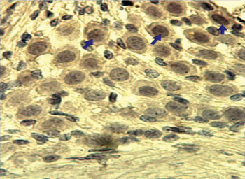
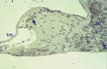
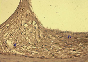
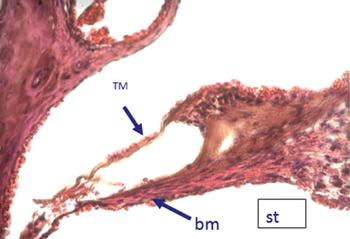
The three different cochlear regions were examined for anti single stranded DNA antibody positivity, with the right ear serving as the control group and the left ear as the treatment group, using light microscopy. Cells positive for anti single stranded DNA monoclonal antibody had brown nuclei, and were observed in the spiral ganglion, organ of Corti and stria vascularis, in all groups (Figures 1 to 5). Non-apoptotic cells had dark blue nuclei when stained with Mayer's H&E, and were seen in the above three cochlear regions, in all groups (Figure 6).
Photomicrograph showing anti single stranded DNA monoclonal antibody positive cells (arrows) in spiral ganglion tissue from the control group. (×100)

Photomicrograph showing anti single stranded DNA monoclonal antibody positive cells (arrows) in the spiral ganglion (sg) and tectorial membrane (tm), within the cochlea of the control group. (×40)

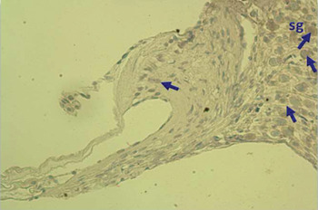
Photomicrograph showing anti single stranded DNA monoclonal antibody positive cells (arrows) in the spiral ganglion (sg) of a dexamethasone-treated ear. (×40)

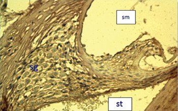
Photomicrograph showing the scala media (sm), scala tympani (st) and spiral ganglion (sg) of a memantine-treated ear. (Anti single stranded DNA monoclonal antibody; ×40)

Photomicrograph showing the spiral ligament (sl) and stria vascularis (sv) of a piracetam-treated ear. (Anti single stranded DNA monoclonal antibody; ×100)

Photomicrograph showing the tectorial membrane (TM), basilar membrane (bm) and scala tympani (st) of a piracetam-treated ear. (H&E; ×40)

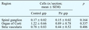
The organ of Corti had a greater number of anti single stranded DNA positive cells, compared with the spiral ganglion and stria vascularis (Tables I to IV).
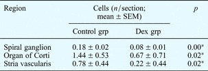
Apoptotic cells in control and dexamethasone groups, by cochlear region

*p < 0.05 vs controls. SEM = standard error of the mean; grp = group; Dex = dexamethasone
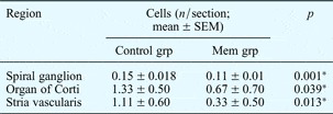
Overall, apoptotic cell numbers were significantly lower in the dexamethasone and memantine groups compared with the piracetam and control groups (p < 0.05). Specifically, we observed a significant decrease in the number of apoptotic cells in the spiral ganglion, organ of Corti and stria vascularis in the dexamethasone group compared with the control group (Figure 3, Table I) (p < 0.05). Similarly, we observed a significant decrease in the number of apoptotic cells in the spiral ganglion, organ of Corti and stria vascularis in the memantine group compared with the control group (Figure 4, Table II) (p < 0.05). We also observed a slight decrease in apoptosis cell numbers in the organ of Corti and stria vascularis in the piracetam group compared with the control group, but this difference was not statistically significant (Figure 5, Table III).
Apoptotic cells in control and memantine groups, by cochlear region

*p < 0.05 vs controls. SEM = standard error of the mean; grp = group; Mem = memantine
Apoptotic cells in control and piracetam groups, by cochlear region

SEM = standard error of the mean; grp = group; Pir = piracetam
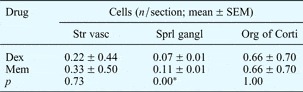
It was concluded that dexamethasone and memantine were more effective than piracetam in preventing apoptosis resulting from ototoxicity. When comparing the dexamethasone and memantine treatment groups, dexamethasone was found to be more effective than memantine in the spiral ganglion, but there was no statistically significant difference in the organ of Corti or stria vascularis (Table IV).
Apoptotic cells in different cochlear regions, by treatment

*p < 0.05, dexamethasone (Dex) vs memantine (Mem). SEM = standard error of the mean; Str vasc = stria vascularis; Sprl gangl = spiral ganglion; Org of Corti = organ of Corti
Discussion
Ototoxicity refers to damage to the cochlea and vestibular organs as a result of exposure to therapeutic and chemical substances.
Cisplatin is believed to cause ototoxicity partly by blocking ion channels in the membranes of outer hair cells, increasing hyperpolarisation and raising the auditory threshold. The study by Peters et al. Reference Peters, Mommersteeg and Heijmen 4 supports this mechanism, by showing that cisplatin ototoxicity is related to malfunction of the antioxidant system, leading to increased lipid peroxidation in the cochlear tissues.Reference Ravi, Somani and Rybak 5
Cisplatin ototoxicity is also believed to develop due to the formation of reactive oxygen radicals in the cochlea, especially free radicals such as superoxide anion, which causes a decrease in intracellular anions.Reference Dehne, Lautermann and Petrat 6 Free radical formation takes place as a result of decreased intracellular glutathion levels and altered antioxidant enzyme activity. Several studies have found that rats undergoing 16 mg/kg cisplatin treatment show a 53 per cent decrease in cochlear glutathion levels compared with controls. Furthermore, Ravi et al. found a 165 per cent increase in malonaldehyde levels, taken as evidence of free radical oxidation of cellular lipids.Reference Rybak, Whitworth and Somani 7 Derangement of the antioxidant defence system causes an increase in lipid peroxidation and therefore leads to apoptosis of hair cells, support cells, the stria vascularis and the auditory nerves.Reference Rybak, Whitworth and Somani 7
Several agents have been shown to have a protective effect against cisplatin ototoxicity, including sodium thiosulphate,Reference Muldoon, Pagel and Kroll 8 diethyldithiocarbamate and d-methionine.Reference Campbell, Meech, Rybak and Hughes 9 Other studies have found a similar effect for tocopherol, vitamin C, melatonin, sodium salicylate, N-acetylcysteineReference Feghali, Liu and Van De Water 10 and lactate.Reference Rybak, Husain and Morris 11 However, the ideal protective agent for intratympanic application has not yet been found.
The current study used an animal model of cisplatin ototoxicity to examine the protective effects of memantine (which blocks N-Methyl-d-Aspartate type glutamate receptors and calcium-sensitive nicotinic acetyl choline receptors), dexamethasone (a proven anti-inflammatory agent) and piracetam (an antioxidant, thrombocyte anti-aggregant and neuroprotective compound).
Steroids (especially prednisolone, dexamethasone and methylprednisolone) have historically been used in the treatment of various ear diseases.Reference Nadel 12 Dexamethasone is now increasingly used intratympanically. Immunohistochemical studies have found high concentrations of dexamethasone in the spiral ligament, basilar membrane, organ of Corti and spiral ganglion.
Glucocorticoids are known to have various physiological effects on the cochlear tissues.Reference Schimmer, Parker, Hardman, Limbird, Molinoff and Ruddon 13 They are commonly used in otology due to their cochlear immunosuppressive and anti-inflammatory functions.Reference Alexiou, Arnold, Fauser, Schratzenstaller, Gloddek and Fuhrmann 14 – Reference Eisenman and Arts 16 For example, nuclear factor κβ (part of the general immune response) regulates the synthesis of several cytokines,Reference Sha, Liou, Tuomanen and Baltimore 17 , Reference Baeuerle and Baltimore 18 and is suppressed by glucocorticoids.Reference Ma, Gee and Lim 19 , Reference Almawi and Melemedjian 20 Glucocorticoid-mediated inhibition of the effect of nuclear factor κβ on specific compartments within the inner ear is thought to be responsible for some of this steroid's effects on hearing loss. In addition to their restorative effects on dysfunctional autoimmunity, corticosteroids also have an effect on ion transport within the stria vascularis of mice.Reference Trune and Kempton 21 The presence of glucocorticoid receptors in a large number of spiral ligament cells supports the potential role of potassium ions in haemostasis. The mechanism of action of glucocorticoids on drug-induced cochlear ototoxicityReference Himeno, Komeda and Izumikawa 22 has also been shown to be applicable to cochlear damage caused by ischaemia,Reference Tabuchi, Oikawa, Uemaetomari, Tsuji, Wada and Hara 23 mechanical damageReference Sekiya, Shimamura, Suzuki and Hatayama 24 and noise.Reference Lamm and Arnold 25 , Reference Takemura, Komeda and Yagi 26
Memantine (3,5-dimethyl-1-adamantane) is an adamantane derivative which blocks calcium-permeable ion channels such as nicotinic acetylcholine receptors or N-Methyl-d-Aspartate-type glutamate receptors. Previous mouse studies have administered it at doses of either 5 mg/kg intraperitoneally or 20 mg/kg subcutaneously. Intratympanic application has not previously been tested. A study of the effect of memantine on cortical neuron cell cultures found the compound to be protective against glutamate toxicity.Reference Oestreicher, Arnold, Ehrenberger and Felix 27
Loss of outer hair cell function is known to be the major cause of the sensorineural hearing loss associated with various noxious insults, including noise and ototoxic agents. A massive influx of calcium ions through nicotinic receptors causes damaging structural changes in the external hair cells. Memantine prevents this influx of calcium ions into the cell, thus protecting the physiological function of the cochlea.Reference Oliver, Ludwig, Reisinger, Zoellner, Ruppersberg and Fakler 28
Piracetam (2-oxo-1-pyrrolidine-acetamine) is a gamma amino butyric acid derivative with low molecular weight. It has anti-inflammatory, anti-apoptotic, cytoprotective and immune modulatory effects. It has been used in sudden hearing loss with favourable results.Reference Gabryel, Adamek, Pudelko, Malecki and Trzeciak 29 The efficiency of intraperitoneal piracetam in treating cochlear radiation damage has been evaluated in guinea pigs with cranial and cervical cancer.Reference Altas, Ertekin, Kuduban, Gündogdu, Demirci and Sütbeyaz 30 In this study, histopathological examination of the cochleae of animals subjected to 60 Gy cranial radiotherapy demonstrated damage to the stria vascularis, spiral ganglion, and inner and outer hair cells; piracetam was shown to reduce this cochlear damage. Another study found that piracetam prevented the ototoxic effects of a cisplatin-gentamicin combination in the auditory tract extending from the cochlea to the midbrain.Reference Altas, Ucuncu, Aktan and Selimoglu 31 These authors have suggested that piracetam acts via rheological effects to increase oxygenation, and via anti-apoptotic effects to prevent apoptosis in healthy cells.
The present study used mouse anti single stranded DNA monoclonal antibody to differentiate apoptotic cells in the stria vascularis, organ of Corti and spiral ganglion. This antibody does not define the double-stranded DNA conformations. It was chosen because it is not affected by internucleosomal DNA fragmentation, it is useful in determining different stages of apoptosis in different cell types, and it is specific for apoptotic cell death. This antibody reacts with deoxycytidine and requires a minimum single-stranded DNA length of 25–30 basepairs for binding.
Our study used light microscopy to count the number of apoptotic cells reacting with this antibody within specific areas of the tissue samples. In the control group, the greatest density of ototoxicity was noted in the organ of Corti of the right cochlea. In the treatment groups, the number of apoptotic cells was reduced in the spiral ganglion, organ of Corti and stria vascularis, compared with their respective control groups. While this reduction was statistically significant for the dexamethasone and memantine groups, it was not significant for the piracetam group. However, we do not believe that this justifies the interpretation that piracetam is ineffective in preventing ototoxicity, as higher doses and greater frequency of use may have improved its efficacy. When the reduction in apoptosis density in the three anatomical regions was compared for the memantine versus dexamethasone groups, dexamethasone was found to have a significantly greater effect in the spiral ganglion, while the differences in the organ of Corti and stria vascularis were not statistically significant. However, this finding alone should not lead to the assumption that dexamethasone is more effective than memantine in preventing apoptosis resulting from cisplatin ototoxicity.
-
• Intratympanic steroids and other compounds are used to treat cochlear disorders
-
• This study evaluated use of intratympanic dexamethasone, memantine and piracetam to reduce cisplatin ototoxicity in rats
-
• Dexamethasone and memantine were better in preventing apoptosis due to ototoxicity
Memantine blocks the passage of calcium ions through N-Methyl-d-Aspartate-type glutamate receptors. Abnormally high intracellular levels of calcium ions induce degeneration of cochlear external hair cells. We believe memantine may be useful in helping prevent damage to the cochlear external hair cells, particularly in the organ of Corti, via binding with glutamate N-Methyl-d-Aspartate receptors and blocking the calcium ion influx which causes cochlear degeneration.