




No CrossRef data available.
Article contents
Stimulation of innate immunity in newborn kids against Cryptosporidium parvum infection-challenge by intranasal/per-oral administration of liposomal formulation of N-L18-norAbu-GMDP adjuvant
Published online by Cambridge University Press: 01 July 2005
Abstract
The effects of a liposomal preparation of lipophilic immunomodulator β-D-GlcNstearoyl-(1-4)-norMurNAc-L-Abu-D-isoGln (N-L18-norAbu-GMDP) were investigated on resistance to Cryptosporidium parvum infection in neonatal kids. The liposomal preparation was administered subcutaneously or intranasally/orally (i.n./p.o.) twice at doses of 100 μg, 200 μg, or 1000 μg per kid pre-infection challenge. The treatment schemes were (i) 72 and 24 h pre-infection challenge, (ii) 24 h pre-infection challenge and 24 h post-infection challenge (oral inoculation with 1×10⁷ oocysts of C. parvum in 5 ml of PBS). Administration of liposomal N-L18-norAbu-GMDP by i.n./p.o. route at the cumulative dose of 2000 μg per kid 72 and 24 h pre-infection challenge, lead to substantially increased clearance of coccidian parasites from various parts of the intestine. On the basis of histological examination, the distribution of cryptosporidia in the intestine and the severity of the infection, treated kids were classified on day 5 as having a strong reduction in infection in comparison to the control group (P<0·05). No cryptosporidia were found on the mucosal surface of treated kids by day 10, while the intestines of the control kids were still infected. All doses and routes of administration were judged effective with respect to suppression of cryptosporidia infections.
Keywords
Information
- Type
- Research Article
- Information
- Copyright
- © 2005 Cambridge University Press
INTRODUCTION
Cryptosporidium parvum, the causative agent of cryptosporidiosis, is classified with the phylum Apicomplexa and belongs to the various genera referred to as coccidia. The parasite primarily infects the microvillus border of the intestinal epithelium and to a lesser extent the extraintestinal epithelia. The infection is self-limiting except in immunodeficient hosts, in which case it may cause protracted and untreatable diarrhoea. Cryptosporidiosis is common in young farm animals (calves, lambs, kids) causing a debilitating disease that can lead to mortality. These infected animals may also play a key role in zoonotic transmission. Avian cryptosporidiosis is an emerging health problem in poultry, associated with respiratory disease in chickens and other Galliformes, or with intestinal disease in turkeys and quails. Owing to an absence of effective drug treatments, the control of cryptosporidiosis relies mainly on the enforcement of hygiene provisions and other good management practice (O'Donoghue, 1995; De Graaf et al. 1999a). Host immune responses that induce protective immunity and contribute to pathogenesis are poorly understood. Recent studies with murine infection models suggest that interferon-γ (IFN-γ) plays a key role in mediating partial protective innate immunity against the infection identified in immuno-compromised mice, and also in mediating the elimination of the coccidia agent by CD4+ T-cells (McDonald, 1999, 2000).
There are 2 target groups for which the development of an immunomodulation strategy against cryptosporidiosis would be justifiable: children and AIDS sufferers in developing countries and newborn ruminants. In the former group, an immunomodulation strategy against cryptosporidiosis might contribute greatly to improvements in public health when control by other preventive measures becomes impossible owing to poverty. Moreover, Cryptosporidium infection is one of the most devastating opportunistic complications of AIDS and should be controlled if for no other reason. In the latter group, the development of an immunomodulation strategy can be justified on the basis of economic losses due to the disease in neonatal ruminants deriving from coccidia present during livestock husbandry in less than sanitary conditions of the sort common in developing countries, and with limited availability of drugs. The development of a vaccine against cryptosporidiosis is a difficult task and many aspects of this problem were reviewed (De Graaf et al. 1999b). For this reason we elected to develop an alternative strategy using our own amphiphilic immunomodulator solubilized by liposome formulation.
Muramyl dipeptide is the smallest active unit of the Mycobacteria cell wall, and has been found to possess a whole range of immunomodulation activities. Several hundred analogues have been synthesized over the years. They stimulate non-specific resistance to microbial infection and tumour cells, and have been shown to have an effect, in vitro, on all cell types of the immune system (Adam and Lederer, 1984). In vivo in experimental infections in mice, muramyl peptide derivatives have also been shown to induce an impressive antiparasital immune response. This was demonstrated convincingly in models of protozoan infections caused by Toxoplasma gondii and Leishmania donovani (Krahnenbuhl et al. 1981; Hockertz et al. 1991). Recently, we developed a lyophilized liposomal formulation of muramyl dipeptide analogue β-D-GlcNstearoyl-(1-4)-norMurNAc-L-Abu-D-isoGln (N-L18-norAbu-GMDP) building upon an extensive literature on the background of phospholipid liposome uses as versatile transport systems for immunomodulatory drugs and as biodegradable slow-release depots for administered drugs (Barratt and Morin, 1995). Here we report on how a liposomal formulation of N-L18-norAbu-GMDP is able to achieve effective immunomodulation of cryptosporidiosis infections in newborn kids that were challenged with C. parvum oocysts by intranasal/oral (i.n./p.o.) administration either pre- or post-administration of liposomal N-L18-norAbu-GMDP.
MATERIALS AND METHODS
N-L18-norAbu-GMDP
The structure of N-L18-norAbu-GMDP derives from peptidoglycan. N-L18-norAbu-GMDP was synthesized according to the procedure of Ledvina et al. (1998). The stearoyl moiety was introduced into the structure to improve incorporation of the molecule into liposomes, which was presumed to represent a suitable carrier system for both subcutaneous and per-oral routes of administration of muramyldipeptide-based immunomodulatory compounds. Structural formulae of peptidoglycan and N-L18-norAbu-GMDP are shown in Fig. 1.

Fig. 1. Derivation of N-L18-norAbu-GMDP from peptidoglycan structure.
Preparation of liposomes
Liposomes (frozen-and-thawed multilamellar vesicles, FTMLV) were prepared from egg-yolk phosphatidylcholine (EPC, purity [ges ]95%), negatively charged lipid 1-palmytoyl-2-oleoyl-sn-glycero-phosphatidylglycerol (POPG) (Avanti Polar Lipids, USA) and N-L18-norAbu-GMDP in molar rate 4[ratio ]16[ratio ]1 by classical lipid film hydration in apyrogenic phosphate-buffered saline (PBS) (Turánek et al. 2003). For application, the liposomes were extruded through a 0·2 μm isopore filter (Millipore) (Turánek et al. 1994). The size and zeta-potential (in PBS) of liposomes were determined by dynamic light scattering (Zetasizer 3000, Malvern Instruments, Malvern, UK).
Inoculum
The source of cryptosporidial oocysts was the faeces of a naturally infected scouring calf (12 days old). Morphologically the oocysts corresponded with the species C. parvum. The faecal material was suspended in 2·5% potassium dichromate and then passed through a series of graded sieves to remove large particles, giving a suspension that was kept at room temperature for 1 week. Oocysts were concentrated by flotation is Sheather's sugar solution and then washed with tap water to remove the flotation solution. To decontaminate the occysts, the suspension was treated with bleach (SAVO, Bochemie, Bohumín, Czech Republic; 1·25% sodium hypochlorite) for 15 min. The purified occysts were stored in 2·5% potassium dichromate at 4 °C for 27 days until used for the inoculation of kids in infection challenge experiments. Before inoculation, the suspension of the oocysts was washed 3 times by centrifugation in sterile phosphate-buffered saline (PBS, pH 7·2) and counted using a haemocytometer.
Experimental design
Forty-two 1-day-old kids (Saanen goat) of both sexes were obtained from a goat-cheese production unit of 110 spring kidding goats known to be free of cryptosporidia infection or other health problems typical of newborn kids. The kids were separated from their dams immediately after the birth. In the first 24 h, they were fed goat colostrum and then transported to an isolator room and fed commercial cow's milk replacer (SANO, Germany) 3 times daily. Before starting the experiment, 4 kids were separated from the herd, placed in a separate stable and served as a non-infective control. The presence of Cryptosporidium oocysts was tested in their excrements and was negative before and during the experiment. Experimental cryptosporidiosis in kids has been described in detail previously (Koudela and Vítovec, 1997).
Treatment and infection challenge
The forty-two neonatal kids were randomly separated into 7 groups (6 animals per group) and all were orally inoculated with 107 C. parvum oocysts at 6 days of age to initiate an infection challenge. In the case of experimental groups C-F, the liposomal formulation of N-L18-norAbu-GMDP was administered subcutaneously or intranasally/per-orally (i.n./p.o.) twice at doses of 100 μg or 1000 μg per kid (500 μl per nostril) at time-intervals of 72 h and 24 h pre-infection challenge. The term i.n./p.o. denotes a route of administration involving intranasal adminstration of liposomal formulation combined with ingestion, hence both intranasal and intestinal mucosae were considered to have been exposed to the liposomal formulation. For the remaining groups G and H, kids were treated with liposomal formulation twice at doses of 200 μg per kid by a combination approach involving first subcutaneous (s.c.) administration (500 μl per kid) followed by i.n./p.o. administration (500 μl per nostril) (or vice versa), at time-intervals of 24 h pre- and 24 h post-infection challenge. Group B acted as an infection control that was not treated with any liposomal formulation. Three kids (group A) were kept in a separate stable and served as an untreated uninfected control. The experimental design is summarized in Table 1.

Assessment of infection
All kids were monitored twice a day for diarrhoea and other clinical signs by observing them. The severity of diarrhoea was scored as 0 for normal, 1 for loose and formless, 2 for semi-fluid, or 3 for watery.
Five days post-inoculation (p.i.) and 10 days, p.i. 3 kids from each experimental group were euthanized with an overdose of a barbiturate (Thiopental®, Spofa, Czech Republic). At necropsy, a complete examination of all organs and tissues was performed. Samples for histopathology were collected immediately after the animals were killed. The first specimen was taken from the ileum within 5 cm from the ileocaecal valve (ostium ileocaecale – OIC). More samples were removed 50 cm from the OIC and then every 50 cm cranially from the OIC so that the last jejunal specimen originated from the duodenum. In the large intestine specimens were collected from the apex caeci, colon near the ansa centralis, and rectum. Additional samples for histology were taken from the abomasum, liver, kidneys, spleen, lung, pancreas, and regional mesenteric lymph nodes. Material was fixed in 10% neutral formalin and processed by routine histological methods. Histological sections were stained with haematoxylin-eosin, and with Wolbach's modification of Giemsa staining. On the basis of such histological examination the distribution of cryptosporidia in the intestine and the degree of infection were classified as follows. Degree 0 – no cryptosporidia found on the mucosal surface; degree 1 – moderate infection, sporadic cryptosporidia distributed on the intestinal surface; degree 2 – medium infection, regularly disseminated cryptosporidia on the intestinal surface; degree 3 – massive infection, most of the epithelial surface covered by cryptosporidia. Microscopical examination of haematoxylin-eosin stained ileal sections was then carried out in order to enumerate cryptosporidia, in various stages of development, that were attached to the enterocytes of 10 villi from the ileum within 5 cm of the ileocaecal valve. The severity of ileal infection was then expressed as the mean number of cryptosporidia per villus.
Statistical analysis
Statistical analyses were carried out using the programme GraphPad PRISM V.3.03 (GraphPad Software Inc., San Diego, CA) where each graphical point represented a degree and distribution of C. parvum infection in the intestine. The severities of ileal infections were analysed by means of an analysis of variance (ANOVA) and comparisons of means (Newman-Keuls test).
RESULTS
Liposomal preparation of N-L18-norAbu-GMDP
Liposomal formulations with N-L18-norAbu-GMDP were prepared by lipid film hydration in PBS followed by extrusion through polycarbonate filters. Liposomes were characterized with a mean size of 178 nm (polydispersity 0·07) and a mean zeta-potential of −30·6 mV (PBS, 25 °C). No side-effects (e.g. intumescence or hyperaemia of the nasal mucosa, oedema and soreness at the site of injection) were found after application of the liposomal preparation.
Histological examination and clinical signs
The distribution and extent of C. parvum infections found in histological sections from the intestine of experimentally inoculated kids is presented in Fig. 2. All doses and all routes of administration of liposomal N-L18-norAbu-GMDP appear to have been effective at suppression of infection, especially at peak infection on day 5 (P<0·05) (Fig. 2). Clearly, the most dramatic effects were observed when liposomal N-L18-norAbu-GMDP was administered i.n/p.o. 72 h and 24 h pre-infection challenge at a cumulative dose of 2000 μg per kid. In this instance, administration led to a considerably increased clearance of coccidian parasites from various parts of the intestine. On the basis of histological examination, the distribution of oocysts in the intestine and the severity of the infection (classified on day 5) were strongly reduced in comparison to the control group. Furthermore, no cryptosporidia were found on the mucosal surface of the treated kids on day 10, while the intestines of the control kids were still infected (Fig. 3). When a lower cumulative dose of 200 μg was employed, liposome administration was still effective, but residual infection was found on day 10. Dose-dependent responses were found for both i.n./p.o. and s.c. routes of administration. Pre- and post-infection applications of liposomal N-L18-norAbu-GMDP were more effective when liposomal preparation was applied first i.n./p.o and then s.c. rather than vice versa (Table 2). According to histological examination of other organs (e.g. abomasum, liver, kidneys, spleen, lung, pancreas, and regional mesenteric lymph nodes), no spreading of C. parvum infection to other organs was found in both control and treated groups. Results from histological examinations correlated well with clinical observations made during the infectious period. The onset of diarrhoea after inoculation of oocytes was more rapid in kids treated with liposomal N-L18-norAbu-GMDP, but the duration of diarrhoea was considerably shorter compared to the control group. The curves reflecting the onset and duration of diarrhoea are presented in Fig. 4. Very similar curves were obtained for kids from different groups treated with liposomal N-L18-norAbu-GMDP, hence only the curve for liposome-treated group D is shown for the sake of clarity in comparison with that of control group B (Fig. 4).

Fig. 2. Distribution and extent of Cryptosporidium parvum infection found in histological sections from the intestine of experimentally infected kids treated with liposomal N-L18-norAbu-GMDP. Detailed description of various experimental groups appears in Table 1. Sampling of the intestine for histological examination is described in the Materials and Methods section under Assessment of infection.

Fig. 3. Comparison of histological sections of the middle jejunum of an infected untreated kid (A) and a kid of group D (B); both were examined 10 days after infection. Cryptosporidial developmental stages on the intestinal surface are indicated by arrows.

Fig. 4. The onset and duration of diarrhoea in the group D and infected control (group B). Classification of severity of diarrhoea is described in the Materials and Methods section.

DISCUSSION
The immunomodulatory effects of muramyl dipeptide, or its analogues, observed in vivo probably result from activation of intraepithelial lymphocytes, which are responsible for a resolution of intestinal infection. For instance, the treatment of mice with muramyl dipeptide appears to enhance resistance to challenge from the obligate intracellular protozoan Toxoplasma gondii (Krahenbuhl et al. 1981). In order to induce this effect, relatively high concentrations of this hydrophilic compound (80 mg/kg) were administered to mice to provoke sufficient immune response. This is probably due to poor penetration of hydrophilic muramyl dipeptide through cell membranes to reach intracellular macrophage receptors (Fogler and Fidler, 1986). In addition, an immunomodulatory preparation based on oat β-glucan particles (1–3 μm) has been found to induce an immune response against another coccidian parasite Eimeria vermiformis. Once again, relatively high doses of this preparation needed to be administered to mice by intragastrical or subcutaneous routes (150 mg/kg i.g., 25 mg/kg s.c.) before prophylactic effects could be seen (Yun et al. 1998).
Liposomes represent well-tolerated drug carriers for various routes of administration. In the case of s.c. administration, liposome particles (>120 nm) may stay at the injection site post-administration and can serve as a slow release drug depot, while smaller liposome particles (<120 nm) may penetrate through the extracellular matrix and reach the lymphatic tissues via the lymphatic capillaries thus delivering the encapsulated drug to the lymphatic tissue macrophages (Oussoren and Storm, 2001). Liposomal formulations of N-L18-norAbu-GMDP were approx. 200 nm in diameter with a few smaller liposome particles present (results not shown). Therefore, we presume in our case that liposomal formulations of N-L18-norAbu-GMDP post s.c.-administration were acting primarily as a slow release depot for N-L18-norAbu-GMDP.
Our data indicate that i.n./p.o.-administration gave the most effective immunomodulation with liposomal N-L18-norAbu-GMDP. The i.n./p.o. route of administration represents a non-invasive approach that is as a result attractive for both veterinary and human applications. However, the mechanism of immunomodulation is perhaps less obvious than for s.c.-administration. Post i.n./p.o. administration, liposomes can promote interactions with mucosal surfaces and thereby aid efficient per-mucosal absorption. Very few studies have been carried out on the mechanism of liposome uptake by mucosal Peyer's patches. Sections of Peyer's patches from experimental animals have been found with M cells harbouring endocytic vesicles containing liposomes. These results indicate that intact liposomes can be endocytosed by M cells and delivered to immune cells in Peyer's patches, so that direct activation of immune cells by the liposome formulation of an immunomodulator seems feasible (Childers et al. 1990). The fate of liposomes in the gastro-intestinal tract depends on their stability towards low pH, physiological concentrations of bile salt and levels of pancreatic lipase. Both N-L18-norAbu-GMDP and other lipids used here for the preparation of FTMLV formulated with N-L18-norAbu-GMDP, are resistant to low pH but not the action of bile acids (unsaturated phospholipids are very bile acid sensitive). This problem could be serious where hydrophilic muramyl dipeptide or its analogues (including norAbur-GMDP) are entrapped in the central aqueous cavity of FTMLVs since any defect in lipid membrane integrity must result in rapid release of liposome encapsulated contents into the surrounding medium.
Conclusions
A liposomal preparation of hydrophobic N-L18-norAbu-GMDP was able to stimulate innate immunity in newborn kids against Cryptosporidium delivering a prophylactic treatment regime. Non-invasive i.n./p.o. administration is most effective and is most favourable for use in both human and veterinary medicine. Further studies on the use of immunomodulatory liposomal formulations of N-L18-norAbu-GMDP for the therapeutic treatment of cryptosporidiosis in newborn ruminants and also for the treatment of immuno-compromised patients are tasks for future research.
This work was supported by a grant the Ministry of Agriculture of the Czech Republic (Grant no. MZE 0002716201 and NAZV, Contract no: QF 3115) and Academy of Sciences (IGA AV ČR No. Z4055905). We also thank IC-Vec Ltd, UK for support.
References
REFERENCES
Adam, A. and
Lederer, E. (
1984).
Muramylpeptides, immunomodulators, sleep factors and vitamins.
Medicinal Research Reviews
4,
111–152.CrossRefGoogle Scholar
Barratt, G. and
Morin, C. (
1995)
Liposomal immunomodulators. In
Liposomes, New Systems and New Trends in their Applications
(ed. Puisieux, F., Couvreur, P., Delattre, J. and Devissaguet, J. P.), pp.
461–506.
Scientific Publications,
Paris.
Childers, N. K.,
Denis, F. R.,
McGee, N. F. and
Michalek, S. M. (
1990).
Ultrastructural study of liposome uptake by M cells of rat Peyer's path, an oral vaccine system for delivery of purified antigen.
Regional Immunology
3,
8–16.Google Scholar
De Graaf, D. C.,
Sspano, F.,
Petry, F.,
Sagodira, S. and
Bonnin, A. (
1999 b).
Speculation on whether a vaccine against cryptosporidiosis is a reality or fantasy.
International Journal for Parasitology
29,
1289–1306.Google Scholar
De Graaf, D. C.,
Vanopdenbosch, E.,
Ortega-Mora, L. M.,
Abbassi, H. and
Peeters, J. E. (
1999 a).
A review of the importance of cryptosporidiosis in farm animals.
International Journal for Parasiotology
29,
1269–1287.Google Scholar
Fogler, W. E. and
Fidler, I. J. (
1986).
The activation of tumoricidal properties in human blood monocytes by muramyl peptides requires a specific intracellular interaction.
Journal of Immunology
136,
2311–7.Google Scholar
Hockertz, S.,
Franke, G.,
Paulini, I. and
Lohmann-Matthes, M. L. (
1991).
Immunotherapy of murine visceral leishmaniasis with murine recombinant interferon-γ and MTPPE encapsulated in liposomes.
Journal of Interferon Research
11,
177–185.CrossRefGoogle Scholar
Koudela, B. and
Vítovec, J. (
1997).
Experimental cryptosporidiosis in kids.
Veterinary Parasitology
71,
273–281.CrossRefGoogle Scholar
Krahenbuhl, J. L.,
Sharma, S. D.,
Ferraresi, R. W. and
Remington, J. S. (
1981).
Effects of muramyl dipeptide treatment on resistance to infection with Toxoplasma gondii in mice.
Infection and Immunity
31,
716–722.Google Scholar
Ledvina, M.,
Zýka, D.,
Ježek, J.,
Trnka, T. and
šaman, D. (
1998).
New effective synthesis of (N-acetyl- and N-stearoyl-2-amino-2-deoxy-beta-D-glucopyranosyl)-(1–>4)-muramoyl-L-2-aminobutanoyl-D-isoglutamine, analogs of GMDP with immunopotentiating activity.
Collection of Czechoslovak Chemical Communications
63,
577–589.CrossRefGoogle Scholar
McDonald, V. (
1999).
Gut intraepithelial lymphocytes and immunity to coccidia.
Parasitology Today
15,
483–487.CrossRefGoogle Scholar
McDonald, V. (
2000).
Host cell-mediated responses to infection with Cryptosporidium.
Parasite Immunology
22,
597–604.CrossRefGoogle Scholar
O'Donoghue, P. (
1995).
Cryptosporidium and cryptosporidiosis in man and animals.
International Journal for Parasitology
25,
139–195.CrossRefGoogle Scholar
Oussoren, CH. and
Storm, G. (
2001).
Liposomes to target the lymphatics by subcutaneous administration.
Advanced Drug Delivery Reviews
50,
143–156.CrossRefGoogle Scholar
Turánek, J. (
1994).
Fast-protein liquid chromatography system as a tool for liposome preparation by the extrusion procedure.
Analytical Biochemistry
218,
352–357.CrossRefGoogle Scholar
Turánek, J.,
Kašná, A.,
Záluská, D. and
Neča, J. (
2003).
Preparation of sterile liposomes by the proliposome – liposome method.
Methods in Enzymology
367,
111–129.CrossRefGoogle Scholar
Yun, Ch-H.,
Estrada, A.,
Van Kessel, A.,
Gajahar, A.,
Redmond, M. and
Laarveld, B. (
1998)
Immunomodulatory effects of oat β-glucan administered intragastrically or parenterally on mice infected with Eimeria vermiformis.
Microbiology and Immunology
42,
457–465.Google Scholar

Fig. 1. Derivation of N-L18-norAbu-GMDP from peptidoglycan structure.

Table 1. Tabular summary of the experimental groups
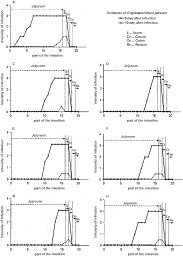
Fig. 2. Distribution and extent of Cryptosporidium parvum infection found in histological sections from the intestine of experimentally infected kids treated with liposomal N-L18-norAbu-GMDP. Detailed description of various experimental groups appears in Table 1. Sampling of the intestine for histological examination is described in the Materials and Methods section under Assessment of infection.
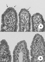
Fig. 3. Comparison of histological sections of the middle jejunum of an infected untreated kid (A) and a kid of group D (B); both were examined 10 days after infection. Cryptosporidial developmental stages on the intestinal surface are indicated by arrows.

Fig. 4. The onset and duration of diarrhoea in the group D and infected control (group B). Classification of severity of diarrhoea is described in the Materials and Methods section.
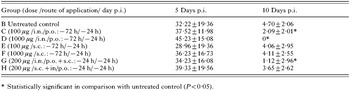
Table 2. Mean number of cryptosporidia per ileal villus in kids
 You have
Access
- 4
- Cited by