Introduction
Adult stem cells have an extensive capacity for multilineage differentiation and replication,Reference Raghunath, Salasinski, Sales, Butler and Seifalian 1 and are thus highly attractive for many tissue engineering applications. Evidence shows that several tissues and organs (e.g. blood, skin and gastrointestinal tract) undergo rapid renewal. Therefore, it is reasonable to assume that postnatal tissues contain a pool of stem cells which initiate such renewal.Reference Robey and Bianco 2 Adult stem cells (e.g. mesenchymal stem cells) can differentiate into a variety of cell types, including chondrocytes, adipocytes and osteoblasts. Multipotent cells have also been identified, derived from bone marrow and from non-marrow tissues such as adipose tissue, teeth and blood.
Adipose-derived stem cells are relatively easy to obtain (from adipose tissue) and to culture, and their extraction is less invasive in comparison with bone marrow derived stem cells.Reference Raghunath, Salasinski, Sales, Butler and Seifalian 1 Considering these characteristics, adipose-derived stem cells may represent a promising source for cell therapy.Reference Zuk, Zhu, Ashjian, De Ugarte, Huang and Mizuno 3 , Reference Yamamoto, Akamatsu, Hasegawa, Yamada, Nakata and Ohkuma 4
Cartilage has several important structural features within joints. Unfortunately, it has poor regenerative capability. However, the low degree of vascularisation in cartilage makes it an ideal target for tissue engineering.Reference Raghunath, Salasinski, Sales, Butler and Seifalian 1 Since mesenchymal stem cells have the ability to differentiate into chondrocytes in a non-vascularised area,Reference Dominici, Hofmann and Horwitz 5 , Reference Bianco, Riminucci, Gronthos and Robey 6 recent studies have focussed on the differentiation of mesenchymal stem cells into chondrocytes for use in cartilage tissue engineering.Reference Raghunath, Salasinski, Sales, Butler and Seifalian 1 Pre-clinical studies have shown that injection of mesenchymal stem cells into damaged tissues is highly plausible both as a treatment for osteoarthritis in rabbits and a means of cartilage regeneration in goats.Reference Csaki, Schneider and Shakibaei 7 , Reference Toghraie, Chenari, Gholipour, Faghih, Torabinejad and Dehghani 8 Interestingly, clinical trials of articular cartilage repair using mesenchymal stem cells have recently begun.Reference Csaki, Schneider and Shakibaei 7
Based on the above observations emphasising the importance of mesenchymal stem cells in regenerative medicine, this study aimed to investigate whether adipose-derived stem cells could differentiate into rabbit ear cartilage. To this end, adipose-derived stem cells were isolated from rabbit fat tissue, cultivated in vitro and injected into a surgically created rabbit ear cartilage defect.
Materials and methods
Adipose-derived stem cell isolation and culture
As described previously,Reference Toghraie, Chenari, Gholipour, Faghih, Torabinejad and Dehghani 8 fragments of subcutaneous adipose tissue were washed with phosphate-buffered saline, minced into small pieces and digested with 0.2 per cent collagenase (Gibco, Grand Island, NY, USA) at 37°C for 2 hours on a shaker. The resultant soup was centrifuged at 400 g for 10 minutes. The pellet, which included the adherent stromal cells, was then carefully placed on Ficoll (Biosera, East Sussex, UK) and centrifuged at 400 g for 30 minutes. The second layer, the stromal vascular fraction, was transferred into a tube and washed with phosphate-buffered saline. The stromal vascular fraction pellet was resuspended in Dulbecco's Modified Eagle's Medium culture medium (Gibco) containing 10 per cent fetal bovine serum (Gibco) and 1 per cent penicillin/streptomycin (Biosera). After 24 hours' culture, non-adherent cells were discarded. The adherent cells were further cultured, changing the medium every 4 days, and were then harvested in passage three, after approximately 30 days of culture.
Animals
We used adult Dutch rabbits, which were obtained from the laboratory animal centre of Shiraz University of Medical Sciences.
The study was approved by the Research Animal Care Committee of Laboratory Animals of Shiraz University of Medical Sciences.
Adipose-derived stem cell injection
We anaesthetised four Dutch rabbits (weight range, 2–2.5 kg) with ketamine (2 mg/kg) and 2 per cent xylocaine (1 mg/kg). In each rabbit, we removed a 2 × 2 cm cartilage plate from the midportion of each auricle, and partially sutured the incision. A catheter was inserted at the excision site, and 0.5 ml of culture medium containing approximately 1010 autologous adipose-derived stem cells was injected. The catheter was then removed and the site completely sutured.
At three, four, five and six months after injection, the auricles were resected and sent for histopathological examination. Specimens were sectioned, stained with haematoxylin and eosin, and assessed for the presence of new cartilage formation and other histopathological changes.
In the control group, three rabbits of the same strain were selected and anaesthetised as in the study group. A 1 × 1 cm cartilage plate was removed from both auricles of each rabbit. Phosphate-buffered saline (0.5 ml) was injected on one side, while the other side was left uninjected. Auricles were resected at two, four and six months after injection and sent for histopathological examination.
Results
Cartilage regeneration in the study group
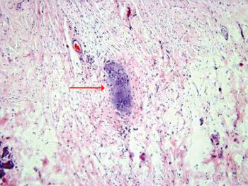
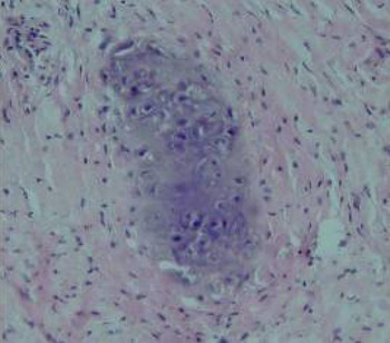
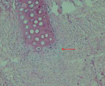
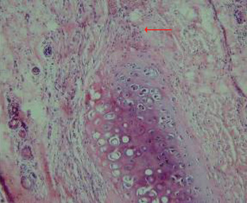
In the four study group rabbits, histopathological examination of auricles removed three months after adipose-derived stem cell injection showed small islands of new cartilage formation at the site of the surgically created defect (Figure 1). The new cartilage was composed of young chondrocytes with scanty, immature, bluish cartilage stroma (Figure 2); these chondrocytes were clustered close together.
Photomicrograph of rabbit auricle three months after injection of stem cells, showing a small island of new cartilage formation within an area from which cartilage has been surgically removed; the arrow indicates young chondrocytes with scant basophilic stroma surrounded by connective tissue cells. (H&E; ×100)

Photomicrograph of rabbit auricle three months after stem cell injection, showing differentiation of injected stem cells into chondrocytes which form an immature mass of new cartilage, located in continuity with a native cartilaginous plate. (H&E; ×200)

In samples removed four months after stem cell injection, we noted the formation of an immature mass of new cartilage, arranged in a line continuous with the native cartilage remnant. In these areas, the cellularity of the new cartilage and the amount of stroma between chondrocytes had increased, compared with specimens obtained three months after injection.
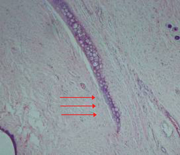
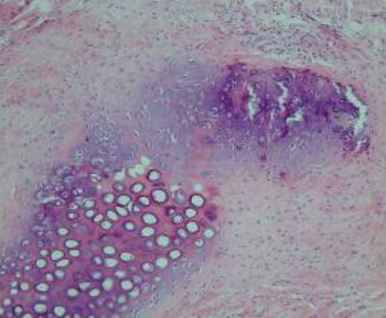
In samples removed five months after stem cell injection, histopathological examination showed that the new cartilage was attempting to form a mature plate to fill the cartilage gap (see Figure 3). At this stage, chondrocytes were well matured with increased stromal component, compared with the four month samples.
Photomicrograph of rabbit auricle five months after stem cell injection, showing new cartilage growing to form a mature plate (arrows) filling the surgically created cartilage defect. (H&E; ×40)

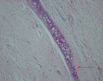
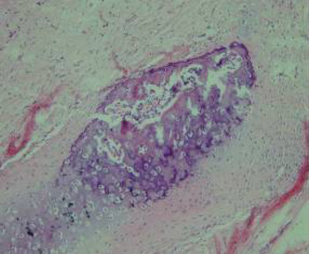
The last samples, taken six months after stem cell injection, showed well-formed, mature cartilaginous plates which completely filled the gap in the native cartilage (Figure 4).
Photomicrograph of rabbit auricle six months after stem cell injection, showing a mature cartilaginous plate (arrow) lying in continuity with native cartilage remnants, completely filling the cartilage defect. (H&E; ×40)

Cartilage regeneration in the control group
In the control group auricles removed four months after intervention, histopathological examination showed no significant difference between the phosphate-buffered saline injected and uninjected auricles.
Auricle specimens removed one month after phosphate-buffered saline injection showed no new cartilage formation.
In auricles removed three and four months after intervention, focal proliferation of chondrocytes was seen only near the edge of the remaining perichondrium. In the area of the defect, fibrous tissue formation was observed (Figures 5–8).
Photomicrograph of control group rabbit auricle one month after cartilage removal and phosphate-buffered saline injection. The incised end of the cartilage plate (arrow) shows no new cartilage formation. (H&E; ×100)

Photomicrograph of control group rabbit auricle one month after cartilage removal (without phosphate-buffered saline injection), showing no evidence of new cartilage formation at the incised end of the cartilage plate. (H&E; ×100)

Photomicrograph of control group rabbit auricle four months after cartilage removal and phosphate-buffered saline injection. There is focal proliferation of chondrocytes and surrounding fibrous tissue, and premature cartilage calcification. (H&E; ×100)
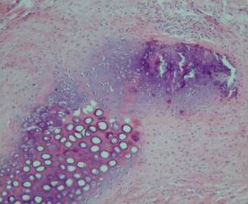
Photomicrograph of control group rabbit auricle four months after cartilage removal (without phosphate-buffered saline injection). As in Figure 7, there is focal proliferation of chondrocytes and fibrous tissue, and premature cartilage calcification. (H&E; ×100)

Discussion
Mesenchymal stem cells isolated from postnatal tissues have a large capacity to replicate and differentiate into different cell types, including chondrocytes.Reference Raghunath, Salasinski, Sales, Butler and Seifalian 1 , Reference Robey and Bianco 2 The importance of cartilage regeneration has been confirmed in various disease conditions.
It has been previously noted that rabbit mesenchymal stem cells show a significantly greater rate of proliferation and cartilage-specific extracellular matrix production in the presence of transforming growth factor β3. Thus, mesenchymal stem cells are potential candidates for cartilage tissue engineering.Reference Park and Na 9
Nöth et al. Reference Nöth, Rackwitz, Heymer, Weber, Baumann and Steinert 10 studied chondrogenic differentiation of human mesenchymal stem cells cultured in collagen type one hydrogel, and reported that mesenchymal stem cells grown in this way show distinct chondrogenic differentiation. These authors also assessed the clinical results of transplantation of autologous bone marrow stromal cells to achieve articular cartilage repair in the human patella, and found that cartilage defects were regenerated with fibrocartilage, enabling a significantly improved patient outcome over long term follow up.
Raghunath et al. Reference Raghunath, Salasinski, Sales, Butler and Seifalian 1 compared the culture and differentiation abilities of adult stem cells and mature chondrocytes in the presence of growth hormones and various biomaterial and bioreactor scaffolds. They found that autologous adult stem cells had far greater potential as cell sources than mature chondrocytes, and did not stimulate an immune rejection response.
-
• In this study, adipose-derived stem cells proliferated and differentiated to fill an in vivo cartilage defect
-
• Adipose tissue is a more appropriate source of mesenchymal stem cells than previously reported sources
-
• Autologous, adipose-derived, mesenchymal stem cells could be used for tissue engineering and regenerative medicine
Wakitani et al. Reference Wakitani, Okabe, Horibe, Mitsuoka, Saito and Koyama 11 used autologous bone marrow derived mesenchymal stem cells to treat human patients with joint cartilage defects. After two-year follow up, there was significantly greater hyaline cartilage formation in the treated group compared with the untreated group.
Finally, Richardson et al. Reference Richardson, Hoyland, Mobasheri, Csaki, Shakibaei and Mobasheri 12 also demonstrated that mesenchymal stem cells are potent candidates for regeneration of articular defects. Interestingly, these authors reported that the newly formed cartilage, derived from mesenchymal stem cells, had the exact physical properties of normal cartilage.
To our best knowledge, the current study is the first to investigate the capacity of adipose-derived stem cells for cartilage differentiation in a surgically created auricular defect. As adipose tissue is abundant and easy accessible, it is hypothesised that this source is more appropriate for mesenchymal stem cell extraction for regenerative medicine applications. In our study, isolated, adipose-derived stem cells grown in a preconditioned, in vivo environment differentiated appropriately into mature chondrocytes, establishing an animal model for cartilage repair. Additionally, the observed growth of chondrocytes through consecutive stages, from new, immature cartilaginous islands to mature, physically palpable cartilage plates, mimicked the normal embryonic cartilage formation in many sites of the body, such as the auricle and the nose. In the control group, repair was quite focal and nonsignificant after four months. Overall, cartilage repair seemed to be significantly enhanced in the study group (receiving injected, adipose-derived stem cells) compared with the control group.
We note that the results of the present study are in consistent with our recent findings on the treatment of osteoarthritis with infrapatellar fat pad derived mesenchymal stem cells in rabbits.Reference Toghraie, Chenari, Gholipour, Faghih, Torabinejad and Dehghani 8
Conclusion
The current study findings indicate that, under specialised in vivo conditions favouring repair with a specific cell maturation line, adipose-derived stem cells can proliferate and differentiate into chondrocytes, enabling chondrogenesis and cartilage defect repair.
We suggest that adipose tissue is a more appropriate source for mesenchymal stem cell extraction, for regenerative medicine applications, than previously described sources. Autologous, adipose-derived, mesenchymal stem cells could represent a promising cell source for tissue engineering and regenerative medicine.
Acknowledgements
This work was funded by grants from Shiraz University of Medical Sciences (grant number 87-4218) and from the Shiraz Institute for Cancer Research (grant number ICR-87-133).