INTRODUCTION
The Trindade and Martin Vaz Oceanic Archipelago (TMV) is the above-water portion of a submarine volcanic chain of about 3–3.5 my rising 5500 m above the seafloor (Marques et al., Reference Marques, Ulbrich, Ruberti and Tassinari1999). Trindade Island and the much smaller Martin Vaz islands are only 49 km apart from each other, but almost 1200 km away from the Brazilian coast and some 4200 km from the nearest African shore. The archipelago is mainly under the influence of the southward flow of the Brazil Current, which runs along the Brazilian coast from about 10°S to 38°S (Stramma et al., Reference Stramma, Ikeda and Peterson1990; Peterson & Stramma, Reference Peterson and Stramma1991; Podesta et al., Reference Podesta, Brown and Evans1991; Stramma, Reference Stramma, Steele, Thorpe and Turekian2001). The influence of the oceanic main current patterns in this zone and the existence of a seamount chain oriented east-west between TMV (whose tops can be as shallow as 50 m [Motoki & Motoki, Reference Motoki and Motoki2013]) and the Brazilian continental shelf, can potentially serve as stepping stones for spillover of shallow-water species and is directly reflected in the taxonomic composition of the TMV invertebrate fauna (Leal & Bouchet, Reference Leal and Bouchet1991; Gasparini & Floeter, Reference Gasparini and Floeter2001; Anker et al., Reference Anker, Tavares and Mendonça2016).
The levels of endemism and species richness in the TMV vary among taxa (Carvalho, Reference Carvalho1950; Oliveira, Reference Oliveira1951; Vannucci, Reference Vannucci1951; Albuquerque & Guille, Reference Albuquerque and Guille1991; Tavares, Reference Tavares1999; Leal, Reference Leal2000; Floeter & Gasparini, Reference Floeter and Gasparini2000; Paiva, Reference Paiva, Lavrado and Ignacio2006; Coimbra et al., Reference Coimbra, Ghilardi, Casetta, Bergue, Mohr, Castro, Costa and Alves2009; Moraes, Reference Moraes2011; Coimbra & Carreño, Reference Coimbra and Carreño2012; Anker et al., Reference Anker, Tavares and Mendonça2016). However, to a certain extent, this may be only a reflection of sample size (McClain, Reference McClain2007), as not only the Brazilian oceanic islands and seamount chains are yet to be intensively sampled, but also many surveys in the area have been and continue to be taxon-oriented.
The current knowledge about the echinoderm diversity of the TMV is restricted to a few studies (Oliveira, Reference Oliveira1951; Krau, Reference Krau1952; Bernasconi, Reference Bernasconi1955b, Reference Bernasconi1957; Brito, Reference Brito1971; Lima-Verde, Reference Lima-Verde1969; Guille & Albuquerque, Reference Guille, Albuquerque, Guille and Ramos1987, Reference Guille and Albuquerque1990; Vadon, Reference Vadon, Guille and Ramos1988; Albuquerque & Guille, Reference Albuquerque and Guille1991). Ophiuroids are the most studied echinoderms, while holothuroids have never been reported from the archipelago. During a 4-year project (ProTrindade/CNPq), five campaigns to the TMV oceanic archipelago were conducted between 2012 and 2015, which resulted in about 605 lots of shallow-water echinoderms, collected mainly by scuba diving and thus forming a solid base for taxonomic assessment of the shallow-water echinoderm fauna of the archipelago. Here we report on 16 echinozoan species, 11 of which represent new records for TMV. Three species are recorded from the south-western Atlantic and Brazilian territorial waters for the first time. Additionally, we studied a few unreported samples from the São Pedro and São Paulo Oceanic Archipelago, a group of small islets rising from the mid-Atlantic ridge located about 1146 km away from Brazil and some 1960 km from the nearest African coast. We take the opportunity to elaborate on the taxonomy of the seven echinoid and the nine holothuroid species reported herein, providing morphological descriptions and illustrations for all of them. We also present morphological comparisons between the studied species and their relevant closest congeners and between insular and coastal specimens, and provide some notes on the echinozoan species composition of the Trindade and Martin Vaz Oceanic Archipelago.
MATERIALS AND METHODS
Most of the material reported herein was collected during 168 scuba diving operations between 4–30 m, at numerous stations around Trindade Island (20°30′S 29°20′W) and two stations off Martin Vaz Islands (20°30′S 28°51′W) (see Anker et al., Reference Anker, Tavares and Mendonça2016: figure 1). Also included in this study are a few unreported samples from the São Pedro and São Paulo Archipelago (20°30′S 29°18′W, also known as Saint Peter and Saint Paul Rocks). All specimens were preserved in 75% ethanol; some were photographed alive in situ and some photographed after preservation.
Holothuroid morphological techniques and terminology are after Rowe & Doty (Reference Rowe and Doty1977) and Samyn et al. (Reference Samyn, VandenSpiegel and Massin2006). Echinoid pedicellariae were classified according to Coppard et al. (Reference Coppard, Kroh and Smith2010). Ossicles, pedicellariae and spines were dried and mounted on metal stubs with double-sided tape, coated with gold and observed with a LEO 440 Scanning Electron Microscope (SEM). Measurements: maximum lengths of holothuroids were obtained from ethanol fixed specimens; maximum sizes of echinoids were accessed from test diameter and size of table ossicles from disc diameter.
Abbreviations: TMV, Trindade and Martin Vaz Archipelago; SPP, São Pedro and São Paulo Archipelago (Saint Peter and Saint Paul Rocks); BOI, Brazilian oceanic islands; ASC, Ascension Island; STH, Saint Helena Island; AST, Ascension and Santa Helena Archipelago; my, million years; spm, specimen.
Acronyms: California Academy of Sciences, San Francisco, USA (CAS-IZ), Museu de Zoologia da Universidade de São Paulo, Brazil (MZUSP), Natural History Museum, London, UK (NHM-UK), National Museum of Natural History, Washington DC, USA (USNM), University of California, Museum of Paleontology, Berkeley, USA (UCMP).
RESULTS
Echinoids and holothuroids from Trindade
A total of 10 families of Echinozoa were sampled, including seven species of echinoids and nine species of holothurians (see Taxonomic List). Seven species of echinoids were found in the Trindade Island (three new records) and three in the Martin Vaz archipelago (all new records). Nine species of holothuroids were found in the Trindade Island and two in the Martin Vaz archipelago (all new records); three species were recorded for the SPP for the first time: Eucidaris tribuloides, Holothuria (Halodeima) manningi and Isostichopus badionotus.
None of the species sampled are endemic to the SPP and TMV. Holothuria (Halodeima) manningi, previously regarded as an Ascension endemic is herein recorded for Trindade Island and the SPP, and is apparently an insular species restricted to the South Atlantic Ocean. Actinopyga agassizii and Pseudoboletia maculata have their first record from the south-western Atlantic, and Euapta lappa from Brazil. The endemic species from Trindade Island, Centrostephanus besnardi Bernasconi and Clypeaster oliveirai Krau as well as dendrochirotids, which are very abundant on the Brazilian coast, were not re-collected there. Known depth range was extended for Holothuria (Halodeima) manningi and Pseudoboletia maculata (see Systematics).
TAXONOMIC LIST
SUBPHYLUM ECHINOZOA
CLASS ECHINOIDEA
Order Cidaroida
Family Cidaridae Gray
Eucidaris tribuloides (Lamarck, Reference Lamarck1816)
Order Diadematoida
Family Diadematidae Gray
Diadema ascensionis Mortensen, Reference Mortensen and Drygalski1909
Order Camarodonta
Family Echinometridae Gray
Echinometra lucunter (Linnaeus, Reference Linnaeus1758)
Family Toxopneustidae Troschel
Pseudoboletia maculata Troschel
Tripneustes ventricosus (Lamarck, 1841)
Order Clyperasteroida
Family Mellitidae
Leodia sexiesperforata (Leske, Reference Leske1778)
Order Spatangoida
Family Brissidae Gray
Plagiobrissus grandis (Gmelin, Reference Gmelin and Gmelin1791)
CLASS HOLOTHUROIDEA
Order Apodida
Family Chiridotidae Östergren
Chiridota rotifera (Pourtalès, Reference Pourtalès1851)
Family Synaptidae Burmeister
Euapta lappa (J. Muller, 1850)
Order Aspidochirotida
Family Holothuriidae Eschscholtz
Actinopyga agassizii (Selenka, Reference Selenka1867)
Holothuria (Cystipus) pseudofossor Deichmann, Reference Deichmann1930
Holothuria (Halodeima) manningi Pawson, Reference Pawson1978
Holothuria (Halodeima) grisea Selenka, Reference Selenka1867
Holothuria (Semperothuria) surinamensis Ludwig, Reference Ludwig1875
Holothuria (Thymiosycia) arenicola Semper, Reference Semper1868
Family Stichopodidae Haeckel
Isostichopus badionotus (Selenka, Reference Selenka1867)
SYSTEMATICS
Class ECHINOIDEA
Order CIDAROIDA Claus, 1880
Family CIDARIDAE Gray, 1825
Genus Eucidaris Pomel, 1883
Eucidaris tribuloides (Lamarck, Reference Lamarck1816)
(Figures 1–3)
Cidarites tribuloides Lamarck, Reference Lamarck1816, p. 56.
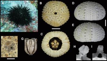
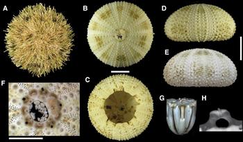
Eucidaris tribuloides (Lamarck, Reference Lamarck1816): A–E, G–H (MZUSP 679); F (MZUSP 651). (A) Specimen from Trindade Island photographed in situ, at 14 m depth; (B) aboral view; (C) oral view; (D) ambulacrum; (E) interambulacrum; (F) detail of the apical disc; (G) Aristotle's lantern; (H) apophysis. Scale bars: B–E, 10 mm; F, 5 mm.

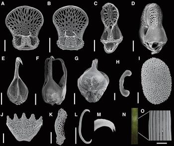
Eucidaris tribuloides (Lamarck, Reference Lamarck1816): A–E, I–L, O (MZUSP 669); F–H (MZUSP 1028); M–N (MZUSP 679). SEM images – (A–B) valves of narrow fistulate globiferous pedicellariae, and (C) detail of the head; valve of broad fistulate globiferous pedicellariae from a specimen (D) from the BOI and (F) from Boipeba island (Brazilian coast); (E, G) detail of the pedicellariae head, respectively; (H) detail of the edge of the pedicellaria depicted in (F); (I) slender tridentate pedicellariae from a specimen from Boipeba island; (J) plate from peristomial membrane; (K–M) tube feet ossicles; and (N–O) detail of a spine. Scale bars: A–B, D, F, I, 150 µm; J, 400 µm; K–O, 50 µm.

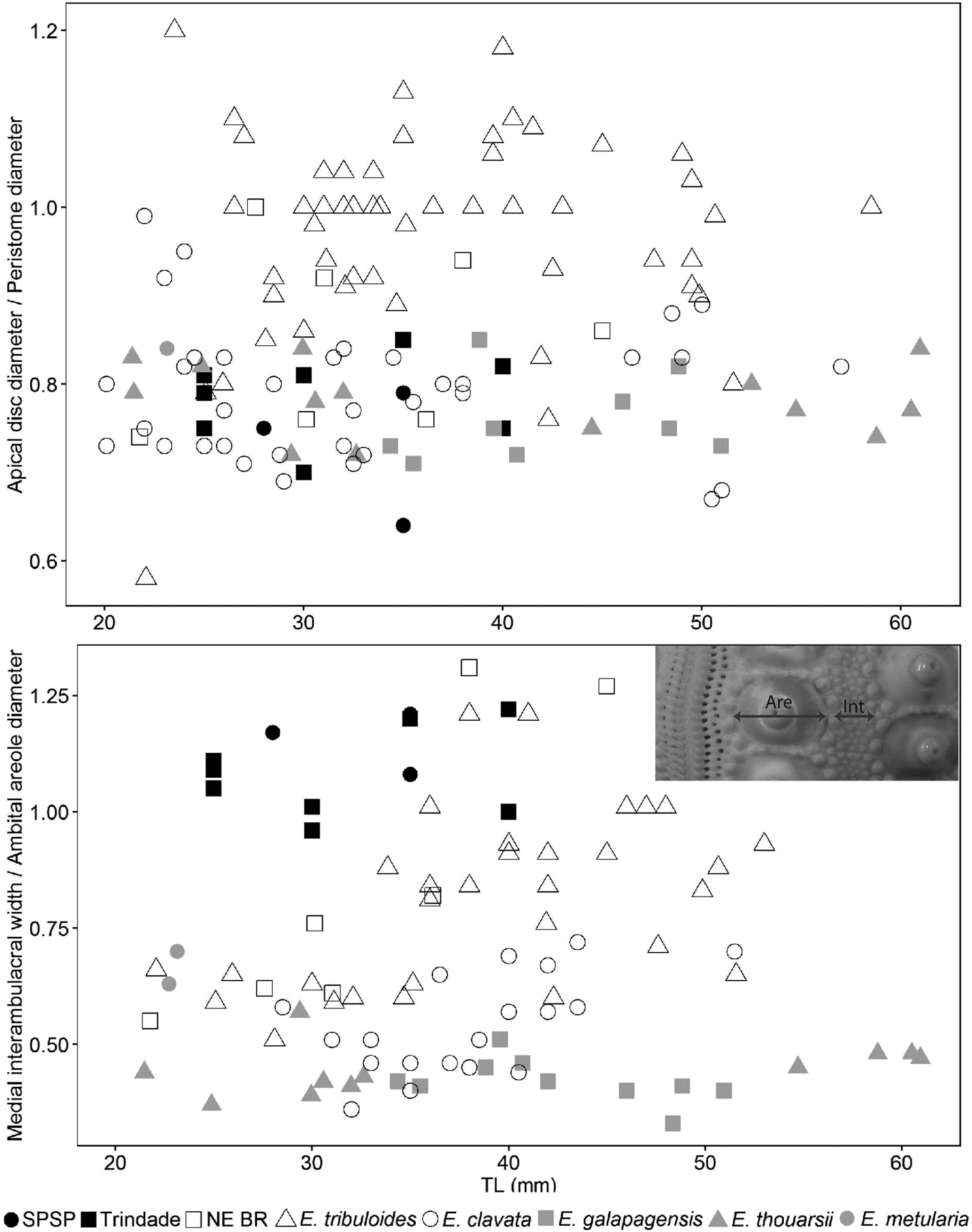
Ratios of structures of Eucidaris specimens plotted against their test diameter. Upper graph: apical disc diameter/peristome diameter ratio; bottom graph: (Int) median interambulacral width/(Are) ambital areole diameter. Measurements taken from E. clavata from AST (data from Pawson (Reference Pawson1978)); E. tribuloides from the Caribbean, West Africa (includes data from Pawson (Reference Pawson1978), and North-east Brazil (Alagoas and Bahia states (NE BR)) (in white), and SPSP and TMV (in black); and E. galapagensis, E. metularia and E. thouarsii from the eastern Pacific (in grey).

Eucidaris clavata Mortensen, Reference Mortensen1928, pp. 408–411, pls. 42, 67, 73, 86.
Cidaris tribuloides – Rathbun, Reference Rathbun1879, p. 143; Agassiz, Reference Agassiz1881, pp. 36–37, pl. 1; Brito, Reference Brito1968, pp. 17–18, pl. 8.
Eucidaris tribuloides – Döderlein, Reference Döderlein1887, p. 42, pl. 9; Mortensen, Reference Mortensen1928, pp. 400–408, pls. 41, 48, 73, 86; Bernasconi, Reference Bernasconi1955a, pp. 52–54, pl. 1.
Eucidaris tribuloides var. africana Mortensen, Reference Mortensen and Drygalski1909, pp. 40–43, pl. 6, 10, 13–16.
Eucidaris tribuloides tribuloides – Serafy, Reference Serafy1979, pp. 15–19, figure 3.
MATERIAL EXAMINED
Martin Vaz Island, ES, Brazil (20°28′–20°31′S 29°18′–20°51′W) – 19 m, 22.1.2012, 1 spm 15 mm long (MZUSP 659); 13 m, 23.7.2013, 3 spms 4.9–16.3 mm long (MZUSP 655); 12 m, 24.7.2013, 2 spms 25 mm long (MZUSP 668); 13 m, 23.8.2013, 4 spms 15–30 mm long (MZUSP 664). Trindade Island, ES, Brazil (20°29′–20°31′S 29°17′–20°20′W) – Andrada beach, 10 m, 17.7.2013, 2 spms 9–24 mm long (MZUSP 666); intertidal, 21.7.2013, 2 spms 15–30 mm long (MZUSP 654, 671). Cabritas beach, 9 m, 14.6.2012, 3 spms 20–40 mm long (MZUSP 690). Calheta beach, 23.7.2011, 1 spm 35 mm long (MZUSP 737); 4 m, 24.6.2012, 1 spm 30 mm long (MZUSP 677); 14 m, 26.6.2012, 3 spm 15–40 mm long (MZUSP 669); 17 m, 8.5.2014, 1 spm 25 mm long (MZUSP 983). Calheta Island, 12 m, 18.6.2012, 2 spms 11–25 mm long (MZUSP 675, 676); 4 m, 14.7.2013, 3 spms 8–25 mm long (MZUSP 658). Crista do Galo beach, 15 m, 21.5.2014, 3 spms 5–10 mm long (MZUSP 990). Enseada da Cachoeira, 12 m, 16–20.6.2012, 3 spms 6–35 mm long (MZUSP 685, 689); 18 m, 4.7.2012, 1 spm 5 mm long (MZUSP 647); 14 m, 9.7.2012, 1 spm 30 mm long (MZUSP 687); 21 m, 12.7.2012, 1 spm 35 mm long (MZUSP 683); 10 m, 9.7.2013, 2 spms 20–30 mm long (MZUSP 667). Enseada de Orelhas, 8 m, 24.1.2012, 1 spm 30 mm long (MZUSP 665); 10 m, 15.6.2012, 5 spms 10–25 mm long (MZUSP 682); 14 m, 30.6.2012, 2 spms 31.5–36 mm long (MZUSP 679); 15 m, 25.6.2012, 1 spm 20 mm long (MZUSP 656); 14 m, 6.7.2013, 6 spms 15–35 mm long (MZUSP 673); 12 m, 4.5.2014, 1 spm 26 mm long (MZUSP 1013); 10 m, 16.5.2014, 2 spms 30–40 mm long (MZUSP 989); 11 m, 21.5.2014, 2 spms 25–30 mm long (MZUSP 986). Enseada Portuguesa, 15 m, 10–15.7.2012, 9 spms 5–35 mm long (MZUSP 650, 663, 688). Farol beach, 13 m, 17.4.2014, 7 spms 10–35 mm long (MZUSP 987, 988); 14 m, 22.4.2014, 1 spm 10 mm long (MZUSP 1016); 13 m, 20.5.2014, 6 spms 10–40 mm long (MZUSP 984). Lixo beach, 23.5 m, 24.6.2012, 4 spms 8–17.5 mm long (MZUSP 653); 25 m, 2.7.2012, 2 spms 25–30 mm long (MZUSP 678). Ponta do Monumento, 12 m, 16.6.2012, 1 spm 25 mm long (MZUSP 651); 19.5 m, 30.6.2013, 1 spm 32 mm long (MZUSP 670). Ponta do Paredão, 19 m, 19.6.2012, 3 spms 6–25 mm long (MZUSP 652); 20 m, 20.6.2012, 4 spms 10–40 mm long (MZUSP 686). Ponta Noroeste, 12 m, 4.7.2012, 1 spm 10 mm long (MZUSP 657). Ponta Norte, 14 m, 18.7.2013, 1 spm 20 mm long (MZUSP 662). Príncipe beach, 19.5 m, 9.7.2013, 1 spm 35 mm long (MZUSP 672). Racha Island, 27 m, 22.6.2012, 5 spms 10–30 mm long (MZUSP 684); 30 m, 12.7.2012, 1 spm 30 mm long (MZUSP 681); 24 m, 13.7.2012, 2 spms 15–35 mm long (MZUSP 691); 25 m, 16.7.2012, 1 spms 15 mm long (MZUSP 661). Secon, 11 m, 23.4.2014, 3 spms 5–8 mm long (MZUSP 1011); 9 m, 12.5.2014, 6 spms 15–30 mm long (MZUSP 985). Tartarugas beach, 9.5 m, 26.4.2012, 1 spm 30 mm long (MZUSP 680).
ADDITIONAL MATERIAL
São Pedro and São Paulo Archipelago, PE, Brazil (20°30′S 29°18′W) – 21.2–5.3.2009, 2 spm 30–35 mm long (MZUSP 735, 736). Ilha Belmonte, intertidal, 27.10.2007, 1 spm 28 mm long (MZUSP 1029).
COMPARATIVE MATERIAL EXAMINED
Eucidaris tribuloides: Alagoas, Brazil (8°40′–10°29′S 35°9′–36°23′W) – 21.11.1998, 4 spms 28–36 mm long (CAS-IZ 15913, 98043). The Bahamas – 1 spm 50 mm long (CAS-IZ 91346); Abaco Island, 8.8.1954, 2 spms 35–42 mm long (CAS-IZ 91363); Gibson Bay – 3 spms 32–48 mm long (UCMP 123405). Bahia, Brazil (12°57′–13°39′S 38°W) – Castelhanos beach, Boipeba, 17.9.2012, 2 spms 38–45 mm long (MZUSP 1028, 1030); Itapuã Beach, Salvador, 29.7.2007, 1 spm 22 mm long (CAS-IZ 175395). Colombia (10°N 75°W) – Cartagena Bay, 3 spms 22–26 mm long (CAS-IZ 91365); La Cieba, 1931, 1 spm 30–34 mm long (UCMP 123406). Gulf of Mexico (24°26′N 81°48′W) – 68 m, 15.1.1885, 3 spms 31–51 mm long (CAS-IZ 91340). Jamaica (18°N 77′W) – 3 spms 30–34 mm long (UCMP 123429). Panama (Atlantic coast) – 6.1913, 1 spm 52 mm long (CAS-IZ 91372). San Pedro Macoris beach, Hispaniola Island, Dominic Republic (18°42′N 69°12′W) – 1.5 m, 4.3.1965, 1 spm 14 mm long (UCMP 123408). St. John Island, Virgin Islands (18°N 64°W) – 4.1958, 1 spm 28 mm long (UCMP 123407). Eucidaris galapagensis Döderlein, Reference Döderlein1887: Galapagos Islands, Ecuador (0°15′S–0°15′N 89°45′–90°15′W) – Academy Bay, Santa Cruz Island, 0–3 m, 24–25.1.1964, 2 spms 46–49 mm long (UCMP 123409). Baltra Island, 2–4.5 m, 12.2.1964, 2 spms 39–51 mm long (UCMP 123410, 123436). Darwin Bay, Genovesa Island, 0–3 m, 1–16.2.1964, 3 spms 25–34 mm long (UCMP 123411, 123437–123438). Plaza Island, 8.2.1964, 1 spm 20 mm long (UCMP 123412). Venedig Lagoon, Santa Cruz Island, 3–4.5 m, 19.2.1964, 1 spm 48 mm long (UCMP 123413). Eucidaris metularia (Lamarck, Reference Lamarck1816): Marianas Islands, Guam, U.S.A. (12–21°N 145°W) – 0.5–1.5 m, 10.8.1992, 1 spm 23 mm long (CAS-IZ 108829). Oahu, Hawaii, USA (21°N 157°W) – 21.7.1939, 1 spm 23 mm long (CAS-IZ 96779). Eucidaris thouarsii (L. Agassiz & Desor, Reference Agassiz and Desor1846): Brasilito Bay, Costa Rica (10°24′N 85°50′W) – 0–12 m, 13.5.1968, 1 spm 31 mm long (UCMP 123428). Colombia (2°57′–6°16′N 77°26′–79°11′W) – Punta Cotudo, 2–27 m, 1–2.5.1968, 1 spm 25 mm long (UCMP 123417). Punta Mono, Cauca, 3–33.5 m, 27.4.1968, 1 spm 21 mm long (UCMP 123418). Culebra Island, Canal Zone, Panama (8°55′N 79°32′W) – 0–4.5 m, 5.4.1968, 2 spm 29–33 mm long (UCMP 123419, 123445). Mexico – Islas Revillagigedo, Clarion Island, 2 spms 44.5–59 mm long (CAS-IZ 91360). Jalisco, Tenacatita Bay, 4.5–12 m, 27.5.1968, 1 spm 22 mm long (UCMP 123420). Los Angeles, Sonora, intertidal, 7.4.1959, 1 spm 32 mm long (UCMP 123440). Puerto Vallarta, intertidal, 5.4.1968, 1 spm 22 mm long (UCMP 123421). Punta Sur, Esmeraldas, Ecuador (1°0′N 79°54′W) – 7.5–12 m, 23.4.1968, 1 spm 30 mm long (UCMP 123416).
DESCRIPTION
Test thick, globular in outline, diameter 5–40 mm. Apical disc (Figure 1F) monocyclic or hemicyclic, ocular plates II and III exsert (ocular plates I, IV and V in contact with elongated periproctal plates); periproctal plates numerous, tubercles tiny, numerous. Peristome about 1.5 larger than apical disc. Perradial zone about 1.5 wider than poriferous zone, two series of secondary tubercles in each half ambulacrum (outer series larger than inner). Interambulacra (Figure 1E) more than twice as wide as ambulacra (Figure 1D); primary tubercle areola at ambitus almost twice width of middle interambulacral region. Peristomial membrane covered with plates. Cidaroid lantern (Figure 1G). Ear-shaped apophyses (Figure 1H). Tubercles perforate, non-crenulate.
Primary spines pencil-like (mean diameter 3.5 mm). Spine shaft with short projections pointing outwards (resembling warts in optical microscope), ending in crown; scrobicular spines short, broad flattened; marginal ambulacral spines short, narrow, flattened. Two types of fistulate globiferous pedicellariae. Narrow type slender, blade margins with numerous small teeth throughout, glandular cavity with large teeth, and terminal tooth often bifurcated or completely split into two (370–520 µm; Figure 2A–C). Broad type slightly curved, broad base, top half of blade with long marginal teeth, else smooth, glandular cavity with large teeth, no terminal tooth (570–690 µm; Figure 2D). Tridentate pedicellariae not found. Ossicles showing as thick plates with small perforations (1200–1320 µm; Figure 2J) in peristomial membrane; thorny rods (115–225 µm; Figure 2L, M) and crowns (220–260 µm; Figure 2K) in the tube feet.
Colour light brown, denuded test green with white tubercles; apical disc plates with transversal dark green stripe across middle of plate forming circle around anus; primary spines brownish, warts dark, crown pale, scrobicular spines creamy with dark brown tips, marginal ambulacral spines apical brown or greenish with dark tips.
DISTRIBUTION
Known from North Carolina (USA) to Rio de Janeiro (Brazil), and Gulf of Guinea; oceanic island distribution: Bermuda, Cape Verde, São Tomé, Fernando de Noronha, ASC, STH, TMV (Clark, Reference Clark1898; Mortensen, Reference Mortensen1928; Bernasconi, Reference Bernasconi1955a; Brito, Reference Brito1968; Lima-Verde, Reference Lima-Verde1969; Pawson, Reference Pawson1978; Serafy, Reference Serafy1979; Brown, Reference Brown2015; present paper). Bathymetric range: 0–800 m (Serafy, Reference Serafy1979).
BIOLOGICAL NOTES
In TMV, specimens were often found in association with brachyuran crabs (Majidae), sponges and algae.
REMARKS
The specimens from the BOI differ from Eucidaris australiae Mortensen, 1950 in having the primary spines ending in a crown and narrower globiferous pedicellariae, from E. metularia by the presence of tubercles widespread in the genital plates, from E. thouarsii by colouration of the scrobicular spines (light with dark tip vs. all dark in E. thouarsii); and from E. galapagensis by the absence of thick spines (5–7 mm). Pawson (Reference Pawson1978) showed that the thickness of the spines in Eucidaris from the Ascension Island increases with the depth, but all E. galapagensis analysed herein, from various depths, had thick spines compared with the other Eucidaris species.
Eucidaris tribuloides vs. Eucidaris clavata Mortensen, Reference Mortensen1928: according to Pawson (Reference Pawson1978), there are two valid Eucidaris species in the Atlantic Ocean, E. tribuloides and E. clavata, both differentiated by the ratio between the apical disc and the peristome width (AW/PW), and by the ratio between the diameter of the primary tubercle's areola and the width of the denuded area of the interambulacrum (DA/WI; both measurements taken at the ambitus). Figure 3 shows the ratios calculated herein combined with data extracted from Pawson (Reference Pawson1978). Only specimens over 20 mm were plotted. The AW/PW ratio (Figure 3) shows that the specimens from the BOI are more similar to E. clavata and to E. thouarsii than to the Caribbean E. tribuloides. Specimens from the Brazilian coast were nested within both groups. Conversely, the DA/WI ratio (Figure 3) shows that the specimens from the BOI are more similar to E. tribuloides than to the other Eucidaris species. However, this similarity is only present in specimens over 35 mm in test diameter because specimens from the BOI did not change the ratio with ontogeny. Because these differences between E. tribuloides and E. clavata are very subtle, we recommend accepting only the species E. tribuloides in the Atlantic Ocean, which is in agreement with the molecular analysis with the COI gene performed by Lessios et al. (Reference Lessios, Kessing, Robertson and Paulay1999).
Variation within E. tribuloides: the shape of the terminal tooth of the small globiferous pedicellariae from the Trindade specimens is slightly different from the terminal tooth of E. tribuloides from the Caribbean; however, data from specimens from the other oceanic islands are needed for further comparison.
Specimens from Boipeba island (Bahia, north-eastern Brazil): two specimens from Bahia analysed for comparison have some morphological differences when compared with other Eucidaris tribuloides specimens: the DA/WI ratio is very high (ratios are 1.25 and above; see Figure 3); slender tridentate pedicellariae are abundant (815–890 µm; Figure 2I) (vs. rare or lacking); and the broad fistulate globiferous pedicellariae have a straight edge (vs. curved), the blade has marginal teeth throughout (vs. only up to the top half), and the glandular cavity has fewer large teeth (1–2 vs.3–4 in each side), and a terminal tooth (300–330 µm; Figure 2F) (vs. terminal tooth lacking). The blades of the broad fistulate globiberous pedicellariae from Eucidaris depicted in Mortensen (Reference Mortensen and Drygalski1909, Reference Mortensen1928) are curved in Eucidaris tribuloides and only denticulated up to their top half in all Eucidaris species; if those drawings are accurate, the pedicellariae from the Boipeba island specimens are unique within the genus. Narrow fistulate globiferous pedicellariae is very similar (445–520 µm).
Intraspecific variation within E. tribuloides could be related to little geographic isolation between the oceanic islands and/or local environmental differences; however, the specimens from Boipeba Island could be a new species and need additional observations. Further molecular studies with more genes and additional specimens from the oceanic and continental islands are necessary to study their connectivity.
Order DIADEMATOIDA Duncan, 1889
Family DIADEMATIDAE Gray, 1855
Genus Diadema Gray, 1825
Diadema ascensionis Mortensen, Reference Mortensen and Drygalski1909
(Figures 4 & 5)
Diadema ascensionis Mortensen, Reference Mortensen and Drygalski1909, pp. 55–58, pls. 7, 16.
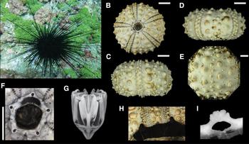
Diadema ascensionis Mortensen, Reference Mortensen and Drygalski1909: A–C, E–F (MZUSP 599); D, I (MZUSP 603); G (MZUSP 600); H (MZUSP 643). (A) Specimen from Trindade Island photographed in situ, at 9.5 m depth; images of the carapace showing (B) aboral view, (C) ambulacrum, interambulacrum of a specimen (D) 39.2 mm long and (E) 50 mm long; detail of the (F) apical disc, (G) Aristotle's lantern, (H) buccal notches, and (I) auricle. Scale bars: B–E, 10 mm; F, 5 mm.

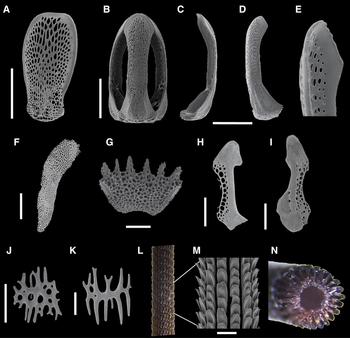
Diadema ascensionis Mortensen, Reference Mortensen and Drygalski1909: A (MZUSP 603); B–E (MZUSP 1003); F, H–K (MZUSP 599); G (MZUSP 600); L–N (MZUSP 590). SEM images – (A) valve of triphyllous pedicellariae; (B–D) valves of narrow rostrate pedicellariae, and (E) detail of the head; (F) plate from peristomial membrane; tube feet ossicles are (G) crown, (H–I) perforated plate, and (J–K) ‘doubled-sided hair-comb’; (L–M) longitudinal view and (N) cross section of the distal portion of a spine. Scale bars: A–D, 100 µm; F, 500 µm; G–K, 100 µm; M, 500 µm.

Diadema setosum – Rathbun, Reference Rathbun1879, p. 143.
Diadema ascensionis – Mortensen, Reference Mortensen1940, pp. 279–281, pls. 48, 54, 61, 73; Tommasi, Reference Tommasi1966, p. 11, figures 3–12; Brito, Reference Brito1968, pp. 19–20, pl. 9; Brito, Reference Brito1971, p. 264; Gondim et al., Reference Gondim, Dias and Christoffersen2013, p. 432, figures 1 & 2.
Diadema antillarum ascensionis – Pawson, Reference Pawson1978, p. 17.
EXAMINED MATERIAL
Martin Vaz Island, ES, Brazil (20°30′S 29°18′W) – 13 m, 23.7.2013, 3 spms 15–25 mm long (MZUSP 641, 644). Trindade Island, ES, Brazil (20°29′–20°31′S 29°17′–20°20′W) – Andrada beach, 10 m, 17.8.2013, 3 spms 25–60 mm long (MZUSP 639, 643). Cabritas beach, 9.5 m, 14.6.2012, 1 spm 40.8 mm long (MZUSP 610). Calheta beach, 16 m, 20.5.2014, 3 spms 10–21 mm long (MZUSP 1018). Calheta Island, 12.6.2012, 1 spm 30 mm long (MZUSP 600). Crista do Galo beach, intertidal, 12.6.2012, 1 spm 40 mm long (MZUSP 640); 12.7.2012, 1 spm 35 mm long (MZUSP 601); 15 m, 21.5.2014, 3 spms 40–50 mm long (MZUSP 1003). Enseada da Cachoeira, 10.5 m, 9.7.2013, 1 spm 31.8 mm long (MZUSP 646). Enseada Portuguesa, 9.5 m, 14.6.2012, 1 spm 16 mm long (MZUSP 638); 15.7.2013, 1 spm 50 mm long (MZUSP 599); 10 m, 18.4.2014, 3 spms 20–25 mm long (MZUSP 1005). Farol beach, 12 m, 15.7.2013, 1 spm 25 mm long (MZUSP 642); 13 m, 17.4.2014, 3 spms 15–40 mm long (MZUSP 1004). Farrilhões beach, 10.5 m, 9.7.2013, 1 spm 35 mm long (MZUSP 646). Secon, 10 m, 29.4.2014, 2 spms 25–65 mm long (MZUSP 1002). Tartarugas beach, 9.5 m, 26.4.2012, 1 spm 15 mm long (MZUSP 739); 2 m, 29.6.2013, 6 spms 15–40 mm long (MZUSP 602, 603, 645).
ADDITIONAL MATERIAL
Fernando de Noronha archipelago, PE, Brazil (3°48′S 32°23′W) – Rata Island, 13.4.1999, 1 spm 73 mm long (MZUSP 604).
COMPARATIVE MATERIAL EXAMINED
Diadema antillarum Philippi, 1845: Discovery Bay, Jamaica – 1 spm 58 mm long (private collection, in Smith & Kroh, Reference Smith and Kroh2011). Colon, Honduras (16°00′N 86°30′W) – 25.5.1975, 1 spm 80 mm long (UCMP 123422). New Providence Island, The Bahamas (25°15′N 77°15′W) – 8.1956, 1 spm 80 mm long (UCMP 123423). Diadema mexicanum A. Agassiz, 1863: Clipperton Island, France (Pacific territory) (10°17′N 109°13′W) – 24.10.1956, 1 spm 55 mm long (UCMP 123424). Isla Montuosa, Panama (7°29′N 82°15′W) – 3–7.5 m, 9.4.1968; 1 spm 55 mm long (UCMP 123425). Santa Cruz Island, Galapagos Islands, Ecuador (0°15′S–0°15′N 89°45′–90°15′W) – 0–3 m, 24–25.1.1964, 1 spm 75 mm long (UCMP 123426). Santa Elena Bay, Ecuador (2°11′S 80°56′W) – 6 m, 17.4.1968, 1 spm 65 mm long (UCMP 123427).
DESCRIPTION
Test circular in outline, low, diameter 10–65 mm. Apical disc (Figure 4F) small, hemicyclic, ocular plates II and III exsert, 1–2 tubercles may occur in ocular plates only. Hydropores of madreporic plate enclosed in crescent moon-shaped area, apparently constrained by arch-shaped depression. Gonopores large, located on distal tip of triangular, wide genital plates; periproct large with tiny plates peripherally. Ambulacra (Figure 4C) slightly elevated, narrow, trigeminate plates composed of pore-pairs in single series, in distinct arcs of three and widening to four pore-pairs adorally. Primary tubercles small, arranged into two parallel series between columns of pore-pairs; secondary tubercles in zigzag series along perradial suture. Interambulacra (Figure D) wide, three series of primary tubercles in specimens over 70 mm in carapace diameter (two series in smaller specimens): outer series parallel to midline of plates; tubercles in middle series smaller, series shorter (only nearby ambitus); tubercles in inner series arranged in single columns. Secondary tubercles scattered, although most concentrated on lateral edges of plates. Peristomial membrane with spineless ambulacral plates, thick and undulating buccal podia. Buccal notches pronounced (Figure 4H). Aulodont lantern (Figure 4G). Auricles fused perradially, lobed edge (Figure 4I). Tubercles perforate, crenulate.
Primary spines long, brittle, with hollow lumen, 22–25 solid wedges; secondary spines smaller, thinner than primary spines. Spine shaft verticillate. Rostrate pedicellariae slender, blade smoothly serrated (double columns), narrow blade tip (335–870 µm; Figure 5B–E); tryphylous pedicellariae with long, broad blade (165–265 µm; Figure 5A); claviform pedicellariae not found. Peristomial membrane ossicles showing as perforated plates of varied shapes (590–1850 µm; Figure 5F); tube feet ossicles of various shapes (perforated plate (230–350 µm; Figure 5H, I), ‘doubled-sided hair-comb’ (90–200 µm; Figure 5J, K), and crown (270–350 µm; Figure 5G)).
Colour black, denuded test and tubercles white; porous region of madreporite and anal cone black; peristomial membrane dirt white with purple-brownish ring around the mouth; primary spines dark, varying from brown to purple (banded white/brownish spines in juveniles), oral spines black.
DISTRIBUTION
Occurs alongside the Brazilian coast, Alagoas probably to Rio de Janeiro (Brazil) (considering that all Brazilian specimens were mistaken for D. antillarum); oceanic island distribution: Rocas Atoll, Fernando de Noronha, ASC, STH, TMV (Mortensen, Reference Mortensen1940; Tommasi, Reference Tommasi1966; Lima-Verde, Reference Lima-Verde1969; Brito, Reference Brito1971; Pawson, Reference Pawson1978; Gondim et al., Reference Gondim, Dias and Christoffersen2013; present paper). Bathymetric range: 0–30 m (Mortensen, Reference Mortensen1940).
BIOLOGICAL NOTES
In TMV, specimens were gregarious, and usually found in reef crevices during the day and also moving around areas with high percentage of green algae cover.
REMARKS
Morphological differences among the Diadema species rival intraspecific differences. Although molecular data support most of the species (e.g. Lessios et al., Reference Lessios, Kessing and Pearse2001), identifying specimens when DNA sequences are not available is very hard. We assigned the specimens from TMV to D. ascensionis based on the absence of broad tridentate pedicellariae, rostrate pedicellariae (narrow tridentate) with broad tip, the number of solid wedges on the spines, and the interambulacra tuberculation pattern.
The broad and narrow types of tridentate pedicellariae are probably not homologous since D. mexicanum has both types. Diadema antillarum has mainly the broad type, while D. ascensionis apparently has only the narrow type. In addition, the number of solid wedges in the spines was higher than the average for D. antillarum (18 solid wedges; Coppard & Campbell, Reference Coppard and Campbell2004). Regarding the tuberculation pattern in the interambulacra, D. antillarum develops the third column of tubercles early on; specimens 60 mm in test diameter already have three columns in each interambulacrum and the inner column is not contiguous. We also analysed specimens from Honduras, and they had the same pattern observed in our specimens. Molecular data have shown that D. ascensionis is nested within D. antillarum, however, the analysis was based on only one gene and it is not possible to infer if both species are indeed separated without a proper analysis to the population level.
The specimens from the BOI differ from D. africanum Rodríguez, Hernández, Clemente & Coppard, 2013 by the shape of the rostrate pedicellariae and the interambulacra tuberculation pattern (three columns in D. africanum), from D. mexicanum by the absence of broad tridentate pedicellariae, from D. palmeri Baker, 1967 by having a hemicyclic apical system (vs. monocyclic), from D. paucispinum A. Agassiz, 1863 in having arch-shaped depression and only 0–2 tubercles on genital plates (vs. 2–6 tubercles), from D. savignyi (Audouin, 1829) in having 22–25 solid wedges on spines (vs. mean of 18), and from D. setosum (Leske, Reference Leske1778) by the absence of long and slender tridentate pedicellariae.
Order CAMARODONTA Jackson, 1912
Family ECHINOMETRIDAE Gray, 1855
Genus Echinometra Gray, 1825
Echinometra lucunter (Linnaeus, Reference Linnaeus1758)
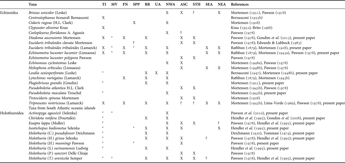
(Figures 6 & 7, Table 1)
Echinus lucunter Linnaeus, 1758, p. 665.
Echinometra lucunter (Linnaeus, Reference Linnaeus1758): A–C, F (MZUSP 619); D–E, G–H (MZUSP 616); I (MZUSP 1290). (A) Specimen from Trindade Island photographed in situ, between 0–1 m depth; images of the carapace showing (B) aboral view, (C) oral view, (D) ambulacrum, and (E) interambulacrum; detail of the (F) apical disc, (G) Aristotle's lantern, and auricle of (H) specimen from the BOI, and (I) from the mainland. Scale bars: B–E, 10 mm; F, 5 mm.

Echinometra lucunter (Linnaeus, Reference Linnaeus1758): A, E–L (MZUSP 623); B–D (MZUSP 616). SEM images – (A) valve of triphyllous pedicellaria; (B–C) valves of fanged globiferous pedicellariae, and (D) detail of the head; (E–H) valves of ophicephalous pedicellariae; (I) plate from peristomial membrane; (J) crown from tube feet; and (K–L) detail of a spine. Scale bars: A, 50 µm; B, 200 µm; C, 150 µm; E–H, 100 µm; I, 200 µm; J–K, 100 µm.

Morphological traits of the test used to distinguish the Echinometra species. Numbers in parentheses indicate rare amounts of pores.

Echinometra lucunter – Mortensen, Reference Mortensen1943b, pp. 357–368, pls. 41–44; Bernasconi, Reference Bernasconi1955a, pp. 62–63, pl. 2; Brito, Reference Brito1968, pp. 21–22, pl. 10.
Echinometra subangularis – Rathbun, Reference Rathbun1879, pp. 143–144.
EXAMINED MATERIAL
Martin Vaz Island, ES, Brazil (20°30′S 29°18′W) – 13 m, 26.6.2013, 1 spm 30 mm long (MZUSP 611); 13 m, 23.7.2013, 3 spm 5–39 mm long (MZUSP 618, 648). Trindade Island, ES, Brazil (20°29′–20°32′S 29°17′–20°20′W) – Andrada beach, intertidal, 5–21.7.2013, 6 spms 5–30 mm long (MZUSP 605, 625); 10 m, 17.7.2013, 5 spms 15–25 mm long (MZUSP 620). Calheta Island, intertidal, 11.6.2012, 4 spms 25–30 mm long (MZUSP 614). Enseada da Cachoeira, 10.5 m, 9.7.2012, 2 spms 35–42 mm long (MZUSP 622); 14 m, 9.7.2014, 1 spm 47 mm long (MZUSP 621). Enseada de Orelhas, 15 m, 25.6.2012, 1 spm 17 mm long (MZUSP 660). Enseada Portuguesa, 12 m, 15–18.7.2013, 4 spms 25–30 mm long (MZUSP 616, 617). Ponta do Monumento, 12 m, 16.6.2012, 3 spms 10–25 mm long (MZUSP 649); 8 m, 2.4.2014, 3 spms 24–37 mm long (MZUSP 1001). Farrilhões beach, 12.5 m, 31.1.2012, 1 spm 10 mm long (MZUSP 608). Secon, 9 m, 12.5.2014, 9 spms 15–35 mm long (MZUSP 1000). Tartarugas beach, 11 m, 1 spm 5 mm long (MZUSP 607); 9.5 m, 26.6.2012, 4 spms 15–25 mm long (MZUSP 609, 740); 14 m, 28.6.2012, 4 spms 10–25 mm long (MZUSP 613); intertidal, 15.7.2012, 2 spms 28.5–37 mm long (MZUSP 619); 2 m, 29.6.2013, 7 spms 20–25 mm long (MZUSP 623); 12 m, 3.7.2013, 1 spm 35 mm long (MZUSP 612); intertidal, 5.7.2013, 4 spms 30–40 mm long (MZUSP 615); 14.5 m, 11.7.2013, 1 spm 10 mm long (MZUSP 606). Tunel beach, 11.6.2012, 2 spms 13–35 mm long (MZUSP 624).
COMPARATIVE MATERIAL EXAMINED
Echinometra lucunter lucunter: Bolivar Province, Colombia (10°N 75°W) – 4 spms 50–54 mm long (CAS-IZ 91207). ‘Gayanca stone reef’, Brazil – 3 spms 30 mm long (CAS-IZ 15938). Jorge Grego Island, Angra dos Reis, RJ, Brazil (coord) – 9.8.2009, 2 spms 44.5–58 mm long (MZUSP 1290). Echinometra insularis H.L. Clark, 1912: Ecuador – 3 spms 40–45 mm long (CAS-IZ 91093). Echinometra mathaei (Blainville, 1825): Midway Island, Pacific Ocean (28°N 177°W) – 2 spms 58 mm long (CAS-IZ 91179). The Philippines – 2 spms 45 mm long (CAS-IZ 189015). Upolu Island, Western Samoa (13°S 171°W) – 2 spms 39 mm long (CAS-IZ 91184). Echinometra oblonga (Blainville, 1825): Islas Revillagigedo, Socorro Island, Mexico (18°N 110°W) – 2–12.5.1925, 2 spms 32–42 mm long (CAS-IZ 91202). Echinometra vanbrunti A. Agassiz1863: Gulf of California, Mexico (22°–24°N 109°–112°W) – Bahia Magdalena, 27.4.1916, 1 spm 45 mm long (CAS-IZ 91232); Cape San Lucas, 2 spms 60 mm long (CAS-IZ 17104).
DESCRIPTION
Test subpentabonal or oval in outline, longer axis along ocular plate I and genital plate 3, diameter 5–47 mm. Apical disc (Figure 6F) monocyclic or hemicyclic, ocular plate V exsert (rarely, ocular plate I also exsert). Plates bearing three or more tubercles, madreporic plates almost entirely covered by hydropores, periproctal plates large, few. Ambulacral plating (Figure 6D) polygeminate, five to six (rarely seven) pore-pairs to a compound plate, arranged in arcs. Poriferous zone a bit larger than perradial zone. Primary tubercles small, arranged into two parallel series between columns of pore-pairs; secondary tubercles in zigzag series along perradial suture. Interambulacra (Figure 6E) with two parallel series of large primary tubercles arranged in midline of plates, one series of smaller primary tubercles near each ambulacrum, one in zigzag along the interradial suture. Peristomial membrane with few dispersed plates. Buccal notches shallow. Camarodont lantern (Figure 6G). Auricles fused perradially, distal portion broad, lobed edge (Figure 6H). Tubercles imperforate, non-crenulate.
Primary spines all cylindrical, medium-sized. Spine shaft with tiny denticulations arranged into transversal groups of four teeth, about 15 µm apart from the next group. Fanged globiferous pedicellariae slender, margins smooth, short lateral unpaired tooth on blade, long terminal tooth (730–1050 µm; Figure 7B–D); ophicephalous pedicellariae robust with irregularly denticulate margins (375–600 µm; Figure 7E–H); triphyllous pedicellariae broad, flat, margin smooth (100–110 µm; Figure 7A); tridentate pedicellariae not found. Ossicles showing as perforated plates in peristomial membrane (800–830 µm; Figure 7I), crowns in tube feet (330–585 µm; Figure 7J), C-shaped ossicles in both (17–43 µm).
Peristomial membrane brownish with dark green rim around the mouth, tube feet green-brownish with pale tips, denuded test beige; primary spines dark purple orally and black aborally, secondary spines greenish to purple (from base to tip); apical plates beige with brownish spots.
DISTRIBUTION
From North Carolina (USA) to Santa Catarina (Brazil) (western Atlantic); from Senegal to Angola (eastern Atlantic); oceanic island distribution: Bermuda, Cape Verde, São Tomé, Fernando de Noronha, ASC, STH, Abrolhos, TMV (Rathbun, Reference Rathbun1879; Agassiz, Reference Agassiz1881; Clark, Reference Clark1898; Mortensen, Reference Mortensen1943b; Pawson, Reference Pawson1978; Hendler et al., Reference Hendler, Miller, Pawson and Kier1995; present paper). Bathymetric range: 0–45 m (Serafy, Reference Serafy1979).
BIOLOGICAL NOTES
In TMV, specimens were gregarious and found in rocky bottoms.
REMARKS
The specimens reported herein are very similar to the coastal populations, but the subpentagonal test shape is unusual and the shape of the auricle is different (Figure 6I). The subspecies Echinometra lucunter polypora Pawson, Reference Pawson1978 was described from AST populations and differs from Echinometra lucunter lucunter by the presence of seven pore-pairs in the arcs (vs. six/five) and the greenish or white colour of the adapical region of the test. However, the number or pore-pairs in the arcs are very variable in the genus Echinometra and molecular analysis with the COI gene does not support the separation of E. lucunter into subspecies (McCartney et al., Reference McCartney, Keller and Lessios2000).
The differences between E. lucunter and the other Echinometra species are summarized in Table 1. We considered E. mathaei (Blainville, 1825) and E. oblonga (Blainville, 1825) as separate species because of morphological differences regarding their auricle and test shape. This separation has also been supported by molecular data (see McCartney et al., Reference McCartney, Keller and Lessios2000; Landry et al., Reference Landry, Geyer, Arakaki, Uehara and Palumbi2003).
Family TOXOPNEUSTIDAE Troschel, 1872
Genus Pseudoboletia Troschel, 1869
Pseudoboletia maculata Troschel, Reference Troschel1869
(Figures 8 & 9)
Pseudoboletia maculata Troschel, Reference Troschel1869, p. 96.
Pseudoboletia maculata Troschel, Reference Troschel1869: A (MZUSP 1027); B–H (MZUSP 1026). (A) Specimen fixed in ethanol; images of the carapace showing (B) aboral view, (C) oral view, (D) ambulacrum, and (E) interambulacrum; detail of the (F) apical disc, (G) Aristotle's lantern, and (H) auricle. Scale bars: B–E, 10 mm; F, 5 mm.

Pseudoboletia maculata Troschel, Reference Troschel1869 (MZUSP 1027). SEM images – (A–B) valves of triphyllous pedicellariae; (C–D) valves of ophicephalous pedicellariae; (E–G) valves of fanged globiferous pedicellariae; (H) C-shaped ossicle; (I) rosette-like plate from peristomial membrane; (J) crown, (K) plate, and (L) C-shaped ossicle from tube feet ([M] detail of the tip of the C-shaped ossicles depicted in (L)), and (K–L) detail of a spine. Scale bars: A–B, 50 µm; C–D, 100 µm; E, 200 µm; F, 100 µm; G, 50 µm; H, 5 µm; I–K, 100 µm; L, 20 µm; O, 250 µm.

Pseudoboletia occidentalis H.L. Clark, Reference Clark1921, pp. 115–118, pl. 2.
Pseudoboletia occidentalis – Mortensen, Reference Mortensen1943a, pp. 538–540.
Pseudoboletia maculata maculata – Pawson, Reference Pawson1978, pp. 17–20.
EXAMINED MATERIAL
Trindade Island, ES, Brazil (20°29′–20°30′S 29°18′–29°20′W) – Enseada de Orelhas, 15 m, 24.10.2014, 2 spms 16–23 mm long (MZUSP 1123). Enseada Portuguesa, 15 m, 10.7.2012, 1 spm 35 mm long (MZUSP 1026). Secon, 9 m, 12.5.2014, 1 spm 55 mm long (MZUSP 1027).
COMPARATIVE MATERIAL EXAMINED
Pseudoboletia atlantica: Prosperous Bay, STH – 27 m, 17.12.1945 (NHM-UK 1945.12.17.8).
Pseudoboletia maculata: Guam, USA – 2 m, 3.1992, 1 spm 73 mm long (CAS-IZ 81252). Lubang Islands, The Philippines (13°50′N 120°05′E) – 1–18 m, 23.5.2014, 1 spm 60 mm long (CAS-IZ 203488). Pseudoboletia indiana (Michelin, 1862): Hawaii, USA – 2 spms 65 mm long (CAS-IZ 91174, 92324); Oahu, 7.5 m, 2 spm 50–60 mm long (CAS-IZ 91339).
DESCRIPTION
Test circular in outline, diameter 35–55 mm. Apical disc small, hemicyclic (Figure 8F), ocular plates II, III and IV exsert. Madreporic plate twice wider than other genital plates; 2–3 tubercles on proximal edge of plates; periproctal plates covering periproct, sometimes bearing tubercle. Ambulacra polygeminate plates composed of arc with four pore-pairs (Figure 8D). Primary tubercles arranged into two parallel series among pore-pairs; tubercles from outer series absent in aboral region. Interambulacra (Figure 8E) with four parallel series of primary tubercles, innermost series smaller, present only at ambitus; secondary tubercles scattered. Peristomial membrane with dispersed small plates, five pairs of buccal plates with spines and pedicellariae. Tip of oral tube feet crenulated. Buccal notches pronounced. Camarodont lantern (Figure 8G). Auricles widened at edges and fused perradially (Figure 8H). Tubercles imperforate, non-crenulate.
All spines cylindrical, small. Fanged globiferous pedicellariae slender, margins smooth, perforations for insertion of adductor muscles widespread on proximal region of blade, terminal tooth present (300–910 µm; Figure 9E–G); ophicephalous pedicellariae robust, broad, margins denticulate, slightly serrate (430–670 µm; Figure 9C, D); triphyllous flat, margin apparently smooth, narrow neck (130–240 µm; Figure 9A, B); tridentate pedicellariae not found. Rosette-like oval ossicles in the peristomial membrane (300–475 µm; Figure 9I); tube feet with crowns (205–555 µm; Figure 9J), perforated plates (265–390 µm; Figure 9K) and thin C-shaped ossicles with bifurcated tips (40 µm; only one ossicle found; Figure 9H); C-shaped ossicles on all pedicellariae (11–30 µm).
Denuded test creamy, brown blotches on aboral region; primary spines with brown/green base, white tips; apical disc plates with brown ring; tube feet and pedicellariae light pink.
DISTRIBUTION
Venezuela, Barbados, Trindade Islands and Indo-West Pacific (Smith & Kroh, Reference Smith and Kroh2011; Alvarado & Solís-Marín, Reference Alvarado and Solís-Marín2013; present paper). Bathymetric range: 9–183 m (Mortensen, Reference Mortensen1943a; present paper).
REMARKS
Pawson (Reference Pawson1978) did an extensive revision of this genus and in this paper we adopt his suggestion that Pseudoboletia occidentalis Clark, Reference Clark1921, is a junior synonym of P. maculata. Pseudoboletia maculata differs from Pseudoboletia atlantica H.L. Clark, 1912 by the presence of four pore-pairs in each arc (vs. five) and by the colouration of the test (sparse dark blotches vs. dark stripes from apical disc to peristome), and from P. indiana in having dark spots on the test (vs. uniform colour). Although known worldwide, this is the first record of P. maculata in the South Atlantic, and the first record of the species for Brazil. This species can easily be mistaken for other toxopneustids, such as Tripneustes ventricosus and Lytechinus variegatus; therefore, we would not be surprised if it occurs elsewhere along the Brazilian coast.
Genus Tripneustes L. Agassiz, 1841
Tripneustes ventricosus (Lamarck, Reference Lamarck1816)
(Figures 10 & 11)
Echinus ventricosus Lamarck, Reference Lamarck1816, p. 44.
Tripneustes ventricosus (Lamarck, Reference Lamarck1816): A–F, H (MZUSP 632); G (MZUSP 633). (A) Specimen from Trindade Island photographed in situ, between 0–1 m depth; images of the carapace showing (B) aboral view, (C) oral view, (D) ambulacrum, and (E) interambulacrum; detail of the (F) apical disc, (G) Aristotle's lantern, and (H) auricle. Scale bars: B–E, 10 mm; F, 5 mm.

Tripneustes ventricosus (Lamarck, Reference Lamarck1816): A, H–I (MZUSP 1020); B–G, J–L (MZUSP 632). SEM images – (A) valve of triphyllous pedicellaria; (B–D) valves of ophicephalous pedicellariae; (E–F) valves of fanged globiferous pedicellariae; (G–H) valves of tridentate pedicellariae; (I) rosette-like plate from peristomial membrane; (J) crown from tube feet; and (K–L) detail of a spine. Scale bars: A, 100 µm; B–H, 200 µm; I–J, 150 µm, L, 100 µm.

Tripneustes ventricosus – Agassiz & Desor, Reference Agassiz and Desor1846, p. 363; Mortensen, Reference Mortensen1943a, pp. 490–498, pls. 33, 34, 37, 38, 56; Tommasi, Reference Tommasi1962, p. 59, figures 1 & 2.
EXAMINED MATERIAL
Trindade Island, ES, Brazil (20°29′–20°31′S 29°18′–29°20′W) – Andrada beach, intertidal, 21.7.2013, 1 spm 40 mm long (MZUSP 632). Cabritas beach, 9 m, 14.6.2012, 2 spms 75–80 mm long (MZUSP 630); 7.5 m, 28.4.2014, 1 spm 75 mm long (MZUSP 998). Calheta beach, 16 m, 29.4.2014, 2 spms 40–55 mm long (MZUSP 991); 17 m, 8.5.2014, 2 spms 30 mm long (MZUSP 995); 16 m, 20.5.2014, 1 spm 20 mm long (MZUSP 1017). Calheta Island, 10 m, 24.7.2011, 1 spm 34 mm long (MZUSP 628); 12 m, 18.6.2012, 1 spm 60 mm long (MZUSP 629); 4 m, 14.7.2013, 1 spm 60 mm long (MZUSP 633). Enseada da Cachoeira, 14 m, 9.7.2012, 1 spm 23.5 mm long (MZUSP 738). Enseada de Orelhas, 14 m, 30.6.2012, 2 spms 35–51 mm long (MZUSP 637). 14 m, 6.7.2013, 5 spms 15–80 mm long (MZUSP 635, 674); 12 m, 18.4.2014, 5 spms 30–70 mm long (MZUSP 999); 14 m, 9.5.2014, 1 spm 77 mm long (MZUSP 1014); 10 m, 21.5.2014, 1 spm 50 mm long (MZUSP 993). Enseada Portuguesa, 14 m, 10.7.2012, 3 spms 30–40 mm long (MZUSP 627); 12 m, 11.7.2012, 4 spms 35–55 mm long (MZUSP 631); 12 m, 15.7.2013, 5 spms 30–70 mm long (MZUSP 636). Farol beach, 13 m, 22.4.2014, 5 spms 50–60 mm long (MZUSP 996); 13 m, 8–20.5.2014, 12 spms 30–60 mm long (MZUSP 994, 1020, 1021). Farrilhões beach, 10 m, 9.7.2013, 2 spms 20–90 mm long (MZUSP 626, 634); 14 m, 5.5.2014, 2 spms 10–15 mm long (MZUSP 1015). Lixo beach, 14 m, 22.4.2014, 1 spm 60 mm long (MZUSP 1012). Ponta do Monumento, 8 m, 2.4.2014, 1 spm 55 mm long (MZUSP 997).
COMPARATIVE MATERIAL EXAMINED
Tripneustes ventricosus: Arraial do Cabo, RJ, Brazil (22°S 42°W) – 1–2 m, 15.10.1998, 1 spm 112.39 mm long (CAS-IZ 116108). Puerto Rico – 1934, 1 spm 71 mm long (CAS-IZ 91159). Tripneustes depressus A. Agassiz, 1863: Isla Danzante, Baja California, Mexico (25°47′N 111°15′W) – 9 m, 2 spms 57–78 mm long (CAS-IZ 101402). Islas Revillagigedo, Clarion Island, Mexico (18°N 114°W) – 19.3.1993, 1 spm 89 mm long (CAS-IZ 103105). Tripneustes gratilla (Linnaeus, Reference Linnaeus1758): Hawaii, USA – Hawaii, 2 spms 60–62 mm long (CAS-IZ 108120); Oahu, 1 spm 111 mm long (CAS-IZ 91164).
DESCRIPTION
Test circular in outline, diameter 10–90 mm. Apical disc (Figure 10F) small, hemicyclic, ocular plates II and III exsert; ocular plate IV may be exsert or not, touching periproct only slightly through an extension between the genital plates. Madreporic plate enlarged, 2–3 tubercles on proximal edge; periproctal plates covering periproct, sometimes bearing a tubercle. Genital plates usually bearing two tubercles, gonopores large. Ambulacra (Figure 10D) with trigeminate plates composed of three columns of pore-pairs; inner column almost straight, pores in middle column arranged diagonally. Primary tubercles arranged in parallel series between columns of pore-pairs; tubercles in inner series larger than in outer series. Secondary tubercles scattered. Interambulacra (Figure 10E) with two parallel series of primary tubercles arranged in midline of plates and scattered tubercles of various sizes. Peristomial membrane naked. Buccal notches pronounced. Camarodont lantern (Figure 10G). Auricles long, widened at edges, fused perradially (Figure 10H). Tubercles imperforate, non-crenulate.
All spines cylindrical, small; spine shaft with tiny vertical projections arranged in horizontal groups of 2–4, about 20 µm apart from next group. Fanged globiferous pedicellariae slender, margins smooth, perforations for insertion of adductor muscles only in half innermost part of proximal region, a terminal tooth present (430–465 µm; Figure 11E, F); ophicephalous pedicellariae robust, margins serrate, denticulate (240–655 µm; Figure 11B–D); tridentate slender, distal cap denticulate, some spines widely separated in middle of valve (370–750 µm; Figure 11G, H); triphyllous pedicellariae broad, flat, upper margin finely serrate (160–205 µm; Figure 11A). Dumbbell-shaped ossicles on all pedicellariae, larger ones curved (9–20 µm), tube feet with crown (340–425 µm; Figure 11J), peristomial membrane with rosette-like round ossicles (255–315 µm; Figure 11I).
Colour dark grey, denuded test white-brownish, tubercles pale; primary spines light grey or pale green with brownish tips, secondary spines grey-purplish; apical plates light grey with dark grey-purplish spots around the gonopores; tube feet and tridentate pedicellariae yellowish, ophicephalous pedicellariae dark brown.
DISTRIBUTION
Occurs from Florida (USA) to São Paulo (Brazil); oceanic island distribution: Fernando de Noronha, ASC (?), STH (?), and Trindade Islands (Mortensen, Reference Mortensen1943a; Tommasi, Reference Tommasi1962; Lima-Verde, Reference Lima-Verde1969; Pawson, Reference Pawson1978). Bathymetric range: 0–55 m (Serafy, Reference Serafy1979).
BIOLOGICAL NOTES
In TMV, specimens were usually solitary and found in rocky bottoms.
REMARKS
The specimens described herein have no apparent morphological variations from specimens from mainland or other islands. Lessios et al. (Reference Lessios, Kane and Robertson2003) found three clades within Tripneustes ventricosus (Caribbean, Brazil and São Tomé) and it would be interesting to sequence molecular data from mid-Atlantic specimens to access the connectivity between these regions.
Only three extant Tripneustes species are recognized as valid worldwide, although the molecular data presented by Lessios et al. (Reference Lessios, Kane and Robertson2003) did not support the Indo-Pacific species. Morphologically, T. ventricosus can be distinguished from T. gratilla in being densely covered with tubercles, especially in the interambulacra; and from T. gratilla and T. depressus in the reduced/absence of plates in the peristomial membrane.
Order CLYPEASTEROIDA A. Agassiz, 1872
Family MELLITIDAE Stefanini, 1912
Genus Leodia Gray, 1851
Leodia sexiesperforata (Leske, Reference Leske1778)
(Figures 12 & 13)
Echinodiscus fexiesperforatus Leske, Reference Leske1778, p. 199.
Leodia sexiesperforata (Leske, Reference Leske1778) (MZUSP 1070). (A) Specimen from Trindade Island photographed in situ, between 16–17 m depth; images of the carapace showing (B) aboral view and (C) oral view; detail of the (D) apical disc, (E) peristome, and (F) periproct; Aristotle's lantern (G) oral and (H) aboral side, and (I–J) detail of individual pyramids. Scale bars: B–C, 20 mm; D–F, 2 mm; G–H, 5 mm; I–J, 1 mm.

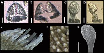
Leodia sexiesperforata (Leske, Reference Leske1778) (MZUSP 1070). Optical microscope images of the (A–D) bidentate pedicellariae, of the spines from the (E) edge of the test, and from the (F) aboral region; and (G) SEM image of the tip of a spine depicted in (F). Scale bars: A–D, 10 µm; G, 100 µm.

Mellita sexforis – A. Agassiz, 1972–1974, p. 141; Rathbun, Reference Rathbun1879, p. 144.
Mellita sexiesperforata – Bernasconi, Reference Bernasconi1947, p. 104.
Leodia sexiesperforata – Mortensen, Reference Mortensen1948b, pp. 429–432, pls. 58, 61, 72; Serafy, Reference Serafy1979, pp. 74–75, figure 32.
EXAMINED MATERIAL
Trindade Island, ES, Brazil (20°29′–20°30′S 29°18′–29°20′W) – Calheta beach, 16–17 m, 20–24.10.2014, 31 spms 39–60 mm long (MZUSP 1070, 1126); 17 m, 4.11.2014, 48 spms 25–70 mm long (MZUSP 1124, 1125). Enseada de Orelhas, 13.9 m, 6.I.2014, 5 spms 47–65 mm long (MZUSP 1190). Racha Island, 25 m, 12.11.2014, 4 spms 60–65 mm long (MZUSP 1191).
COMPARATIVE MATERIAL EXAMINED
Leodia sexiesperforata: Boipeba, BA, Brazil (13°36′S 38°53′W) – Bainema beach, 17–31.1.2015, 17 spms 55–83.5 mm long (MZUSP 1198).
DESCRIPTION
Test pentagonal in outline, posterior edge sharp, flattened, 25–70 mm long. Six long, narrow lunules; anal lunule's proximal margin right after end of petals. Apical disc monobasal (Figure 12D), four gonopores (G5 missing), hydropores spread throughout apical disc, ocular plates small. Petals broad, short (1/3 of the ambulacrum), petal III longest. Both columns of pore-pairs bowed, closing at end; poriferous zone broad, fully tuberculated, up to seven primary tubercles per row. One to two extra pore-pairs beyond petals. Peristome small, circular (Figure 12E). Clypeasteroid lantern with prominent, strongly lamellar pyramid wings, supra-alveolar processes located far from tip (Figure 12G–J). Periproct (Figure 12F) small, longer than wide, on first interambulacral plate, slightly closer to peristome than to anal lunule. Food grooves bifurcate near peristome, broadens, and narrow down again towards posterior margin of test, bifurcating again. Oral tube feet extend into interambulacra. Test fully tuberculated, tubercles larger on oral ambulacrum than on interambulacrum. Tubercles perforate, crenulate.
Spines thin, small, mainly of three types: straight spines of varying sizes orally (largest between peristome and periproct), curved spines on peristome and at ambitus, swab-shaped spines aborally. Bidentate pedicellariae with broad serrate blade, short base on aboral region (32–38 µm; Figure 13A–D).
Specimen blackish in vivo, test pale in ethanol; spines pale.
DISTRIBUTION
From South Carolina (USA) to Rio La Plata (Argentina); oceanic island distribution: Bermuda Island and Trindade Islands (H.L. Clark, Reference Clark1898; Bernasconi, Reference Bernasconi1947; Mortensen, Reference Mortensen1948b; present paper). Bathymetric range: 0–490 m (A. Agassiz, Reference Agassiz1972–1974; Serafy, Reference Serafy1979).
BIOLOGICAL NOTES
In TMV, specimens were found in soft bottoms (medium-grained sand).
REMARKS
Leodia sexiesperforata is the only extant species of the genus. Some intraspecific variations to coastal Brazilian specimens include the shape of the test (subpentagonal to round in coastal specimens) and the location of the anal lunule (placed more proximally, usually between posterior petals, in coastal specimens).
Order SPATANGOIDA L. Agassiz, 1840
Family BRISSIDAE Gray, 1855
Genus Plagiobrissus Pomel, 1883
Plagiobrissus grandis (Gmelin, Reference Gmelin and Gmelin1791)
(Figures 14 & 15)
Echinus grandis Gmelin, Reference Gmelin and Gmelin1791, p. 3200.
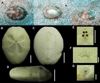
Plagiobrissus grandis (Gmelin, Reference Gmelin and Gmelin1791): A (MZUSP 1084); B, G (MZUSP 1289); C–F (MZUSP 1117). (A) Specimen from Trindade Island photographed in situ, at 16 m depth; images of the carapace showing (B) aboral view, (C) oral view and (D) lateral view of the body; and detail of the (E) apical disc, (F) peristome and (G) periproct. Scale bars: B–D, 20 mm; E–G, 5 mm.

Plagiobrissus grandis (Gmelin, Reference Gmelin and Gmelin1791) (MZUSP 1289). SEM images – (A–B) valves of triphyllous pedicellariae; (C) valve of rostrate pedicellaria; (D–E) valves of tridentate pedicellariae; (F–G) valves of long globiferous pedicellariae; (H–I) rods from tube feet; and (K–L) detail of a spine. Scale bars: A–B, 30 µm; C–E, 50 µm; F–G, 100 µm; H–J, 20 µm; K, 200 µm.

Metalia pectoralis – A. Agassiz, 1881, p. 199.
Plagiobrissus grandis – Mortensen, Reference Mortensen1951, pp. 496–368, pls. 40–41, 63; Serafy, Reference Serafy1979, pp. 98–99, figure 43.
EXAMINED MATERIAL
Trindade Island, ES, Brazil (20°30′S 29°18′W) – Calheta beach, 16 m, 24.10.2014, 9 spms 58–76 mm long (MZUSP 1084); 17 m, 4.11.2014, 7 spms 60–100 mm long (MZUSP 1117, 1289).
COMPARATIVE MATERIAL EXAMINED
Plagiobrissus grandis: The Bahamas (24°02′–25°01′N 74°29′–77°24′W) – Nassau, 1 spm 195 mm long (NHM-UK 87.4.25.6); San Salvador Island, 3 m, 23.10.1983, 1 spm 87 mm long (USNM E32634). Florida, USA – 1 spm 148 mm long (USNM E13737); 5–11 m, 1964, 1 spm 87 mm long (USNM E36776). Panama (9°24′N 79°51′W) – Galeta Island, 10.7 m, 30.9.1980, 1 spm 97 mm long (USNM E36447).
DESCRIPTION
Test oval in outline, flattened, 58–90 mm in length. Frontal groove slight but present, more developed in larger specimens. Apical disc ethmolytic (Figure 14E), four gonopores and genital plate 2 separates oculars III and IV. Petals almost 180 degrees in younger specimens, angle diminishing in largest specimen. Paired petals deeply sunk; anterior petal slightly sunk. First half of inner columns of paired petals less developed than outer columns; distal plates in petals occluded. Interporiferous zone much narrower than poriferous zones. Peripetalous fasciole indented in interambulacral 2 and 3, subanal fasciole shield-shaped, anal and subanal fascioles confluent. Primary tubercles inside peripetalous fasciole developed. Periproct large, tear-shaped (Figure 14G), truncate. Peristome broad, kidney shaped (Figure 14F); labrum short, in contact with both sternal plates. Sternal plates parallel, very long, narrow, with an elevated midline, completely tuberculated. Phyllodes well developed, with four to five pores in each half ambulacrum. Tubercles perforate, crenulate.
Spines thin; large inside peripetalous fasciole, around periproct and on oral region (except on plastron); small elsewhere. Long globiferous pedicellariae triangular, robust, with dense stereom (510–700 µm; Figure 15F, G), located near periproct. Rostrate pedicellariae with short denticulate blade, long and narrow neck, smooth margins, broad base (350–400 µm; Figure 15C). Tridentate pedicellariae near petals, blade narrow, serrate, base broad (165–270 µm; Figure 16D, E). Triphyllous pedicellariae near peristome, blade long, serrate margin, narrow neck, base short (100–150 µm; Figure 15A, B). Regular globiferous and ophicephalous pedicellariae not found. Phyllopodia ossicles showing as thin rods, straight or slightly curved, sometimes bifurcated at middle or at tips (35–100 µm); flat perforated plates (50–95 µm). Respiratory tube feet with rods only (40–140 µm; Figure 15H, I).
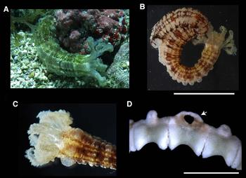
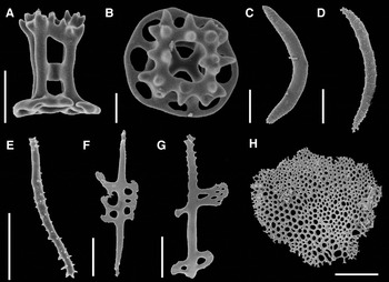
Chiridota rotifera (Pourtalès, Reference Pourtalès1851): A, C (MZUSP 944); B (MZUSP 946); D (MZUSP 973). Specimen fixed in ethanol: (A) lateral view of the body, (B) miliar granules (indicated by the arrow) on the posterior region of the body, (C) detail of the anterior region (arrow indicates warts (aggregations of wheel) in tentacles and body wall), and (D) detail of tentacles and calcareous ring (indicated by the arrow). Scale bars: A–B, 5 mm; D, 1 mm.

Colour in vivo: aboral surface of specimen dark brown with white patches, oral surface with pale spines, phyllopodia red, long globiferous pedicellariae dark brown. Test white in ethanol.
DISTRIBUTION
Occurs from Florida (USA) to São Paulo (Brazil); oceanic island distribution: Trindade Island (Mortensen, Reference Mortensen1951; Brito, Reference Brito1962; present paper). Bathymetric range: 1–210 m (Serafy, Reference Serafy1979).
BIOLOGICAL NOTES
In TMV, specimens were found in soft bottoms, usually completely buried in medium-grained sand.
REMARKS
The specimens from the BOI differ from the Caribbean specimens in having plates with similar width in each column of the anterior interambulacral (when comparing same-sized specimens from both regions). The inner column of the Caribbean specimens is much narrower than the outer column. Also, the tubercles are slightly larger on the specimens from the Caribbean. This variation is very slight and does not justify description of a new species.
Plagiobrissus grandis differs from P. africanus (Verrill, 1871) by the absence of large primary tubercles close to the frontal ambulacrum (there are medium-sized tubercles in the specimens from the BOI), and by the shape of the periproct, which is obliquely truncate and not visible in oral view (vs. vertically truncate and visible in oral view); it differs from Plagiobrissus (Rhabdobrissus) costae (Gasco, 1876), P. (R.) jullieni (Cotteau, 1889) and P. (R.) pacificus (Clark, 1940) in having sunk petals (vs. flush petals).
Class HOLOTHUROIDEA
Order APODIDA Brandt, 1835
Family CHIRIDOTIDAE Östergren, 1898
Genus Chiridota Eschscholtz, 1829
Chiridota rotifera (Pourtalès, Reference Pourtalès1851)
(Figures 16 & 17)
Synapta rotifera Pourtalès, Reference Pourtalès1851, p. 15.
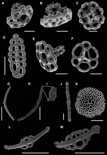
Chiridota rotifera (Pourtalès, Reference Pourtalès1851): (MZUSP 944). SEM and optical microscope images of the ossicles – wheels from body wall in (A) ventral, (B) dorsal and (C) lateral view; (D) early developmental stage of a wheel; (E) C-shaped ossicles from body wall; (F–H) rods from tentacles. Scale bars: A–D, 20 µm; E, 10 µm; F–H, 50 µm.

Chiridota rotifera – Ludwig, Reference Ludwig1881, pp. 116–117.
Toxodora ferruginea – Verrill, Reference Verrill1882, p. 220; Clark, Reference Clark1907, p. 126; Deichmann, Reference Deichmann1930, p. 213.
Chiridota ferruginea – Deichmann, Reference Deichmann1940, pp. 228–229.
EXAMINED MATERIAL
Trindade Island, ES, Brazil (20°29′–20°31′S 29°18′–20°20′W) – Enseada de Orelhas, 14 m, 9.5.2014, 2 spms 1–2 cm long (MZUSP 945). Farol beach, 13 m, 8.5.2014, 2 spms 0.5–2 cm long (MZUSP 942); 12 m, 8.4.2014, 3 spms 1–2 cm long (MZUSP 937); 13 m, 8–22.4.2014, 4 spms 2–3 cm long (MZUSP 938–941). Farrilhões beach, 12 m, 4.4.2014, 1 spm 1 cm long (MZUSP 946). Secon, 12 m, 12.4.2014, 1 spm 3.5 cm long (MZUSP 944).
DESCRIPTION
Body vermiform (Figure 16A), maximum length 3.5 cm. Body wall transparent, miliar granules posteriorly (Figure 16B). Twelve white, digitate (fingerlike) tentacles subdivided into five pairs of digits (Figure 16C). Calcareous ring with 12 pieces, radial pieces perforated on top for insertion of radial nerves (Figure 16D). Ten to 20 Polian vesicles.
Body wall wheels (30–100 µm; Figure 17A–D) with six spokes each, inner rim serrated, margins smooth; C-shaped ossicles (20–60 µm; Figure 17E) with few small projections mainly at tips. Tentacles with wheels as in body wall (30–70 µm); slightly curved rods (30–90 µm; Figure 17F) with some projections at tips (rarely along the body); bi or tri-radiated, curved rods, branched at ends rods (40–60 µm; Figure 17G, H).
DISTRIBUTION
Occurs from Florida to Brazil (to São Paulo); oceanic island distribution: Trindade Island (Deichmann, Reference Deichmann1930; Tommasi, Reference Tommasi1969; Hendler et al., Reference Hendler, Miller, Pawson and Kier1995; present paper). Bathymetric range: from the intertidal down to 360 m (Alvarado & Solís-Marín, Reference Alvarado and Solís-Marín2013).
BIOLOGICAL NOTES
In TMV specimens found in association with calcareous algae.
REMARKS
Eight species of Chiridota are known from the Atlantic Ocean, of which only C. rotifera, C. marenzelleri Perrier, 1904 and C. pisanii Ludwig, 1887 have been recorded from the South Atlantic so far.The tentacle morphology is the main character to distinguish C. rotifera from its Atlantic congeners: C. rotifera differs from C. peloria Deichmann, Reference Deichmann1930, C. ferruginea (Verrill, Reference Verrill1882), C. laevis (Fabricius, 1780) and C. heheva (Pawson & Vance, 2004) in the numbers of digits (10 vs. 14, 10–12, 4–6, 20, respectively), and can be separated from C. hydrothermica (Smirnov & Gebruk, 2000) in having finger-like tentacles (vs. lobe-like). Regarding the South Atlantic species, C. rotifera differs from C. pisanii in the number of Polian vesicles (7 vs. 10–12), and differs from C. marenzelleri by the presence of stellate ossicles. Clark (Reference Clark1907) recommended a thorough comparison between C. rotifera and C. ferruginea as both species share similar ossicles and overlap in the number of tentacles digits. No morphological differences were observed between coastal and oceanic insular specimens of C. rotifera.
Family SYNAPTIDAE Burmeister, 1837
Genus Euapta Östergren, 1898
Euapta lappa (Müller, Reference Müller1850)
(Figures 18 & 19)
Synapta lappa Müller, Reference Müller1850, pp. 134–135.
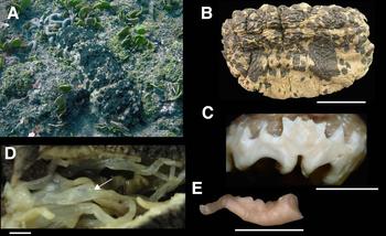
Euapta lappa (Müller, Reference Müller1850) (MZUSP 289). (A) Specimen from Trindade Island photographed in situ, at 14 m depth, and (B) fixed in ethanol; (C) detail of pinnate tentacles; (D) calcareous ring, arrow indicates place for insertion of radial nerves. Scale bars: B, 5 cm; E, 2 mm.

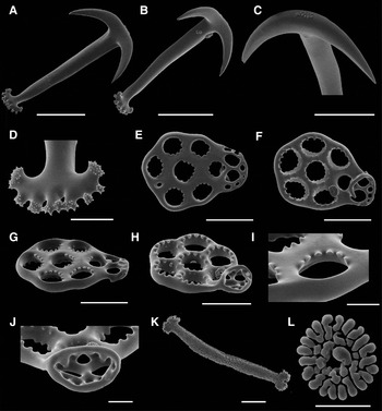
Euapta lappa (Müller, Reference Müller1850) (MZUSP 289). SEM images of the ossicles – anchors from body wall in (A) dorsal and (B) ventral view, (C) detail of minute tubercles on anchor vertex, and (D) detail of branched anchor stock; anchor plates from body wall in (E–F) dorsal and (G–H) ventral view; detail of (I) dentate holes and of (J) anchor handle; (K) rods and (L) rosettes from tentacles. Scale bars: A–C, 100 µm; D, 20 µm; E–H, 100 µm; I–L, 20 µm.

Synapta polii Ludwig, Reference Ludwig1875, p. 80.
Euapta lappa – Clark, Reference Clark1907, p. 73, pl. 4; Clark, Reference Clark1924, pp. 464–465, pl. 1; Heding, Reference Heding1928, pp. 136–137, pl. 8.
Euapta tobagoensis – Heding, Reference Heding1928, pp. 133–136, pl. 8.
EXAMINED MATERIAL
Trindade Island, ES, Brazil (20°29′S 29°19′W) – Enseada Portuguesa, 14 m, 10.7.2012, 12 spms 3–15 cm long (MZUSP 289, 982); 12 m, 15.7.2013, 10 spms 3–12 cm long (MZUSP 317, 318). Farol beach, 13 m, 8–17.4.2014, 26 spms 1.5–25 cm long (MZUSP 976, 980, 981); 14 m, 22–27.4.2014, 2 spms 13–20 cm long (MZUSP 977, 979); 13 m, 8.5.2014, 3 spms 6–11 cm long (MZUSP 978).
COMPARATIVE MATERIAL EXAMINED
Euapta lappa: Bacia do Espírito Santo, ES, Brazil, (20°26′S 39°44′W) – 1350 m, 9.2010, 2 spms 0.5–1 cm long (MZUSP 290). Great Sound, Long Island, Bermudas – 2 spms 5–6 cm long (USNM 25001). Euapta godeffroyi (Semper, Reference Semper1868): Baja California, Mexico – intertidal, 11.1.1959, 1 spm 8 long (CAS 192451); 17.12.1960, 3 spms 10 cm long (CAS 105518); 3.1889, 1 spm 7 cm long (USNM E 2437); 15 m, 30.4.1888, 1 spm 6 cm long (USNM E 2431).
DESCRIPTION
Body vermiform (Figure 18A, B) with brown, beige stripes, maximum length 25 cm. 15 pale pinnate tentacles (Figure 18C). Calcareous ring (Figure 18D) with 15 pieces and radial pieces, perforated at top for insertion of radial nerves. Many Polian vesicles, madreporite single.
Body wall anchors (200–360 µm; Figure 19A–C) with smooth arms, vertex with minute knobs (about 12), stock branched (8–9), minute knobs at ends (60–100 µm; Figure 19D). Anchor plates oval (200–300 µm long, 130–180 µm wide; Figure 19E–J), margins smooth, undulating; anterior region broad, seven dentate holes, three in midline, two in each side (denticles forming circles in both ventral and dorsal sides); bridge smooth, well developed; posterior region narrow with two large and 5–6 marginal holes. Tentacles with spiniform rods (100–300 µm; Figure 19K), slightly branched ends, and rosettes (10–40 µm; Figure 19L).
DISTRIBUTION
Occurs from the Gulf of Mexico to Brazil (Espírito Santo); oceanic island distribution: ASC, STH and Trindade Islands (Pawson, Reference Pawson1978; Hendler et al., Reference Hendler, Miller, Pawson and Kier1995; present paper). Bathymetric range: intertidal to 1350 m (Hendler et al., Reference Hendler, Miller, Pawson and Kier1995; Alvarado & Solís-Marín, Reference Alvarado and Solís-Marín2013).
BIOLOGICAL NOTES
In TMV this species has been found near rhodolites.
REMARKS
There are four valid species of Euapta worldwide: E. lappa differs from E. magna Heding, Reference Heding1928 in the presence of webbed digits on the tentacles, and from E. tahitiensis Cherbonnier, 1955 in the shape of the anchor plate. Euapta lappa and E. godeffroyi were historically separated by their pattern of geographic distribution (West-Indian and Indo-Pacific, respectively). Clark (Reference Clark1924) distinguished them by the size of their anchors and anchor plates (larger in E. lappa), while Heding (Reference Heding1928) found differences in the shape of their anchors.
This is the first record of E. lappa from the Brazilian coast. The specimens from the BOI differ from the specimens from Ascension islands in having larger anchors and anchor plates. No other morphological difference was found between them.
Order ASPIDOCHIROTIDA Grube, 1840
Family HOLOTHURIIDAE Burmeister, 1837
Genus Actinopyga Bronn, 1860
Actinopyga agassizii (Selenka, Reference Selenka1867)
(Figures 20 & 21)
Mülleria agassizii Selenka, Reference Selenka1867, pp. 311.
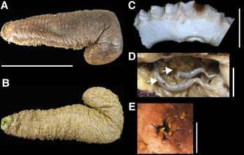
Actinopyga agassizii (Selenka, Reference Selenka1867) (MZUSP 293). (A) Specimen from Trindade Island photographed in situ, depth not recorded; (B) calcareous ring; (C) top view of mouth showing numerous tentacular ampullae (indicated by a yellow arrow in (D)) and a mottled brown Polian vesicle (indicated by a white arrow in (D)); and (E) detail of anal teeth. Scale bars: B, 5 mm; C, 1 mm; D, 5 mm; E, 3 cm.

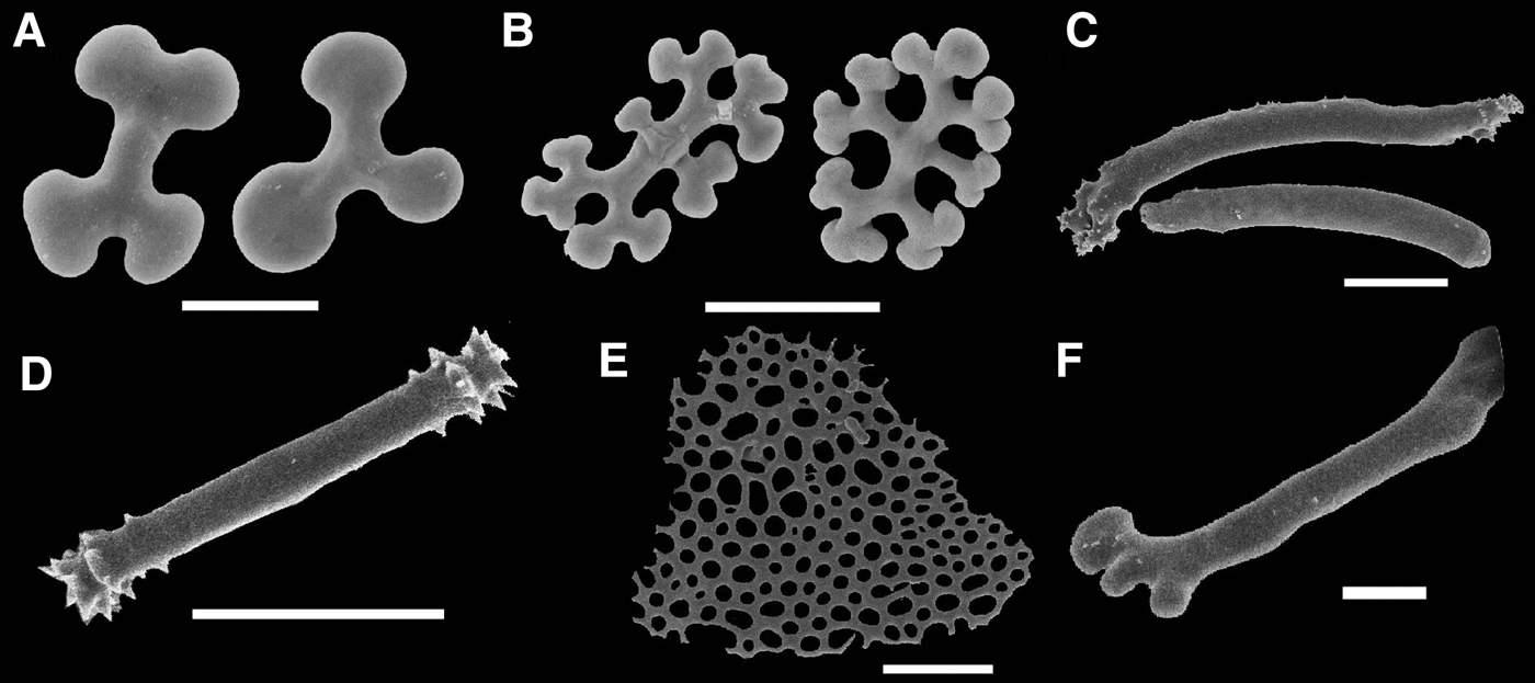
Actinopyga agassizii (Selenka, Reference Selenka1867) (MZUSP 293). SEM images of the ossicles – rosette-like elements from body wall: (A) ‘dog biscuit’-shaped and (B) branched rods; (C–D) rods from tentacles; and (E) end plate and (F) rod from tube feet. Scale bars: A–B, 20 µm; C–E, 100 µm; F, 20 µm.

Actinopyga agassizii – Hendler et al., Reference Hendler, Miller, Pawson and Kier1995, pp. 282–284, figures 157, 180.
EXAMINED MATERIAL
Martin Vaz archipelago, ES, Brazil (20°30′S 29°18′W) – Martin Vaz Island, 13 m, 23.7.2013, 13 spms 9–15 cm long (MZUSP 325, 327). Trindade Island, ES, Brazil (20°29′–20°31′S 29°17′–20°20′W) – Andrada beach, 10 m, 17.7.2013, 1 spm 14 cm long (MZUSP 326). Calheta beach, 12 m, 16.6.2012, 4 spms 15–17 cm long (MZUSP 292); 18.6.2012, 1 spm 14 cm long (MZUSP 293); 14 m, 26.6.2012, 1 spm 12 cm long (MZUSP 420); 12 m, 18.7.2012, 2 spms 13–15 cm long (MZUSP 723); 3.7.2012, 4 spms 9 cm long (MZUSP 305); 16 m, 28.4.2014, 6 spms 15–28 cm long (MZUSP 957, 960); 15 m, 29.4.2014, 1 spm 15 cm long (MZUSP 966); 12 m, 14.5.2014, 1 spm 11 cm long (MZUSP 954); 16 m, 20.5.2014, 1 spm 11 cm long (MZUSP 953). Enseada da Cachoeira, 9 m, 8.8.2013, 1 spm 14 cm long (MZUSP 418). Enseada de Orelhas, 10 m, 15.6.2012, 4 spms 12–15 cm long (MZUSP 304); 10 m, 6.7.2013, 1 spm 14 cm long (MZUSP 414). Farol beach, 12 m, 17.7.2013, 1 spm 3 cm long (MZUSP 320); 13 m, 22.4.2014, 4 spms 4–28 cm long (MZUSP 957, 963). Farrilhões beach, 31.1.2012, 1 spm 12 cm long (MZUSP 294); 12.5 m, 31.1.2012, 1 spm 17 cm long (MZUSP 297); 16 m, 2.7.2012, 2 spms 15 cm long (MZUSP 303). Lixo beach, 14 m, 22.4.2014, 2 spms 15–20 cm long (MZUSP 964). Pedra da Garoupa, 10 m, 1 spm 3 cm long (MZUSP 316). Rachada Island, 27 m, 2.6.2012, 1 spm 12 cm long (MZUSP 302). Tartarugas beach, 12.7.2013, 1 spm 12 cm long (MZUSP 415).
DESCRIPTION
Body thick (Figure 20A), without distinct warts dorsally or laterally, maximum length 28 cm. Body wall mottled brown, orange or yellow. 20–30 large yellow peltate tentacles. Calcareous ring (Figure 20B) without posterior projections; radial and interradial plates square-shaped, united along all length. Radial plates with two notches, three short projections in anterior region, base slightly curved. Interradial plates with three anterior projections (middle one bifurcated), straight base. Tentacular ampullae large, numerous (20–30; Figure 20C), mottled brown Polian vesicle (Figure 20D). Five conspicuous, white, calcareous teeth surrounding anus (Figure 20E).
Body wall rosette-like ossicles varying from simple ‘dog biscuit’ shapes (20–60 µm; Figure 21A) to complex dichotomously branched rods (20–40 µm; Figure 21B). Tentacles with two types of rods: curved and spiniform rods with perforated ends (150–200 µm; Figure 21C) and straight rods, smooth along their length and spiniform tips (40–70 µm; Figure 21D) (40–800 µm). Tube feet with perforated end plate (200–400 µm; Figure 21E); rods minutely branched, smooth along length, tips branched (30–100 µm; Figure 21F).
DISTRIBUTION
Occurs from South Carolina (USA) to the Caribbean; oceanic island distribution: Bermuda, TMV (Pawson et al., Reference Pawson, Pawson and King2010; present paper). Bathymetric range: intertidal up to 54 m (Pawson et al., Reference Pawson, Pawson and King2010).
BIOLOGICAL NOTES
Found in association with the pearlfish Carapus sp.
REMARKS
According to Panning (Reference Panning1944), the genus Actinopyga has about 18 valid species subdivided into three main groups: ‘Act. lecanora’, characterized by having simple and smooth rosettes, which occasionally have lateral extensions and swollen ends; ‘Act. echinites’, characterized by the narrowing and the elongation of the rosettes from the ventral body wall; and the ‘obesa-gruppe’, characterized by the presence of rough and often branched rods. Actinopyga agassizii belongs to the ‘Act. echinites’ group, and is the only species of the genus that occurs in the western Atlantic.
Actinopyga agassizii has been mentioned from Porto Seguro (Brazil) in a checklist without any characterization or illustration (see Magalhães et al., Reference Magalhães, Martins and Alves2005). This is, therefore, the first formal record of this species from the South Atlantic. SEM images of ossicles, images of specimens in situ and of internal morphology are provided.
Genus Holothuria Linnaeus, 1767
Holothuria (Cystipus) pseudofossor Deichmann, Reference Deichmann1930
(Figures 22 & 23)
Holothuria pseudofossor Deichmann, Reference Deichmann1930, pp. 57–58, figures 9–14.
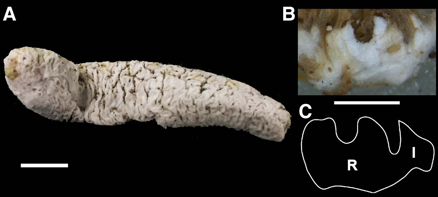
Holothuria (Cystipus) pseudofossor Deichmann, Reference Deichmann1930 (MZUSP 1305). (A) Specimen fixed in ethanol, (B) photograph and (C) drawing of the calcareous ring (‘R’, radial plate, ‘I’, interradial plate). Scale bars: A, 1 cm; B, 2 mm.

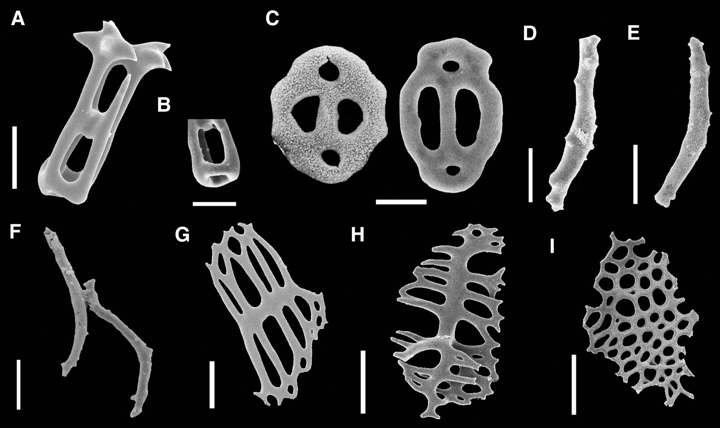
Holothuria (Cystipus) pseudofossor Deichmann, Reference Deichmann1930 (MZUSP 1305). SEM images of the ossicles – tables from body wall in (A) bottom, (B) lateral and (C) top view; (D) button from body wall; tables from ventral region in (E) lateral and (F) top view; (G–H) large curved rods ((I) detail of the tip of the rod depicted in (H) showing tiny perforation), and (J) small straight rod from tentacles; (K) end plate and (L–M) rods from tube feet. Scale bars: A–C, 20 µm; D, 50 µm; E–F, 20 µm; G–H, 50 µm; J, 10 µm; K–M, 100 µm.

Holothuria pseudofossor ― Deichmann, Reference Deichmann1954, pp. 389, 394, figure 66, 29–34.
Holothuria (Cystipus) pseudofossor ― Rowe, Reference Rowe1969, pp. 157; Tommasi, Reference Tommasi1974, pp. 2, figure 1; Borrero-Pérez et al., Reference Borrero-Pérez, Benavides-Serrato and Diaz-Sanchez2012, pp. 182–183; Prata et al., Reference Prata, Manso and Christoffersen2014, pp. 138–140, Figure 5.
EXAMINED MATERIAL
Trindade Island, ES, Brazil (29°30′S 29°20′W) – Racha Island, 27 m, 29.7.2015, 1 spm 6 cm long (MZUSP 1305).
DESCRIPTION
Body elongate (Figure 22A). Tube feet throughout whole body, maximum length 10 cm. Colour white in ethanol. Ten peltate tentacles. Calcareous ring simple, delicate, without posterior projections (Figure 22B, C). Radial plates rectangular-shaped, anterior region with one large notch, posterior region curved. Interradial plates with triangular anterior region, curved base. Internal organs were degraded and body was filled with sediment.
Body wall tables four-pillared (50–70 µm; Figure 23A–C), disc with one large central hole, eight marginal oval perforations, disc margin undulated, knobs bifurcated, adjacent to marginal perforations; spire knobbed, ending in 15–30 teeth projected laterally and upwards; buttons elongated, knobbed (100–160 µm; Figure 23D), margins undulated, 12–14 perforations of unequal size arranged into two parallel rows; middle bar knobbed, usually projecting beyond one extremity (rarely beyond both extremities, or not projecting at all). Ventral region of body with four-pillared tables, disc with one central hole, 6–8 marginal perforations, disc margin undulated, spire tall, several projections at tip (spire height: 40–70 µm, disc diameter: 60–70 µm; Figure 23E, F). Tentacles with two types of spiniform rods: large curved rods with pointed projections along outer side and at tips, inner side smooth, tips sometimes with tiny perforations (100–180 µm; Figure 23G–I); small straight rods with coarse projections along whole ossicle (40–70 µm; Figure 23J). Tube feet end plate (100–240 µm; Figure 23K) with large perforations, smaller perforations on edges; rods with 4–10 central perforations, 2–3 perforations at tips (100–200 µm; Figure 23L, M).
DISTRIBUTION
Occurs from the Gulf of Mexico to São Paulo (Brazil); oceanic island distribution: Trindade Island (Tommasi, Reference Tommasi1974; Alvarado & Solís-Marín, Reference Alvarado and Solís-Marín2013; present paper). Bathymetric range: 3–370 m (Alvarado & Solís-Marín, Reference Alvarado and Solís-Marín2013).
REMARKS
There are three species of Holothuria (Cystipus) in the Atlantic Ocean: H. (C.) pseudofossor differs from H. (C.) cubana Ludwig, Reference Ludwig1875 by the absence of ‘large supporting plates’ (sensu Deichmann, Reference Deichmann1930) in the tube feet and the simple morphology of the table spire (vs. heavily knobbed spires sometimes ending in a sphere with blunt teeth; note that simpler tables are also found in this species, and these are probably an earlier developmental stage of the tables), and from H. (C.) occidentalis Ludwig, Reference Ludwig1875 in having simple flattened knobbed buttons (vs. complex ovoid knobbed buttons sometimes folded and/or with protuberances).
No morphological differences were observed between specimens from the BOI and the Brazilian coast. However, ontogenetic variation, which has already been documented for Caribbean specimens (Deichmann, Reference Deichmann1930; Ortiz-Gómez et al., Reference Ortiz-Gómez, Borrero-Pérez, Zea, Harris, Böttger, Walker and Lesser2006), was found. The specimens described by Tommasi (Reference Tommasi1974; length = 2.4 cm) and Prata et al. (Reference Prata, Manso and Christoffersen2014; length = 4 cm) from Paraíba (North-east of Brazil) have buttons and tables with undeveloped knobs, and the teeth at the top of their tables are also undeveloped.
Holothuria (Halodeima) grisea Selenka, Reference Selenka1867
(Figures 24 & 25)
Holothuria grisea Selenka, Reference Selenka1867, p. 328, pl. 18.
Holothuria (Halodeima) grisea Selenka, Reference Selenka1867 (MZUSP 1057). (A) Specimen fixed in ethanol; detail of calcareous ring (B) before and (C) after bleach treatment. Scale bars: A, 6 cm; B–C, 5 mm.

Holothuria (Halodeima) grisea Selenka, Reference Selenka1867 (MZUSP 1057). SEM images of the ossicles: body wall tables in (A) lateral, (B) top and (C) bottom view; (D–F) different developmental stages of body wall buttons; rods from (G–M) tentacles, and (N) the tube feet. Scale bars: A, 20 µm; B–C, 10 µm; D–F, 20 µm; G–N, 100 µm.

Holothuria unicolor Selenka, Reference Selenka1867, p. 329, pl. 18.
Holothuria (Holothuria) grisea – Panning, 1935, p. 31, figure. 23; Cutress, Reference Cutress1996, pp. 51–55, figures 5 & 6; Pawson et al., Reference Pawson, Pawson and King2010, p. 36, figure 29; Prata et al., Reference Prata, Manso and Christoffersen2014, pp. 132–134, figure 3.
EXAMINED MATERIAL
Trindade Island, ES, Brazil (20°30′S 29°18′W) – Calheta beach, 5 m, 10.4.2014, 1 spm 12 cm long (MZUSP 1057).
COMPARATIVE MATERIAL EXAMINED
Holothuria (Halodeima) grisea: North-eastern Brazilian coast – Cairu, BA, 17.9.2012, 1 spm 11 cm long; Maceió, AL, intertidal, 31.5.2011, 1 spm 10 cm long (MZUSP 540). Natal, RN, intertidal, 16.11.2009, 1 spm 14 cm long (MZUSP 534). Porto Seguro, BA, 1 spm 14 cm long (MZUSP 563). South-eastern Brazilian coast – Paraty, RJ, 19.9.1960, 1 spm 12 cm long (MZUSP 531). São Sebastião, SP, 2 m, 31.10.2013, 1 spm 14 cm long (MZUSP 422). South Brazilian coast – Balneário da Penha, SC, 1 spm 13 cm long (MZUSP 552).
DESCRIPTION
Body cylindrical, (Figure 24A) warts distinct, flattened ventrally; maximum length 12 cm. Body wall colour varying from grey to brownish; tube feet yellow, covering body, more abundant in ventral region forming a sole. 20–25 black peltate tentacles. Calcareous ring (Figure 24B, C) short, simple, without projections. Radial plates square-shaped, anterior region with single-notched, posterior region curved. Interradial plates with triangular anterior region, base curved.
Body wall tables four-pillared (30–80 µm; Figure 25A–C), one central, 9–10 marginal perforations; disc with 12–14 marginal spines, spire with 16 spines (4 groups of 4) at tip; buttons oval (30–60 µm; Figure 25D–F), 2–4 central and 2–6 marginal perforations. Tentacles with two types of rods: curved rods, smooth along length, spiniform at tips (70–120 µm; Figure 25G); complex rods with many perforations, sometimes highly ramified (140–600 µm; Figure 2H–M). Tube feet with end plate (400–600 µm), rods smooth (90–300 µm; Figure 25N), 5–6 perforations at tips.
DISTRIBUTION
Western Atlantic: from Florida to Brazil; oceanic island distribution: ASC and Trindade Islands (Pawson, Reference Pawson1978; Hendler et al., Reference Hendler, Miller, Pawson and Kier1995; present paper). Bathymetric range: intertidal down to 25 m (Prata et al., Reference Prata, Manso and Christoffersen2014).
BIOLOGICAL NOTES
In TMV was found in rocky bottoms, near Actinopyga agassizii.
REMARKS
There are four Holothuria (H.) species in the western Atlantic: H. (H.) grisea differs from H. (H.) manningi Pawson, Reference Pawson1978 by the morphology of the body wall tables (Knobbed in H. (H.) manningi); and from H. (H.) floridana (Pourtalès, Reference Pourtalès1851) and H. (H.) mexicana Ludwig, Reference Ludwig1875 by the number of Polian vesicles (1–3 vs. 2–3 and 1, respectively) and in having free stone canals (vs. stone canals forming lateral tufts).
Holothuria (H.) grisea is an amphi-Atlantic species, occurring along the entire Brazilian coast. Probably because of its wide geographic distribution, this species is thought to be a ‘species-complex’. However, thorough morphological comparisons are required to properly address this issue. The specimens from the BOI differ from the specimens from the Brazilian coast only in the shape of the tentacle rods, which are actually more similar to the rods from Bahamas specimens (see Pawson, Reference Pawson1978); and from the specimens from Ascension by the larger size of their tables and buttons.
Holothuria (Halodeima) manningi Pawson, Reference Pawson1978
(Figures 26 & 27)
Holothuria (Halodeima) manningi Pawson, Reference Pawson1978, p. 26, pl. 11.
Holothuria (Halodeima) manningi Pawson, Reference Pawson1978 (MZUSP 301). (A) Dorsal and (B) ventral view of specimen fixed in ethanol; (C) calcareous ring; (D); arrows indicate the Polian vesicles; and (E) detail of anal papillae. Scale bars: A–B, 10 cm; C–D, 2 cm; E, 1 cm.

Holothuria (Halodeima) manningi Pawson, Reference Pawson1978 (MZUSP 301). SEM images of the ossicles – body wall tables in (A) lateral view and (B) detail of reduced disc; (C) buttons from body wall; (D–F) rods from tentacles; and (G–H) flattened plates and (I) end plate from tube feet. Scale bars: A–B, 20 µm; C, 10 µm; D–F, 20 µm; G–I, 50 µm.

EXAMINED MATERIAL
Trindade Island, ES, Brazil (20°31′S 29°17′W) – Tartarugas beach, 12 m, 1 spm 17 cm long (MZUSP 463).
ADDITIONAL MATERIAL EXAMINED
São Pedro e São Paulo archipelago, PE, Brazil (20°30′S 29°18′W) – Ilha Belmonte, 22.9.1979, 1 spm 22 cm long (MZUSP 725); 27.10.2007, 1 spm 19 cm long (MZUSP 306); 05.3.2009, 1 spm 19 cm long (MZUSP 301).
DESCRIPTION
Body cylindrical (Figure 26A–B), brown maximum length 22 cm. 20–24 olive green peltate tentacles. Calcareous ring (Figure 26C) robust, square-shaped plates lacking posterior projections, united along length. Radial plates three-notched, four thick anterior projections, slightly curved base. Interradial plates with one notch and two thick anterior projections. Tentacular ampullae numerous, one Polian vesicle, dark-spotted (Figure 26D). Tube feet sparsely scattered dorsally, more numerous ventrally, but not forming conspicuous sole. Anus with five thick papillae (Figure 26E).
Body wall tables (40–70 µm; Figure 27A) with reduced smooth discs (20–30 µm; Figure 27B), long spire surrounded by 20 projections at top; two small perforations at tips of buttons (15–30 µm; Figure 27C), two large middle perforations. Tentacles with minute spiniform rods (50–70 µm; Figure 27D–F), often curved and very scarce. Tube feet with flat perforated plates (80–200 µm; Figure 27G, H), and end plates (100–200 µm; Figure 27I).
DISTRIBUTION
Ascension (type locality), SPP and Trindade Islands (Pawson, Reference Pawson1978; present paper). Bathymetric range: up to 12 m (Pawson, Reference Pawson1978; present paper).
REMARKS
Holothuria (H.) manningi can be distinguished from all other Holothuria (Halodeima) species in having tables with reduced discs, buttons with four perforations, and flat perforated plates. No morphological differences were found between the specimens from the BOI and the specimens from Ascension Island. Surprisingly, the ossicles (i.e. tables and flat perforated plates) of Holothuria (H.) manningi show greater similarity to the Indo-Pacific species H. (H.) nigralutea O'Loughlin, 2007 and H. (H.) edulis Lesson, 1830 (see O'Loughlin et al., Reference O'Loughlin, Paulay, VandenSpiegel and Samyn2007).
This is the first record of this species in the western Atlantic Ocean.
Holothuria (Semperothuria) surinamensis Ludwig, Reference Ludwig1875
(Figures 28 & 29)
Holothuria surinamensis Ludwig, Reference Ludwig1875, p. 111, figure 27.
Holothuria (Semperothuria) surinamensis Ludwig, Reference Ludwig1875 (MZUSP 1058). (A) Specimen fixed in ethanol; (B) peltate tentacles; (C) calcareous ring; (D) madreporite. Scale bars: A, 5 cm; C, 2 mm; D, 1 mm.

Holothuria (Semperothuria) surinamensis Ludwig, Reference Ludwig1875 (MZUSP 1058). SEM images of the ossicles – body wall tables in (A) lateral, (B) bottom, and (C) top view; (D–G) rods from tentacles; (H) plate, (I–J) rods, and (K) end plate from tube feet. Scale bars: A–C, 20 µm; D, 10 µm; E, 20 µm; F–G, 100 µm; H, 50 µm; I–K, 100 µm.

Holothuria surinamensis – Deichmann, Reference Deichmann1930, pp. 63–64, figures 12–15, 19.
Holothuria (Semperothuria) surinamensis – Rowe, Reference Rowe1969, pp. 135–136, figure 6.
Semperothuria surinamensis – Deichmann, Reference Deichmann1958, p. 303.
EXAMINED MATERIAL
Trindade Island, ES, Brazil (20°29′–20°31′S 29°18′–20°20′W) – Calheta beach, 16 m, 28.4.2014, 1 spm 30 cm long (MZUSP 958). Enseada de Orelhas, 14 m, 5.4.2014, 1 spm 10 cm long (MZUSP 951). Principe beach, 14 m, 23.4.2014, 2 spms 18–24 cm long (MZUSP 959). Secon, 10 m, 29.4.2014, 2 spms 2.5–3 cm long (MZUSP 947, 948). Secon, 9 m, 12.5.2014, 1 spm 12 cm long (MZUSP 1009). Farrilhões beach, 12 m, 04.4.2014, 2 spms 16–26 cm long (MZUSP 1058).
COMPARATIVE MATERIAL EXAMINED
Holothuria (Semperothuria) surinamensis: Angra dos Reis, RJ, Brazil – 2.1956, 4 spms 6–8 cm long (MZUSP 518). Vila Velha beach, 21.7.1996, 1 spm 8 cm long (MZUSP 521).
DESCRIPTION
Body cylindrical (Figure 28A), maximum length 30 cm. Body wall light brown, 20 pale peltate tentacles (Figure 28B). Calcareous ring (Figure 28C) simple, short, without projections. Radial plates square-shaped, one central notch, base curved. Interradial plates small, triangular, slightly curved at base, united to radial plates at base. One stone canal, head oblong, Polian vesicle very long, madreporite long, filamentous (Figure 28D). Tube feet scarce dorsally, abundant ventrally.
Body wall tables four-pillared (30–70 µm; Figure 29A–C), usually cylindrical, with reduced or absent discs, 10–16 projections in double layers on top of spire. Tentacles with two types of rods: minute curved rods, often branched at tips, some projections along length (40–70 µm; Figure 29D, E); large curved rods smooth along length, tips spiniform, sometimes perforated with tiny holes (100–600 µm; Figure 29F, G). Tube feet with flattened plates, undulating margins, two large middle perforations, small perforations elsewhere (100–350 µm; Figure 29H); straight rods with solid midline, perforations at tips and/or along margins (100–200 µm; Figure 29I, J); end plate with several small perforations (400–600 µm; Figure 29K).
DISTRIBUTION
Occurs from Bermuda to Southern Brazil; oceanic island distribution: Trindade Island (Clark, Reference Clark1898; Tommasi, Reference Tommasi1969; Pawson et al., Reference Pawson, Pawson and King2010; present paper). Bathymetric range: intertidal down to 42 m (Pawson et al., Reference Pawson, Pawson and King2010).
REMARKS
Seven species are recognized in the subgenus Holothuria (Semperothuria): Holothuria (S.) surinamensis differs from H. (S.) imitans Ludwig, Reference Ludwig1875 (circumtropical) and H. (S.) languens Selenka, Reference Selenka1867 (eastern Pacific) in the number of spines at top of table (10–16 vs. 8 and 8), in the morphology of the perforated plate-like rods from the tube feet (undulating vs. dentate outline); from H. (S.) flavomaculata Semper, Reference Semper1868 (Indo-Pacific) and H. (S.) roseomaculata Kerr, 2013 (western Pacific) in the morphology of the rods from the tube feet (smooth vs. spiniform); and from H. (S.) cinerascens (Brandt, 1835) and H. (S.) granosa Cherbonnier, 1988 (Indo-Pacific) in the presence of perforated plates in the tube feet.
No difference was observed between species from the BOI and from the Brazilian coast. According to Cutress (Reference Cutress1996), the body wall tables vary over ontogeny in H. (S.) surinamensis: in young specimens (up to 3 cm), tables have large discs with several perforations (7–8) while in adults tables have reduced discs with a single perforation. This same pattern was observed in the young specimens of H. (S.) surinamensis studied herein.
Holothuria (Thymiosycia) arenicola Semper, Reference Semper1868
(Figures 30 & 31)
Holothuria arenicola Semper, Reference Semper1868, p. 61, pls. 20, 30, 35.
Holothuria (Thymiosycia) arenicola Semper, Reference Semper1868 (MZUSP 949). (A) Specimen from Trindade Island photographed in situ, at 10 m depth (the colour may have been affected by light gradients), and (B) fixed in ethanol; (C) peltate tentacles; (D) calcareous ring; (E) white arrow indicates tentacular ampullae attached to the calcareous ring (red arrow). Scale bars: B, 5 cm; C, 2 cm; D, 2 mm; E, 1 mm.

Holothuria (Thymiosycia) arenicola Semper, Reference Semper1868 (MZUSP 949). SEM images of the ossicles – tables from body wall in (A) top and (B) lateral view; (C) button from body wall; (D–F) rods from tentacles; (G–H) rods and (I) end plate from tube feet. Scale bars: A–C, 20 µm; D–H, 50 µm; I, 100 µm.

Bradtothuria arenicola – Deichmann, Reference Deichmann1958, p. 290, pl. 1.
Holothuria arenicola – Deichmann, Reference Deichmann1930, p. 66, pl. 4.
Holothuria (Holothuria) arenicola – Panning, 1935, pp. 88–89, figure 73.
Holothuria (Thymiosycia) arenicola – Rowe, Reference Rowe1969, p. 147; Cutress, Reference Cutress1996, pp. 84–88, figures 24 & 25.
EXAMINED MATERIAL
Martin Vaz archipelago, ES, Brazil (20°30′S 29°18′W) – Martin Vaz Island, 13 m, 26.6.2013, 1 spm 12 cm long (MZUSP 611); 20 m, 23.7.2013, 1 spm 9 cm long (MZUSP 648). Trindade Island, ES, Brazil (20°29′–20°32′S 29°17′–20°20′W) – Andrada beach, intertidal, 5–21.7.2013, 2 spms 11–15 cm long (MZUSP 323, 416). Calheta beach, 23.7.2011, 1 spm 15 cm long (MZUSP 310); 16 m, 20.5.2012, 1 spm 20 cm long (MZUSP 952); 14 m, 26.6.2012, 2 spms 8–12 cm long (MZUSP 296, 421); 14 m, 16.9.2013, 12 spms 8–13 cm long (MZUSP 413); 4 m, 18.5.2014, 4 spms 4–7 cm long (MZUSP 973, 974). Enseada da Cachoeira, 10 m, 9.7.2013, 1 spm 11 cm long (MZUSP 417). Enseada de Orelhas, 15 m, 20.6.2012, 1 spm 11 cm long (MZUSP 313). Enseada Portuguesa, 14 m, 10.7.2012, 4 spms 9–12 cm long (MZUSP 312); 10 m, 18–23.4.2014, 28 spms 3–12 cm long (MZUSP 972, 975). Farilhões beach, 11 m, 4.7.2012, 1 spm 9 cm long (MZUSP 308). Farol beach, 13 m, 22.4.2012, 1 spm 8 cm long (MZUSP 950); 12 m, 17.7.2013, 1 spm 8 cm long (MZUSP 321); 13 m, 22.4.2014, 1 spm 5 cm long (MZUSP 962). M beach, 15 m, 8.7.2013, 1 spm 16 cm long (MZUSP 322). Pedra da Garoupa, 12 m, 16.7.2013, 1 spm 13 cm long (MZUSP 324). Ponta Noroeste, 14 m, 27.1.2012, 1 spm 9 cm long (MZUSP 309). Ponta Norte, 12 m, 17.7.2013, 1 spm 13 cm long (MZUSP 313); 11 m, 1.4.2014, 2 spms 6–16 cm long (MZUSP 956). Secon, 10 m, 29.4.2014, 1 spm 10 cm long (MZUSP 949). Tartarugas beach, 14 m, 06.7.2012, 1 spm 9 cm long (MZUSP 311).
DESCRIPTION
Body cylindrical (Figure 30A, B), maximum length 20 cm. 20 small peltate tentacles (Figure 30C). Body brown or beige with brown spots, tentacles white. Calcareous ring (Figure 30D) short, delicate, tentacular ampullae numerous (Figure 30E), two Polian vesicles. Tube feet scattered throughout body.
Body wall tables four-pillared (50–100 µm; Figure 31A, B), subcircular, smooth disc perforated by one central, 4–8 marginal hole, spire ending in 8–12 spines, buttons elongated (60–80 µm; Figure 31C), three pairs of abundant elongated perforations. Tentacles with straight, curved rods, smooth along length, some spines at tips (80–120 µm; Figure 31D–F). Tube feet rods (130–180 µm; Figure 31G, H) with solid midline, 4–6 marginal perforations; end plate (200–300 µm; Figure 31I) with large perforations.
DISTRIBUTION
Occurs from the Gulf of Mexico to Brazil (Western Atlantic) and in the Gulf of California (eastern Pacific); oceanic island distribution: ASC, STH and TMV (Pawson, Reference Pawson1978; Hendler et al., Reference Hendler, Miller, Pawson and Kier1995; Solís-Marín et al., Reference Solís-Marín, Arriaga-Ochoa, Laguarda-Figueras, Frontana-Uribe and Durán-González2009; present paper). Bathymetric range: intertidal to 121 m (Solís-Marín et al., Reference Solís-Marín, Arriaga-Ochoa, Laguarda-Figueras, Frontana-Uribe and Durán-González2009).
REMARKS
Holothuria (T.) arenicola is considered a circumtropical species and surprisingly, there are only few differences in the size and shape of the ossicles throughout its geographic range (Deichmann, Reference Deichmann1930). There are three species of Holothuria (Thymiosycia) in the Atlantic Ocean: H. (T.) arenicola differs from H. (T.) impatiens Forsskål, 1775 (and Holothuria (T.) thomasi Pawson & Caycedo, 1980 by the morphology of the body wall table discs (circular vs. square-shaped and irregular outline, respectively).
The specimens from the BOI differ from the specimens from Ascension Island in having larger buttons and tables. Body wall ossicles composition in aspidochirotids changes over ontogenic; buttons of H. (T.) arenicola, for instance, are not present in younger specimens (Deichmann, Reference Deichmann1930; Pawson, Reference Pawson1978; Cutress, Reference Cutress1996). In the specimens studied herein, body wall buttons were present only in specimens larger than 3 cm.
Family STICHOPODIDAE Haeckel
Genus Isostichopus Deichmann
Isostichopus badionotus (Selenka, Reference Selenka1867)
(Figures 32 & 33)
Stichopus badionotus Selenka, Reference Selenka1867, p. 316, pl. 18.
Isostichopus badionotus (Selenka, Reference Selenka1867) (MZUSP 936). (A) Specimen from Trindade Island photographed in situ, at 11.5 m depth, and (B) fixed in ethanol; (C) calcareous ring; (D) tentacular ampullae indicated by arrow; and (E) detail of the madreporite. Scale bars: B, 5 cm; C–E, 5 mm.

Isostichopus badionotus (Selenka, Reference Selenka1867) (MZUSP 936). SEM images of the ossicles – tables from body wall in (A) lateral and (B) top view; (C–E) rods from tentacles; (F–G) rods and (H) end plate from tube feet. Scale bars: A, 20 µm; B, 5 µm; C–D, 200 µm; E, 50 µm; F–G, 100 µm; H, 200 µm.

Isostichopus badionotus – Deichmann, Reference Deichmann1958, pp. 279–280; Hendler et al., Reference Hendler, Miller, Pawson and Kier1995, p. 280, figures156, 187; Cutress, Reference Cutress1996, pp. 105–113, figures 37–40.
Stichopus badionotus – Deichmann, Reference Deichmann1930, p. 80, pl. 5.
EXAMINED MATERIAL
Trindade Island, ES, Brazil (20°29′–20°31′S 29°18′–20°20′W) – Calheta beach, 12 m, 18.6.2012, 1 spm 15 cm long (MZUSP 291); 14 m, 26.6.2012, 1 spm 14 cm long (MZUSP 295); 12 m, 14.5.2014, 1 spm 12 cm long (MZUSP 955); 15 m, 29.4.2014, 1 spm 20 cm long (MZUSP 965); 16 m, 12.5.2014, 1 spm 15 cm long (MZUSP 968); 17 m, 28.4.2014, 4 spms 15–26 cm long (MZUSP 935, 967, 969, 971). Enseada de Orelhas, 10 m, 16.5.2014, 1 spm 14 cm long (MZUSP 970). Enseada Portuguesa, 11 m, 23.4.2014, 1 spm 19 cm long (MZUSP 933). Farol beach, 13 m, 22.4.2014, 1 spm 4 cm long (MZUSP 961); 14 m, 22.4.2014, 2 spms 5–8 cm long (MZUSP 932). Farrilhões beach, 17 m, 20.6.2012, 1 spm 13 cm long (MZUSP 307). Ponta Norte, 11.5 m, 1.4.2014, 1 spm 12 cm long (MZUSP 936).
ADDITIONAL MATERIAL EXAMINED
São Pedro and São Paulo Archipelago, PE, Brazil (20°30′S 29°18′W) – Ilha Belmonte, 1980, 1 spm 14 long (MZUSP 727).
DESCRIPTION
Body thick (Figure 32A, B), warts distinct dorsally and laterally, maximum length 26 cm. 20 brown peltate tentacles. Body wall molted dark brown, mustard. Calcareous ring (Figure 32C) robust, plates are united throughout. Radial plate square-shaped, strongly dipping posteriorly, projections short. Interradial plates with triangular apex, curved base. One Polian vesicles; one stone canal; large tentacular ampullae (Figure 32D); madreporite long, filamentous (Figure 32E). Dorsal and ventral surfaces sharply defined by lateral rim of conspicuous papillae. Ventral surface flat, covered with numerous cylindrical tube feet organized into three rows.
Body wall tables four-pillared (30–45 µm; Figure 33A, B), disc with one central hole, 8–9 marginal perforations, margin smooth, undulating, spire ending in about 24 teeth; C-shaped and S-shaped ossicles (50–60 µm). Tentacles with two types of rods: curved and thick rods with minute spines (300–800 µm; Figure 33C, D), nearly straight and thin rods with large spines (50–120 µm; Figure 33E). Tube feet rods (200–400 µm; Figure 33F, G) with perforations in central region, sometimes at the tips; end plates (300–600 µm; Figure 33H) large, with small perforations.
DISTRIBUTION
Gulf of Mexico to Brazil (western Atlantic), and Gulf of Guinea (eastern Atlantic); oceanic island distribution: SPP, ASC and Trindade Islands (Pawson, Reference Pawson1978; Hendler et al., Reference Hendler, Miller, Pawson and Kier1995; present paper). Bathymetric range: intertidal up to 65 m (Hendler et al., Reference Hendler, Miller, Pawson and Kier1995).
REMARKS
Isostichopus is composed by three species: I. badionotus differs from I. macroparentheses (Clark, Reference Clark1922a) in having smaller C-shaped ossicles and from I. fuscus (Ludwig, Reference Ludwig1875) in having tables with circular outline (vs. squarish). According to Clark (Reference Clark1922b) and Deichmann (Reference Deichmann1930), the three species cannot be distinguished from one another using external morphology.
No differences were observed among the specimens from the BOI, Ascension Island and the Brazilian coast. According to Pawson (Reference Pawson1978) and Cutress (Reference Cutress1996), the ossicles change throughout ontogeny in Isostichopus. Similarly, in the Brazilian specimens of I. badionotus tables decreased in width and height with increasing size and C-shaped ossicles, absent in juveniles, were present in larger specimens.
DISCUSSION
Zoogeographic notes
A total of 16 echinozoan species are reported herein from the TMV, and SPP. Of these, 15 (78.9%) are also known from the western Atlantic coast, 4 (21%) are eastern Atlantic species, 1 (5%) is a so far restricted insular species and 16 (84%) are amphi-Atlantic in distribution. The Trindade Island is home to two endemic echinoids, Centrostephanus besnardi Bernasconi, 1955 and Clypeaster oliveirai Krau, Reference Krau1952, none of them found in the present survey. Centrostephanus besnardi and C. oliveirai are said to inhabit depths greater than 30 m (Krau, Reference Krau1952; Bernasconi, Reference Bernasconi1955b) and have not been reported again since they were described, probably because they live in depths less accessible to scuba divers. Although the depth range for C. besnardi is acually unknown (Bernasconi, Reference Bernasconi1955b), it supposedly inhabits deep waters as do its congeners known from depths beyond 40 m (Pawson & Miller, Reference Pawson and Miller1983). Clypeaster oliveirai was caught at 50 m.
The holothuroid Actinopyga agassizii and the echinoid Pseudoboletia maculata are new records from the south-western Atlantic, whereas the holothuroid Euapta lappa is recorded from the Brazilian territorial waters for the first time. The echinoid Eucidaris tribuloides tribuloides is first recorded from the SPP. The echinoids Leodia sexiesperforata, Plagiobrissus grandis and Pseudoboletia maculata are first records from Trindade, whereas Diadema ascensionais, Eucidaris tribuloides and Echinometra lucunter are recorded from Martin Vaz for the first time. The nine holothuroid species reported herein are all first records from Trindade.
That all 16 echinozoan species known from the TMV also occur in at least one of the oceanic islands of the south Atlantic (SPP, Atol das Rocas, Fernando de Noronha, Ascension Island, Saint Helena Island, Cape Verde Archipelago, and islands of the Gulf of Guinea) is an indicator of larval pressure and of the role of the oceanic islands as stepping stones in the dispersal of the shallow-water benthic fauna (Table 2).
Geographic distribution of echinozoan species so far known from the tropical southern Atlantic oceanic islands. Abbreviations and symbols as following: ASC, Ascension Island; BR, continental coast and shelf of Brazil, including Abrolhos Archipelago; FN, Fernando de Noronha Archipelago; MV, Martin Vaz Archipelago; NEA, North-east Atlantic (continental coast and shelf of West Africa, from Morocco to Equatorial Guinea, Canary Islands, Cape Verde Archipelago, São Tomé and Príncipe Island, and West European coast); NWA, North-west Atlantic (North Carolina to Florida and Texas, Gulf of Mexico, Caribbean Sea, Bahamas and Bermuda); SPP, São Pedro and São Paulo Archipelago; SEA, South-east Atlantic (continental coast and shelf of West Africa, from Gabon to South Africa); STH, Saint Helena Island; TI, Trindade Island; UA, continental coast and shelf of Uruguay and Argentina (including Falkland/Malvinas Islands). X, Previously known record; *, New record; ?, Record to be confirmed.

Dendrochirotids and crinoids were not found in the TMV and ASP, nor in the AST (Pawson, Reference Pawson1978, present paper) even though they are the most diverse shallow-water holothuroids in the Brazilian coast (Alvarado & Solís-Marín, Reference Alvarado and Solís-Marín2013). Different life history strategies (e.g. reproductive mode, larval duration, ecology and dispersal potential) will affect dispersal and endemicity of species (Floeter & Gasparini, Reference Floeter and Gasparini2000; Leal, Reference Leal2000). The absence of dendrochirotids and crinoids in the BOI are probably due to the lack of a planktonic larval stage (Pawson, Reference Pawson1978). An additional explanation is the lack of suitable habitat. Many dendrochirotid species (e.g. Coronatum baiensis Martins & Souto and Ocnus braziliensis (Verrill)) live preferably under rocks; however, loose rocks were scarce in the TMV.
Unexpectedly, despite the prevalence of the trans-Atlantic South Equatorial Current (Colling, Reference Colling2001), which brings warm waters from West Africa towards Brazil, therefore facilitating the spillover of eastern Atlantic species westward, the echinozoan fauna of the TMV has a strong western Atlantic component (78.9%), whereas the effect of the eastern Atlantic component on the insular echinozoan fauna of TMV is too low (21%). This is consistent with the results found by Anker et al. (Reference Anker, Tavares and Mendonça2016) from a dataset of 120 alpheid shrimp species. Indeed, about 20% of the echinoid and holothuroid species found along the Brazilian coast are also found in Trindade and Martin Vaz, supporting the suggestion that TMV (and other Brazilian oceanic islands) is to a great extent a subset of the Brazilian shallow-water fauna in oceanic domain (Edwards & Lubbock, Reference Edwards and Lubbock1983; Floeter & Gasparini, Reference Floeter and Gasparini2000; Anker et al., Reference Anker, Tavares and Mendonça2016).
Based on our morphological observations and from the number of subspecies described so far, there is little morphological variation between the conspecific apodid and aspidochirotid holothuroids throughout the Atlantic. Their similarity could be either the result of the relative simplicity of body organization and/or their great effectiveness as dispersers. Actually, we found slight morphological differences between conspecifics from the TMV and the coast; however, we preferred to be conservative and not describe new species based on slight differences that could be a result of environmental plasticity. Whether molecular data can provide further resolution and help in clarifying the separation of TMVs and mainland populations is unclear. Indeed, two echinoids have been described from Ascension Island based on slight morphological variation from coastal specimens (Echinometra lucunter polyplora and Eucidaris clavata). In both cases, molecular data (although restricted to the COI gene) have not fully supported the proposed separation (Lessios et al., Reference Lessios, Kessing, Robertson and Paulay1999; McCartney et al., Reference McCartney, Keller and Lessios2000). Although quite far from the coast, the TMV are connected to the mainland by seamounts, which probably contribute to the species richness on these islands. At about 250 km apart and with peaks at 50–120 m of depth, the six major seamounts act as stepping stones between the mainland and the islands for shallow-water species (Leal & Bouchet, Reference Leal and Bouchet1991; Peterson & Stramma, Reference Peterson and Stramma1991; Floeter & Gasparini, Reference Floeter and Gasparini2000; Pinheiro et al., Reference Pinheiro, Mazzei, Moura, Amado-Filho, Carvalho-Filho and Braga2015; Anker et al., Reference Anker, Tavares and Mendonça2016). Even though these seamounts cannot support species restricted to very shallow habitats, the connectivity between the coast and TMV was possibly facilitated by the low sea level during the Pleistocene glaciations (Andrade et al., Reference Andrade, Dominguez, Martin and Bittencourt2003; Coimbra & Carreño, Reference Coimbra and Carreño2012). This connectivity has most probably influenced species spillover and, not surprisingly, resulted in the low level of endemism of echinoids and holothuroids in the TMV. True, a moderate to high level of endemism has been documented for other marine organisms (e.g. prosobranch gastropods, reef fishes, sponges; Leal, Reference Leal2000; Moraes & Muricy, Reference Moraes, Muricy, Custódio, Lôbo-Hajdu, Hajdu and Muricy2007; Floeter et al., Reference Floeter, Rocha, Robertson, Joyeux, Smith-Vaniz, Wirtz, Edwards, Barreiros, Ferreira, Gasparini, Brito, Falcón, Bowen and Bernardi2008; Pinheiro et al., Reference Pinheiro, Mazzei, Moura, Amado-Filho, Carvalho-Filho and Braga2015 – but see Coimbra & Carreño, Reference Coimbra and Carreño2012 (ostracods)), but whether this is only a reflection of sample size (e.g. McClain, Reference McClain2007) is yet to be clarified.
The Trindade and Martin Vaz Oceanic Archipelago has rather harmonic faunal elements (Holdgate, Reference Holdgate1960; Carlquist, Reference Carlquist1974; Whittaker et al., Reference Whittaker, Rigal, Borges, Cardoso, Terzopoulou, Casanoves, Guilhaumon, Ladle and Triantis2014), harbouring only two species with disharmonic geographic distribution patterns: Holothuria (Thymiosycia) arenicola (also known from the Eastern Pacific) and Pseudoboletia maculata (also occurring in the Indo-West Pacific).
ACKNOWLEDGEMENTS
We are grateful to the anonymous reviewers. We thank Erica Clites (UCMP), Jon Norenburg (USNM) and Rich Mooi (CAS) for granting us access to the echinoderm collections, and Lara Guimarães (MZUSP) for technical support with SEM images.
FINANCIAL SUPPORT
LM thanks FAPESP for providing financial support by way of a doctoral fellowship (201318202-9). MT thanks CNPq (403940/2012-5) for financial support to the ProTrindade Project and a technical grant given to JBM, and CNPq (301806/2010-1) and PETROBRAS (4600224970) for funding studies on the systematics and taxonomy of marine invertebrates. This is contribution 2 of the ProTrindade Marine Invertebrate Project.