Introduction
Yoldiidae are protobranchiate bivalves with high diversity in deep-sea environments, totalling 171 described species (Huber, Reference Huber2015; MolluscaBase eds 2024). This family taxonomy is primarily based on shell morphology and hinge characteristics (Verrill and Bush, Reference Verrill and Bush1897; Allen and Hannah, Reference Allen and Hannah1986; Allen et al., Reference Allen, Sanders and Hannah1995; Hoffman et al., Reference Hoffman, Heugten and Lavaleye2009; Benaim et al., Reference Benaim, Viegas and Absalão2011). Species of Yoldiidae with uncertain generic assignments are often placed in Yoldiella (Verrill and Bush, Reference Verrill and Bush1897), which has become a provisional taxonomic repository (La Perna, Reference La Perna2004). Yoldiella is the most speciose genus, with 107 recorded species worldwide (MolluscaBase eds 2021), 11 of which have been reported in Brazil (Esteves, Reference Esteves1984; Allen et al., Reference Allen, Sanders and Hannah1995; Allen, Reference Allen2008; Benaim and Absalão, Reference Benaim and Absalão2011a). Considerable variation in hinge morphology and shell shape among many Yoldiella species often leads to uncertainty in distinguishing intraspecific from interspecific variation, particularly as juvenile stages frequently exhibit markedly different morphology compared to adults (Ockelmann and Warén, Reference Ockelmann and Warén1998; La Perna, Reference La Perna2008).
Benaim and Absalão (Reference Benaim and Absalão2011a) described eight species of Yoldiella for Brazil, including three new records and four newly described species in the Campos Basin, Rio de Janeiro, among these were Yoldiella lapernoi Benaim and Absalão (Reference Benaim, Viegas and Absalão2011) and Yoldiella paranapuensis Benaim and Absalão (Reference Benaim, Viegas and Absalão2011). Benaim and Absalão (Reference Benaim and Absalão2011b) used morphometric analyses using 26 variables from both shell morphology and hinge plates to differentiate these same eight species. Later, Benaim et al. (Reference Benaim, Viegas and Absalão2011) conducted additional morphometric analyses, incorporating hinge characters (tooth height and position) for three morphologically similar species: Y. paranapuensis, Y. lapernoi, and Yoldiella aff. jeffreysi.
Despite the advances achieved through successive morphometric studies of Brazilian Yoldiella species, the original descriptions of Y. paranapuensis and Y. lapernoi leave uncertainties regarding their taxonomic distinction. This study reevaluates the validity of Y. paranapuensis using morphometric approaches, analysing the same suite of morphological characters (particularly hinge plate and shell outline features) examined by Benaim and Absalão (Reference Benaim and Absalão2011a, Reference Benaim and Absalão2011b) and Benaim et al. (Reference Benaim, Viegas and Absalão2011), but with a distinct analytical framework.
Material and methods
Examined material
All examined specimens of Yoldiella (Verrill and Bush, Reference Verrill and Bush1897) were sourced from the collection of the Biological Institute of the Federal University of Rio de Janeiro, along with type material deposited in the following institutional collections: the Museu Nacional/UFRJ (MNRJ, Rio de Janeiro, Brazil), the Museu de Paleontologia Irajá Damiani Pinto (MP-UFRGS, Porto Alegre, Brazil), the National Museum of Natural History, Smithsonian Institution (USNM, Washington, USA), the Muséum National d’Histoire Naturelle (MNHN, Paris, France), and the Natural History Museum (NHMUK, London, United Kingdom).
Taxonomic identifications of non-type specimens were conducted through direct comparison with type material and/or original descriptions and illustrations of all Yoldiella species recorded from Brazilian waters. To reassess the distinction between Y. lapernoi and Y. paranapuensis, valves from 25 specimens of each species were selected, drawn from both non-type material and type series (including holotypes and paratypes). Specimen selection prioritized optimal shell preservation, with particular attention to those exhibiting intact hinge plates.
Valve photography was conducted using a Nikon D3200 DSLR camera equipped with a Carl Zeiss Plan S 1.0 × lens, capturing both inner and outer surfaces with an included scale reference. During initial screening, no distinction was made between right and left valves due to their equivalve condition (Benaim and Absalão, Reference Benaim and Absalão2011a); specimens were subsequently selected for analysis based on hinge plate preservation. Prior to Elliptical Fourier Descriptor (EFD) analysis, left valves were digitally mirrored horizontally to standardize orientation across specimens. All examined materials were catalogued using ‘v’ to denote disarticulated valve counts, with asterisks (*) identifying lots selected for statistical analysis (see Supplementary Data). Character nomenclature follows the standardized terminology of Oliver et al. (Reference Oliver, Holmes, Killeen and Turner2016).
Statistical analysis
Specimens used in statistical analysis include the type series and selected non-type specimens of Y. lapernoi and Y. paranapuensis. Two variables were selected and extracted by a single observer (AMAP): the shell length (L) was measured using the open-source software ImageJ (Rasband, Reference Rasband1997), and the total number of teeth on the hinge plate was counted under a stereomicroscope. To assess the statistical support for the taxonomic distinction between the analysed species, two regression-based approaches were employed. First, a Generalized Linear Model (GLM) with Poisson dependent variable (Nelder and Wedderburn, Reference Nelder and Wedderburn1972) was applied to verify whether species differ in overall size and the growth rate of the number of teeth along the hinge plate. The interpretation followed the classical acceptance-rejection reasoning of the null hypothesis, using the p-value. Second, a Bayesian model selection framework (Jeffreys, Reference Jeffreys1998) was applied, comparing two competing linear regression models: (i) M1, assuming all specimens belong to a single species, and (ii) M2, assuming the data derive from two distinct species. The Bayesian approach penalizes unnecessary model complexity (Jefferys and Berger, Reference Jefferys and Berger1992) and provides a direct quantification of support for each model. Although both methods provide different interpretations for the probabilities, they should be equivalent. The Bayesian approach provides more flexibility for changing the dependent variable modelling. To determine whether the groups differ solely in shell size, a Mann–Whitney U test was conducted. A non-parametric approach was preferred since the dependent variable is not normally distributed. Finally, to investigate shape variation from a geometric morphometric perspective, shell outlines were analysed using EFDs (Kuhl and Giardina, Reference Kuhl and Giardina1982). Leveraging the absence of small structures on the shell outline, we used the first five components |cn|2 (where n is the Fourier mode) of the power spectrum for a group comparison using the Kruskal–Wallis H-test. We also provided the same comparison using a summary of all coefficients using a Principal Components Analysis. All coefficients are provided in the supplementary data. The analysis was performed using the Python programming language, along with the libraries numpy (Harris et al., Reference Harris, Millman, van der Walt, Gommers, Virtanen, Cournapeau, Wieser, Taylor, Berg, Smith, Kern, Picus, Hoyer, van Kerkwijk, Brett, Haldane, Del Río, Wiebe, Peterson, Gérard-Marchant, Sheppard, Reddy, Weckesser, Abbasi, Gohlke and Oliphant2020), pandas (McKinney, Reference McKinney2010), pymc3 (Abril-Pla et al., Reference Abril-Pla, Andreani, Carroll, Dong, Fonnesbeck, Kochurov, Kumar, Lao, Luhmann, Martin, Osthege, Vieira, Wiecki and Zinkov2023), pingouin (Vallat, Reference Vallat2018), matplotlib (Hunter, Reference Hunter2007), and pyefd (Kuhl and Giardina, Reference Kuhl and Giardina1982).
Results
Statistics
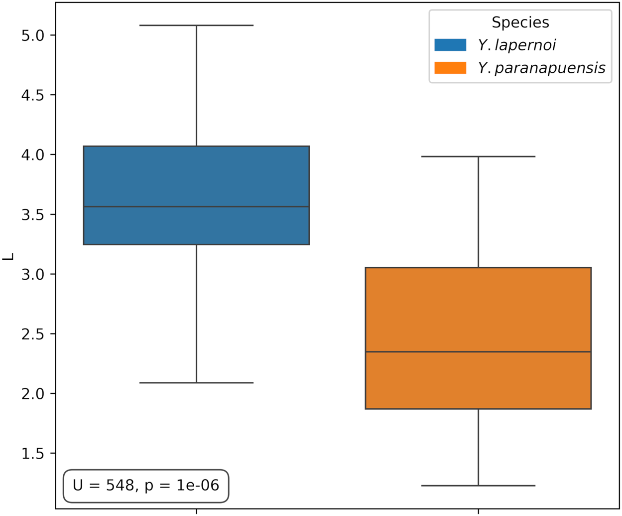
A considerable size difference was observed between the species, with Y. lapernoi significantly larger than Y. paranapuensis (Figures 1 and 2). This difference is confirmed by the Mann–Whitney U test, which indicates a statistically significant difference between the two samples (U = 548, p-value = 10−⁶).
Boxplot comparing shell length between Yoldiella paranapuensis and Yoldiella lapernoi. The Mann–Whitney U test revealed a significant size difference between the two taxa (U = 548, p-value = 10−⁶), rejecting the null hypothesis that they belong to the same population sample.

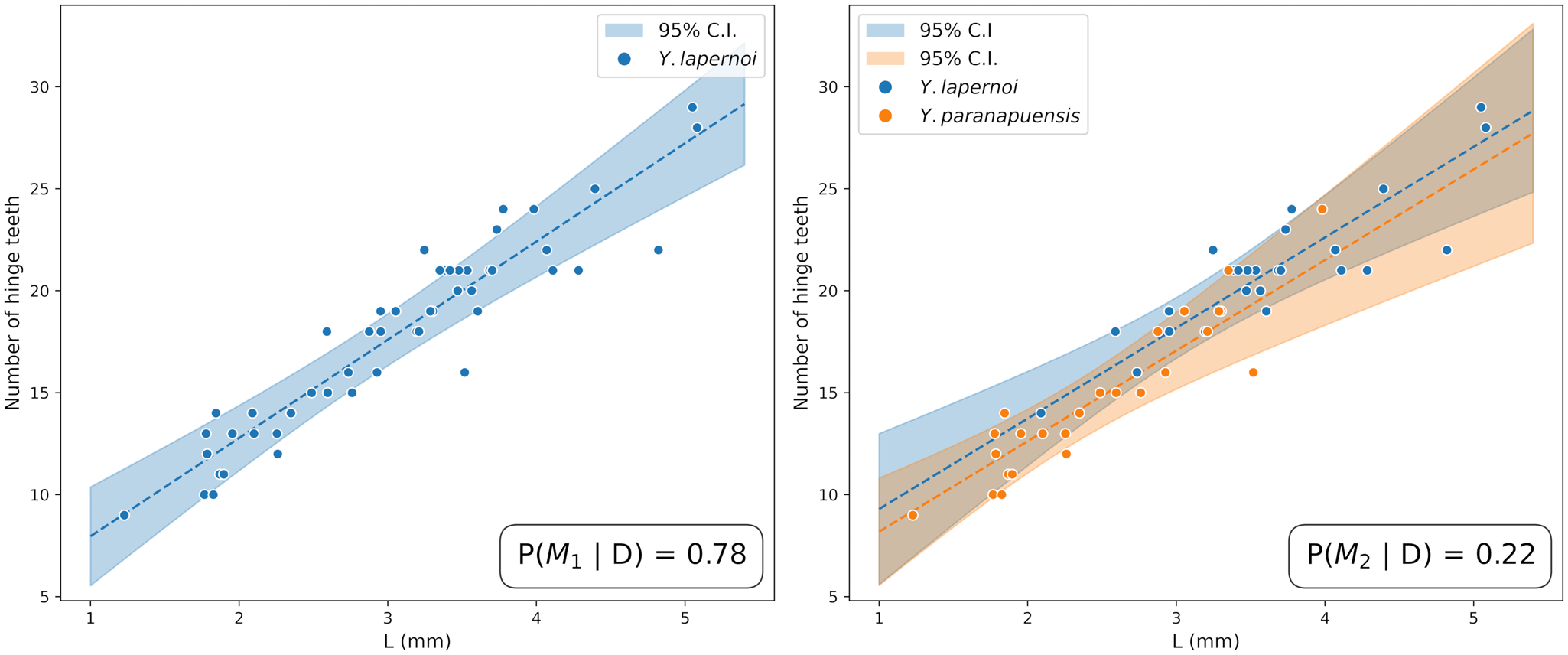
Bayesian regression analysis for the correlation between the number of hinge teeth and valve length. A comparison between model M1 (single species) and model M2 (two species) favoured the single-species model with a 78% degree of belief, indicating it is 3.5 times more probable than M2. The results suggest that shell size is the main factor governing the variation in the number of teeth.

In frequentist analysis, the growth rate showed no statistical support for the separation of the species (F = 0.012, p-value = 0.91). A GLM test using shell length as a covariate, however, showed a significant positive correlation, with a direct relationship between the number of teeth on the hinge plate and the valve length (F = 198.37, p-value <10−1⁷). The same results were found in the Bayesian approach (Figure 2), which also showed a positive correlation, indicating a direct relationship between the number of teeth on the hinge plate and the valve length. This result favours the M1 model, which attributes a degree of belief of 78% to this hypothesis, making it 3.5 times more probable than the M2 model. The extra parameters introduced in the two-species model are not supported by the data.
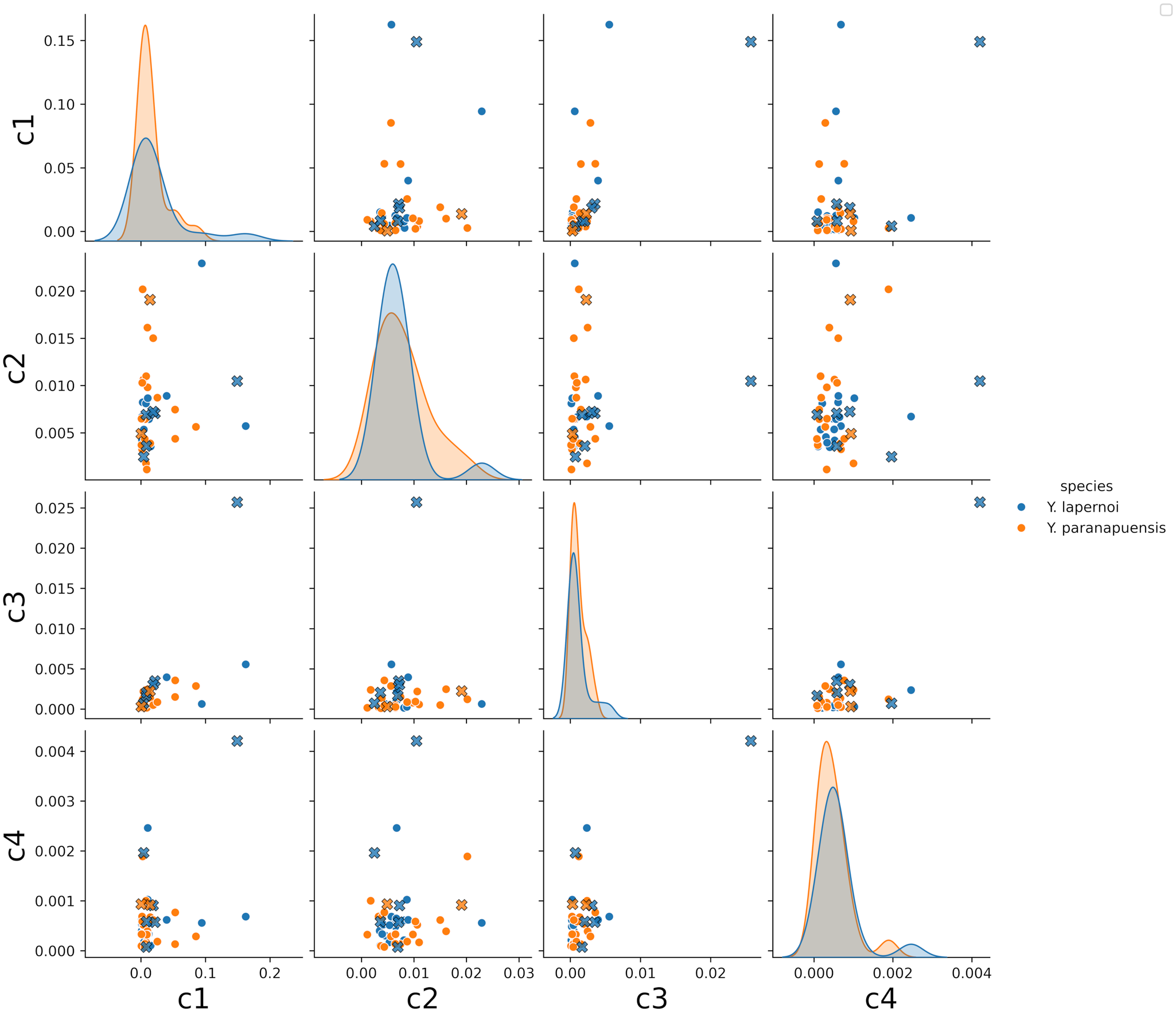
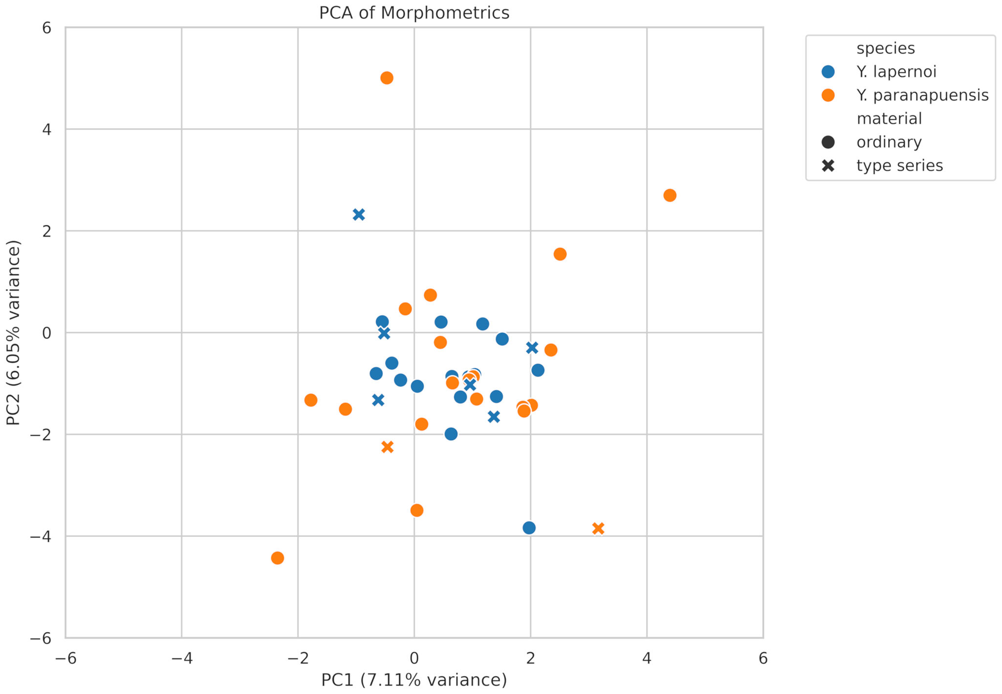
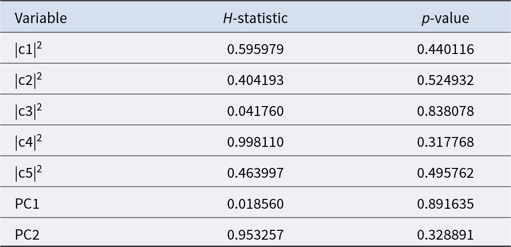
Elliptical Fourier Analysis (EFA) revealed no distinction in shell outline between Y. lapernoi and Y. paranapuensis. The Kruskal–Wallis H-test showed no significant difference in the elliptical Fourier coefficients (Table 1), either for the first five power spectrum coefficients (Figure 3) or for the principal components (Figure 4). Specimens of both species, including the type series, exhibited a high degree of overlap in morphospace, indicating that the observed variation in shell contour is continuous and not diagnostic at the species level. This overlap suggests that shell outline morphology, as captured by EFA, does not support the recognition of these taxa as separate species.
Elliptical Fourier Analysis (EFA) scatterplot illustrating the overlap in shell outline between the species. The plot demonstrates that there is no distinction in shell shape between Yoldiella paranapuensis and Yoldiella lapernoi based on the first five power spectrum components. The type series specimens overlap with the general morphological variation.

Principal Component Analysis (PCA) scatterplot of all Elliptical Fourier descriptors. The plot demonstrates a high degree of overlap in morphospace, suggesting that shell shape is not a diagnostic character for distinguishing between the two species.

Summary of the Kruskal–Wallis H-test statistic for the first five coefficients of the Elliptical Fourier Analysis and the principal components. The results demonstrate the absence of statistically significant differences in shell contour morphology between the species for all analysed coefficients

Systematics
Family Yoldiidae Dall, 1908
Genus Yoldiella A. E. Verrill & K. J. Bush, 1897
Yoldiella lapernoi Benaim & Absalão, 2011
(Figures 3F-L)
Yoldiella lapernoi Benaim & Absalão, 2011: 515-517, figs. 2A-B, G, J; Cavallari et al. (Reference Cavallari, Dornellas and Simone2016: 34); Oliveira et al. (Reference Oliveira, Pimenta, Figueira and Absalão2018: 109–110).
Yoldiella paranapuensis Benaim & Absalão, 2011: 517–518, figs. 3A-B, E-H; Cavallari et al. (Reference Cavallari, Dornellas and Simone2016: 41); Oliveira et al. (Reference Oliveira, Pimenta, Figueira and Absalão2018: 110).
Yoldiella sp1: Benaim and Absalão (Reference Benaim and Absalão2011a: 1666, fig. 1A, 4, 5); Benaim et al. (Reference Benaim, Viegas and Absalão2011: 40, figs. 1A-B).
Yoldiella sp2: Benaim and Absalão (Reference Benaim and Absalão2011a: 1666–1669, fig. 1B, 4, 5); Benaim et al. (Reference Benaim, Viegas and Absalão2011: 40, fig. 1C-D).
Distribution
Only known from Brazil, Rio de Janeiro state, Campos Basin.
Redescription
Shell small (max height: 3.5 mm; max length: 5.1 mm), inequilateral, inflated, narrowly subovate, rostrated, white, opaque. Umbones inflated, slightly anterior to midline, opisthogyrous. Antero-dorsal margin slightly convex, smoothly descending to meet antero-ventral margin, forming a broadly rounded anterior margin. Postero-dorsal margin almost straight, descending, posterior margin subrostrated, postero-ventral margin slightly convex (in sub-adults) or forming slight sinuosity (in adults). Surface with smooth commarginal striae, becoming more conspicuous, irregularly spaced towards the ventral margin. Hinge plate slightly thickened, almost straight in sub-adults, angulated in adults, anterior plate thicker than posterior. Hinge taxodont, with chevron-shaped teeth, the same number of teeth on anterior and posterior plates (max number of teeth: 29), the new teeth rising first on the posterior plate during development. Resilifer drop-shaped. Protoconch smooth, with 100–270 μm.
Remarks
Benaim and Absalão (Reference Benaim and Absalão2011a) considered the specimen from the Mediterranean, in the Calabria region, figured as Yoldiella striolata in La Perna (Reference La Perna2008: 17; Figure 2E and F), a misidentification of Y. lapernoi. Nevertheless, the figured specimen has a sharper umbo, a convex and more inclined postero-dorsal margin, and the anterior margin has an angulation break, whereas Y. lapernoi has a more inflated and less acute umbo, the postero-dorsal part is straight and less inclined, and the anterior margin has a smooth and constant curve. None of the individuals analysed in the present study exhibited the characteristics mentioned for the specimen of Y. striolata in La Perna (Reference La Perna2008: 17; Figure 2E and F); therefore, the record of Y. lapernoi to the Calabria region is here disregarded.
Among the Yoldiella species reported from the Atlantic, Y. lapernoi is distinguished from Y. orstomi Cosel, 1995 by its more inflated shape, a less acute posterior margin, and a sinuosity along the postero-ventral margin.
Among Yoldiella species reported from Brazilian waters, Y. riograndensis (Esteves, Reference Esteves1984) and Y. lapernoi share the same narrowly subovate shape. However, Y. riograndensis can be distinguished by its low, anteriorly positioned umbo, postero-dorsal margin angle less pronounced, a triangular resilifer, and posterior teeth aligned in a straight line.
Discussion
Hinge teeth and shell outline are the most commonly employed characters in Yoldiella taxonomy, making them particularly crucial for species with limited information regarding their general anatomy, such as Y. lapernoi and Y. paranapuensis (Allen and Hannah, Reference Allen and Hannah1986; Verrill and Bush, Reference Verrill and Bush1897; Allen et al., Reference Allen, Sanders and Hannah1995; Benaim et al., Reference Benaim, Viegas and Absalão2011).
The argument for a distinction between Y. paranapuensis and Y. lapernoi, as presented in Benaim and Absalão (Reference Benaim and Absalão2011b) and Benaim et al. (Reference Benaim, Viegas and Absalão2011), is controversial. The original descriptions of both species already provide insights towards their synonymy due to ambiguous or very similar diagnoses. For instance: i) the ratio between hinge plate length to height (H/L), is described as ‘0.69’ for the former and ‘0.71’ for the latter, a minimal deviation of only 0.02 that can easily be explained as a measurement artefact, ii) the resilifer shape is identical, described as ‘drop-shaped’ for adult specimens of both species, iii) while the length of the tooth row is cited as diagnostic, the hinge plate and tooth rows are not symmetrically rectangular, which leads to distinct lengths depending on the measurement location. Furthermore, there is ambiguity about which region was measured in both species, as no information is provided on whether the comparison refers to the anterior or posterior hinge plate of Y. lapernoi.
We propose Yoldiella paranapuensis as a junior synonym of Y. lapernoi, since both represent distinct developmental stages within the same ontogenetic series. Our analysis revealed a significant difference in shell length between the two taxa (Figure 1). In such cases, the reliance on linear morphometric variables is heavily influenced by size, which can artificially categorize different ontogenetic stages as separate groups or, as in the case of Y. lapernoi and Y. paranapuensis, as different species. When we controlled for shell length as a covariate, no statistically significant difference in the number of hinge teeth was observed between Y. lapernoi and Y. paranapuensis (GLM, F = 0.012, p-value = 0.91; see Figure 2). We also found a direct relationship between the number of teeth on the hinge plate and valve length. Our analysis showed that once size-related variation was accounted for, the apparent distinction between the two taxa, based on tooth count, was no longer supported, indicating that these differences are a consequence of ontogenetic stages rather than species-level distinctions.
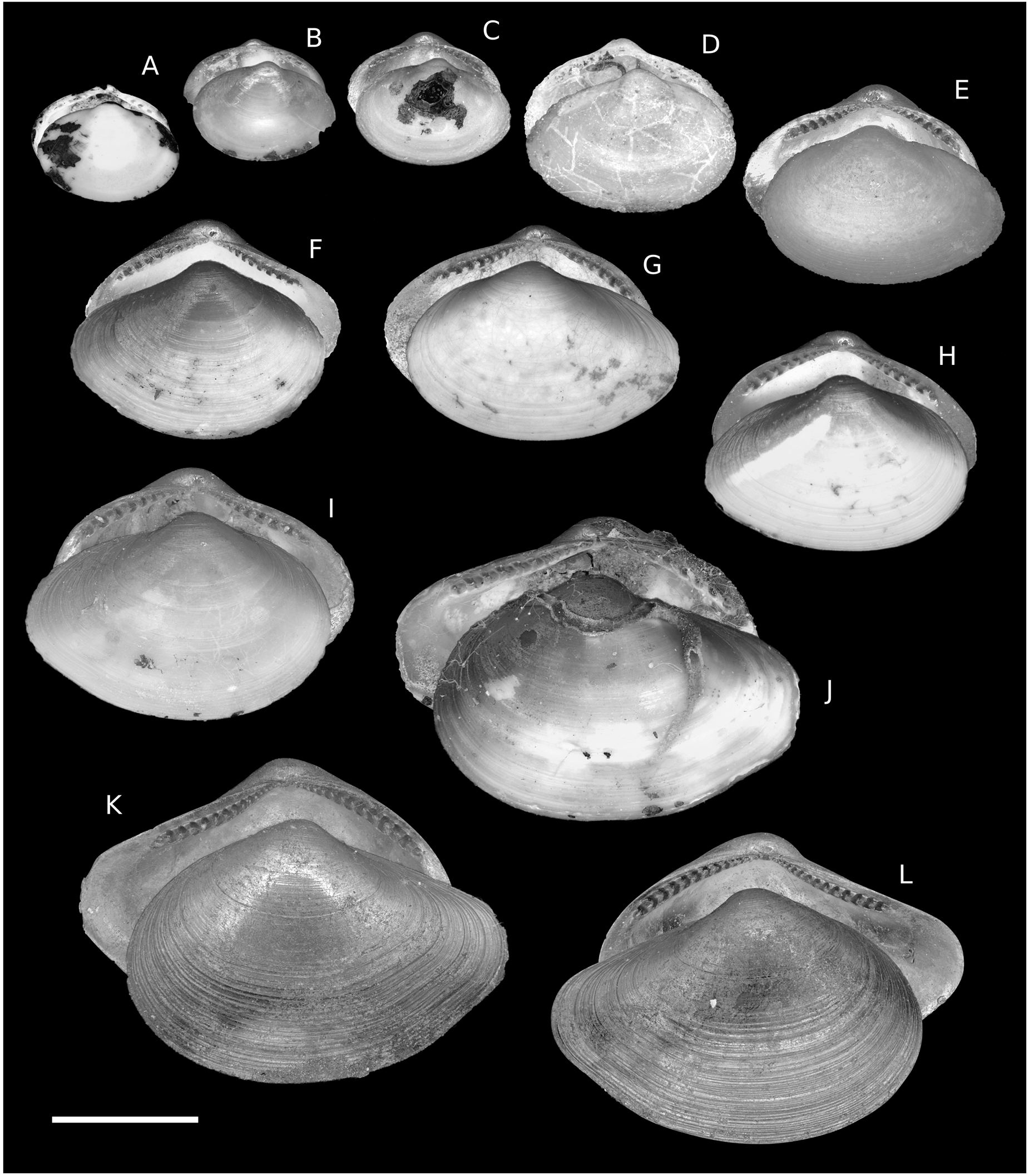
EFA was used to capture shape variation among specimens because it effectively eliminates the influence of size. This method summarizes comprehensive shape information independently of the ontogenetic stage, creating a more reliable framework for evaluating taxonomic distinctions by converting morphological shape characteristics into quantifiable data (Crampton, Reference Crampton1995). EFA is a robust technique for analysing differences in shell shape, particularly in taxa that lack homologous landmarks, such as Yoldiella. After examining the shell outlines of specimens at different ontogenetic stages using EFA, we found no significant variation between Y. paranapuensis and Y. lapernoi (Figure 3). There is a morphological change of the conchological characters during development, as exemplified by the complete type series of both species (Figure 5). Juveniles, referred to as Y. paranapuensis in Benaim and Absalão (Reference Benaim and Absalão2011a), as Yoldiella sp. 2 by Benaim and Absalão (Reference Benaim and Absalão2011b) and Benaim et al. (Reference Benaim, Viegas and Absalão2011), possess a broadly subovate shape and develop a rostrum during later stages, turning into the typical subovate form of Y. lapernoi. Concomitant with this development, the concentric striae become more conspicuous near the ventral margin, the hinge plate appears curved due to a change in growth orientation, and a slight sinuosity develops in the posteroventral margin, attributed to the development of the rostrum. In fact, there is no distinction between the holotype of Y. paranapuensis and the two smallest paratypes of Y. lapernoi (Figure 5E–G).
Ontogenetic series composed of the entire type series of Yoldiella paranapuensis (paratypes A–D, holotype E) and Yoldiella lapernoi (paratypes F–K, holotype L), providing evidence for a morphological continuum. Each valve is represented in both internal (i.e. hinge plate) and external views. The progression from juvenile to adult illustrates a change in shell shape, from a broadly subovate form to the typical subovate, rostrate form of Y. lapernoi. Concomitantly, morphological characters are progressively acquired during development, such as the conspicuous concentric striae, the curved hinge plate, and a slight sinuosity in the posteroventral margin. (A) IBUFRJ 18748; (B) IBUFRJ 18748; (C) IBUFRJ 18748; (D) IBUFRJ 18748; (E) MNRJ 15201; (F) IBUFRJ 18759; (G) IBUFRJ 18758; (H) IBUFRJ 18756; (I) IBUFRJ 18760; (J) IBUFRJ 18757; (K) IBUFRJ 18762; (L) MNRJ 15200. Scale bar: 2 mm.

When considering the shell outline, Benaim and Absalão (Reference Benaim and Absalão2011b) and Benaim et al. (Reference Benaim, Viegas and Absalão2011) disregarded shape variation associated with the ontogenetic stage by not distinguishing between juvenile and adult specimens. In addition, the excessive use of size-correlated variables, exacerbated by an overreliance on proportions, the absence of partitioned datasets for training, and the inconsistent identification of homologous hinge characters (e.g., the measured ‘highest tooth’ varies among individuals depending on how it is counted from the valve edge to the centre) may have produced biased results. Such methodological and analytical shortcomings are consistently observed across the three referenced studies. Collectively, these issues compromise morphological comparisons and obscure the morphometric relationships among specimens at different ontogenetic stages.
The analysis of shell characters, such as hinge structure and overall valve shape, has already proven effective in resolving taxonomic issues (Chrpa and Oleinik, Reference Chrpa and Oleinik2015; De Lima et al., Reference De Lima, Mello and Oliveira2024). For many groups, it remains the only feasible approach, especially for taxa such as Yoldiella and other deep-sea species inhabiting extreme depths (>700 m). In such cases, the only available material is often held in scientific collections and typically lacks soft parts. This practice reflects a historical tradition of preserving only the shells, and partly a result of logistical constraints during large-scale expeditions (such as those responsible for most specimens examined in this study), which commonly preserved animals in formalin, thereby precluding the use of molecular analyses. Therefore, prioritizing molecular approaches at the expense of shell-based morphometric analyses overlooks the continued relevance and utility of morphological data, particularly given the vast number of specimens preserved in zoological collections, which highlights the need for complementary methodologies. When rigorously applied, morphometric analyses remain a reliable and accessible tool for species delimitation, contributing significantly to biodiversity documentation.
Conclusion
Our analysis demonstrates that Y. paranapuensis is a juvenile stage of Y. lapernoi, supporting their synonymy. This case illustrates how morphometric approaches, even with advanced statistical tools, can overemphasize minor differences and potentially inflate the number of described species when developmental stages are not adequately accounted for.
Supplementary material
The supplementary material for this article can be found at https://doi.org/10.1017/S0025315425100969.
Acknowledgements
We are very grateful to Tom White and the staff of Imaging Department of Natural History Museum for the photographs of Yoldiella specimens, to Virginie Héros and the Mollusques Collection of the Muséum National d’Histoire Naturelle, for providing information regarding the collection’s specimen lots, to Laboratório de Entomologia – IB – UFRJ, for allowing access to digital camera, to Leonardo Santos and Fernando Erthal for access to material from Irajá Damiani Pinto Museum of Paleontology, to Alexandre Dias Pimenta, for access to material from Museu Nacional/UFRJ, to Natalia Benaim and Diniz Viegas, who provided useful discussions and access to raw morphometric data, to Yohanny Melo and Ariel La Pasta for critical suggestions on the first draft of this manuscript, and to the two anonymous reviewers for their insightful comments and constructive suggestions.
Author contributions
Conceptualization, methodology, validation, formal analysis, investigation, data curation, writing – review and editing, writing – original draft: A.M.A.-P., T.C.d.L, V.B.B.M, and C.D.d.C.O. All authors have read and agreed to the current version of the manuscript.
Funding
A.M.A.-P. was financially supported by the Coordenação de Aperfeiçoamento de Pessoal de Nível Superior – Brasil (CAPES) – Finance Code 001.
Competing interests
The authors declare that they have no conflicts of interest in relation to this work.
Data
The authors confirm that the data supporting the findings of this study are available within the article and its supplementary materials.