Introduction
In vitro fertilization (IVF) is vastly employed worldwide (Meczekalski et al. Reference Meczekalski, Szeliga, Podfigurna, Miechowicz and Adashi2020). Treatments are generally based on ovarian superovulation with gonadotropins, followed by oocyte extraction and embryo development in the laboratory. Embryos are then transferred a few days after fertilization (fresh transfer), or frozen and transferred in a subsequent cycle. Treatment is aimed at maximizing oocyte yield, and in the presence of multiple growing follicles, often entails high supra-physiological oestradiol levels. High oestradiol levels are associated with increased risk for ovarian hyperstimulation syndrome. Therefore, in the presence of high levels, a decision to electively freeze all embryos is often considered (Mourad et al. Reference Mourad, Brown and Farquharn.d.). Yet, for candidates for fresh transfer, a building body of evidence in literature now points to a higher risk of adverse events associated with implantation in the setting of high oestradiol levels. These may include lower live birth rates according to some (Ying et al. Reference Ying, Lu, Zhang, Arhin, Hou, Wang, Wu, Lu and Tang2021) (but not all (Kosmas et al. Reference Kosmas, Kolibianakis and Devroey2004)). Lower birth weights (Kohl Schwartz et al. Reference Kohl Schwartz, Mitter, Amylidi-Mohr, Fasel, Minger, Limoni, Zwahlen and von Wolff2019; Liu et al. Reference Liu, Kuang, Wu, Feng, Lyu, Wang, Sun and Sun2017; Pereira et al. Reference Pereira, Reichman, Goldschlag, Lekovich and Rosenwaks2015, Reference Pereira, Elias, Christos, Petrini, Hancock, Lekovich and Rosenwaks2017) and additional placental-mediated complications (Farhi et al. Reference Farhi, Ben-Haroush, Andrawus, Pinkas, Sapir, Fisch and Ashkenazi2010; Imudia et al. Reference Imudia, Awonuga, Doyle, Kaimal, Wright, Toth and Styer2012) have been more persistently associated with excess oestradiol levels at embryo transfer. The underlying pathophysiology is not well understood.
In light of these potential risks associated with high oestradiol levels during stimulation, a ‘freeze all’ approach may potentially minimize suboptimal outcomes. The assumption for such an approach is that in a subsequent frozen-thawed transfer cycle, oestradiol levels will be lower, and thus any adverse effects on the endometrium minimized. Indeed, frozen cycles are generally associated with a lower rate of low-birth-weight neonates and placental complications (other than preeclampsia) (Maheshwari et al. Reference Maheshwari, Pandey, Amalraj Raja, Shetty, Hamilton and Bhattacharya2018). Yet, this approach does not consider an effect of high oestradiol levels during stimulation on the oocyte and any possible effects on subsequent embryo development. If such an affect exists, via metabolomic or epigenetic for instance, embryos created in the presence of high oestradiol levels could also carry these risks forward to subsequent frozen embryo transfers (FETs). The benefit of freezing all embryos in such case needs investigation.
When examining frozen–thawed embryo transfer cycles following IVF cycles with high oestradiol concentrations, it was reported that implantation and pregnancy rates were not impaired (Yu Ng Reference Yu Ng2000). Less studies examined later obstetrical complications of ongoing pregnancies.
Past studies of obstetric outcomes of singleton pregnancies following FET, demonstrated an inverse association between oestradiol levels during stimulation and neonatal weights (Cai et al. Reference Cai, Liu, Xu, Liu, Jiang, Li, Sha and Ren2019) (Tarlatzi et al. Reference Tarlatzi, Venetis, Sassi, Devreker, Englert and Delbaere2021) while in an additional similar study design, no such association was found (Huang et al. Reference Huang, Lu, Lin, Chen, Gao, Lyu, Cai and Kuang2020).
Our aim was to investigate the effect of high oestradiol levels during ovarian stimulation on obstetric outcomes and placental pathologies in subsequent FET.
Materials and methods
Setting and patients
This retrospective cohort study was conducted on deliveries between 2009 and 2017 at the Royal Victoria Hospital, McGill University’s primary obstetrical teaching centre. During the study period, hospital policy required histological examination of all placentas, irrespective of delivery complications or pregnancy history. Inclusion criteria were singleton live births ≥ 24 weeks gestation following stimulated IVF cycles with autologous oocytes and programmed FET cycles. Exclusion criteria included all fresh embryo transfers, natural-cycle FETs, in vitro maturation, preimplantation genetic testing and clinical pregnancies of more than one embryo (as seen on first ultrasound).
Data collection
For eligible cases, data were extracted from computer files including – baseline patient characteristics, pre-treatment testing, treatment parameters, obstetric outcome, neonatal outcomes and placental assessment. The McGill University Health Centre Ethics Committee approved the study on March 12, 2018 (MUHC-2019-5026). Informed consent was waived by the review board, and the study adhered to relevant institutional guidelines and regulations.
Design
High oestradiol levels were defined as ≥10,000 pmol/L on the trigger day. Outcomes of pregnancies from the FET succeeding the reference stimulation cycle were compared between two groups: (1) high oestradiol levels during stimulation and (2) controls with lower oestradiol levels. A subanalysis was conducted for subjects with oestradiol levels >12,000 pmol/L compared to controls.
Treatment protocols
Ovarian stimulation was carried out using Follicle Stimulating Hormone, with or without Luteinizing Hormone. The dosing was determined at the discretion of the treating physician. The following medications were used: Follitropin alfa (Gonal-f®) Follitropin beta (Puregon®), HP-hMG (Menopur®) and Lutropin alfa (Luveris®).
For antagonist protocols, GnRH antagonists Cetrorelix (Cetrotide®) or Ganirelix (Orgalutran®) were used. For agonist protocols GnRH agonist Buserelin (Superfact®) was used, as outlined in prior work [17]. Monitoring included ultrasound scans and serum oestradiol measurements. Serum oestradiol samples were collected between 8 AM and 10 AM on the trigger day. Oestradiol measurements were performed using the ADVIA Centaur Enhanced Estradiol Assay on the IMMULITE 2000 system (Siemens).
Ovulation triggering was done by hCG (250 mcg, Ovidrel®) or GnRH agonist Triptorelin (Decapeptyl®) once two leading follicles reached 18 mm, with oocyte retrieval performed 36 hours later.
For programmed FETs, oestradiol valerate (6 mg/day orally) was initiated on cycle day 2–3. Endometrial thickness was assessed after 10–12 days. Progesterone (200 mg vaginally, three times/day, or 100 mg intramuscular) was initiated if the endometrial lining was ≥7 mm. Single day 3 or day 5 embryo transfer was performed, though more embryos were transferred in select cases based on maternal age, embryo quality or previous failed transfers.
Placental pathology
A single specialized perinatal pathologist, blinded to patient history and treatment details, conducted all placental examinations at the Royal Victoria Hospital pathology department. Placentas underwent standard gross and microscopic assessments following institutional protocols. Macroscopic evaluation included weight, size and examination of the umbilical cord, membranes, foetal surface, maternal surface and placental parenchyma. Placental sections (umbilical cord, membranes and random parenchymal tissue) were fixed and stained with Hematoxylin and Eosin. Histological findings were categorized per Amsterdam Placental Workshop Group guidelines [18,19].
Statistical analysis
Data analysis was performed using Epi Info version 7.0 (CDC, Atlanta, GA). Continuous variables were summarized as mean ± standard deviation (SD) or median with range/IQR and compared using the Student’s t-test or Mann-Whitney U test, as appropriate. Categorical data were presented as frequencies (percentages) and analyzed using chi-square or Fisher’s exact tests. A two-tailed p-value of <0.05 was considered statistically significant.
Results
A total of 308 live singleton deliveries following FET met inclusion criteria and were analyzed, 114 in the high oestradiol group and 194 in the control group (lower serum oestradiol group).
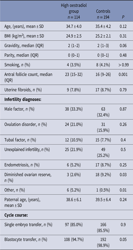
Baseline characteristics were comparable between the groups (Table 1). A significantly higher antral follicle count was noted in the high oestradiol group, p < 0.001 (as expected per study design, as oestradiol level correlate to follicle number). Among infertility aetiologies, a diagnosis of diminished ovarian was significantly less common in the high oestradiol group, 2.6% vs. 9.2%, p = 0.03 as expected.
Demographic and IVF cycle characteristics of the high oestradiol and control groups

SD = standard deviation, n = Number, BMI = body mass index, pre-gestational.
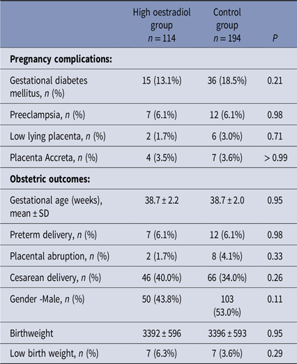
Pregnancy outcomes are presented in Table 2. No differences were found in adverse obstetric outcomes including rates of preeclampsia, preterm birth and gestational diabetes. Neonatal weight was similar between the groups, as was the rate of low-birth-weight neonates.
Obstetric outcomes of the high oestradiol and control groups

SD = standard deviation, IQR = interquartile range, n = Number.
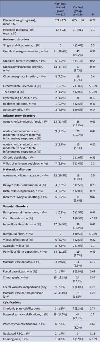
Table 3 describes placental findings in the high oestradiol and control groups. No differences were noted in gross placental and cord abnormalities, inflammatory, maturation disorder and vascular lesions between the study groups.
Placental findings in the high oestradiol and control groups

In a sub analysis (hereby described and presented as a table), study outcomes were compared between patients with very high oestradiol levels, ≥12000 pmol/L (n = 69) and control with lower levels (n = 239). Similarly, no differences were found in clinical outcomes and placental findings.
Discussion
Our aim was to investigate the effect of high oestradiol levels during ovarian stimulation on obstetric outcomes and placental findings of subsequent frozen transfer cycles. No adverse clinical or placental outcomes were observed in association with prior high oestradiol exposure. Thus, our findings support the notion that prior adverse outcomes described in the literature in regard to fresh embryo transfer with a high oestradiol level may stem from an adverse effect on the uterus/endometrium itself, and not an oocyte/embryonic effect.
Previous studies have explored the association between high oestradiol levels during stimulation and pregnancy outcomes with fresh embryo transfer. Transfer in the presence of high oestradiol levels has been repeatedly associated with adverse obstetric outcomes, such as small for gestational age newborns and lower birth weights (Farhi et al. Reference Farhi, Ben-Haroush, Andrawus, Pinkas, Sapir, Fisch and Ashkenazi2010; Imudia et al. Reference Imudia, Awonuga, Doyle, Kaimal, Wright, Toth and Styer2012; Kohl Schwartz et al. Reference Kohl Schwartz, Mitter, Amylidi-Mohr, Fasel, Minger, Limoni, Zwahlen and von Wolff2019; Liu et al. Reference Liu, Kuang, Wu, Feng, Lyu, Wang, Sun and Sun2017; Pereira et al. Reference Pereira, Reichman, Goldschlag, Lekovich and Rosenwaks2015, Reference Pereira, Elias, Christos, Petrini, Hancock, Lekovich and Rosenwaks2017), a higher rate of pregnancy-induced hypertension (Imudia et al. Reference Imudia, Awonuga, Doyle, Kaimal, Wright, Toth and Styer2012) and premature rupture of membranes (Farhi et al. Reference Farhi, Ben-Haroush, Andrawus, Pinkas, Sapir, Fisch and Ashkenazi2010). As all these adverse outcomes share a common placental origin, it may be speculated that endometrial changes in response to high oestradiol levels, with subsequent defective placentation, may explain the clinical associations observed. Indeed, this theory has been supported by placental changes in mice that underwent superovulation (de Waal et al. Reference de Waal, Vrooman, Fischer, Ord, Mainigi, Coutifaris, Schultz and Bartolomei2015) and by genetic changes related to remodelling, demonstrated in endometrial samples of oocyte donors following superovulation (Senapati et al. Reference Senapati, Wang, Ord, Coutifaris, Feng and Mainigi2018). Similarly, in baboons infused with high oestrogen levels in early gestation, maternal blood pressure was increased, and changes to uteroplacental flow were demonstrated later in gestation (Aberdeen et al. Reference Aberdeen, Bonagura, Harman, Pepe and Albrecht2012), suggesting a suppressive role of high oestradiol on spiral artery remodelling. Recently, our group has also demonstrated a higher rate of placental cord abnormalities associated with excessive oestradiol levels following stimulation (Ganer Herman et al. Reference Ganer Herman, Volodarsky-Perel, Ton Nu, Machado-Gedeon, Cui, Shaul and Dahan2022).
Nevertheless, another plausible explanation of adverse outcomes with high oestradiol levels may relate to the effect on the developing oocyte and embryo. Initially speculated to result from an embryotoxic effect and resultant chromosomal abnormality, recent good-quality data does not support an association between oestradiol levels and embryo euploidy (Irani et al. Reference Irani, Canon, Robles, Maddy, Gunnala, Qin, Zhang, Xu and Rosenwaks2020) and live birth rates (Irani et al. Reference Irani, Canon, Robles, Maddy, Gunnala, Qin, Zhang, Xu and Rosenwaks2020; Kosmas et al. Reference Kosmas, Kolibianakis and Devroey2004). Yet, this does not rule out the possibility of genetic or metabolomic alterations related to subsequent placentation, and clinical observations in the literature are inconsistent. While Cai et al found a negative association between peak oestradiol levels during stimulation and birthweights of singletons conceived through subsequent FET cycles (Cai et al. Reference Cai, Liu, Xu, Liu, Jiang, Li, Sha and Ren2019), Huang et al did not demonstrate such an association in a large cohort of 8501 deliveries (Huang et al. Reference Huang, Lu, Lin, Chen, Gao, Lyu, Cai and Kuang2020). Our findings agree with the latter group, with the additional insight of placental examination. The inclusion of placental analysis allowed us higher sensitivity to assess the effects of prior high oestradiol exposure on placentation, as some clinical differences may only manifest after significant placental damage. Thus, our findings support a lack of effect of high oestradiol levels on developing embryos that are frozen for future transfer.
Study strengths include its unique inclusion of placental analysis, in the setting of a single-centre analysis. It also investigated a period in which placental analysis was performed for all deliveries irrespective of pregnancy complications, thereby minimizing selection bias. In addition, all examinations were performed by a single pathologist, blinded to the patient-assisted reproductive history, and findings were described according to contemporary classification.
Study limitations must be acknowledged. These mainly stem from the study’s retrospective design, as not all data of interest were available for analysis, including patients past adverse obstetric outcomes, which influence incidence in following pregnancies. In addition, our assumption was that frozen cycles would entail lower oestradiol levels than prior stimulated cycles as reported by other groups (Li et al. Reference Li, Ruan, Zhu, Yang, Zhu and Luo2022), yet we did not routinely obtain oestradiol measurements as a part of patient follow-up in frozen cycles. Furthermore, we included only programmed FET cycles in our study, which were the main frozen transfer mode at our institution during the study period. Yet is possible that natural cycles, which entail even lower oestradiol levels, are associated with improved outcomes and placental findings. The employment of such cycles following stimulation with high oestradiol levels remains to be investigated in a larger study. Finally, we must consider the inherent selection bias that may be associated with the inclusion of relatively good prognosis patients who are candidates for freezing in the study group. This means that our conclusions may not be generalized to intermediate oestradiol levels (we cannot assume a linear correlation of the effect of oestradiol). Thus, we cannot automatically extrapolate to patients with few embryos available, in whom the rare but theoretical possibility of embryos not surviving freezing must be weighed against any theoretical advantages of freezing.
A post hoc sample power analysis was performed. Based on the differences in the main outcome of low birth weight found between the groups, a total sample size of 2517 patients would be required to provide significance with an 80% power and alpha of 0.05. As such, our sample was underpowered to certain differences, yet this remains serves as a first study with such an investigation to undertake placental evaluation. Furthermore, the data may serve as an addition to a meta-analysis on the subject given the few existing similar studies on pregnancy complications.
In conclusion, our study demonstrates similar obstetric outcomes and placental findings in FET cycles after stimulation with high and lower oestradiol levels. These conclusions possibly shed light on previous findings in the literature of adverse outcomes associated with fresh transfer in the presence of high oestradiol levels and support a direct adverse effect on the uterus. Accordingly, no adverse effects on embryo development were demonstrated in subsequent FET transfers. Thus, our findings offer justification for the consideration of freezing, to optimize obstetric outcomes. Further large studies are required to understand better the implications of high oestradiol on embryo alterations and the role on pregnancy complications and placental modifications.
Funding statement
None to declare.
Competing interests
The authors declare no conflict of interest, including any financial, personal or professional interests.
Ethical standards
The McGill University Health Centre Ethics Committee approved the study on March 12, 2018 (MUHC-2019-5026). Informed consent was waived by the review board, and the study adhered to relevant institutional guidelines and regulations.