Partially or totally anomalous pulmonary venous connection of the right lung to the inferior caval vein is almost always associated with hypoplasia of the right lung and aberrant systemic arterial supply to the right lower lung. As these three abnormalities occur together in a recognizable pattern, and the anomalous pulmonary vein resembles a Turkish sword, the combination is usually described as the scimitar syndrome.1 The so-called “scimitar sign”, however, has also been recognized in a few cases where the right pulmonary vein had a scimitar-like course, but was normally connected to the left atrium.2 Goodman et al. introduced the term ‘meandering right pulmonary vein’ for such cases.3 Also reported are cases where a scimitar vein connects to both the inferior caval vein and the left atrium, for which Pearl4 coined the term “scimitar variant”. Many have speculated that these lesions are related.5 We describe here a case with a meandering right pulmonary vein, horseshoe lung and left pulmonary arterial sling. Our case provides further evidence to support the notion that the meandering right pulmonary vein is a variant of the scimitar syndrome.
Case report
A 15-month-old female was admitted to our hospital because of respiratory distress. At 11 months of age, she developed recurrent infections of the lower respiratory tract. She experienced frequent choking on ingestion of solid food. At admission, she was tachypnoeic, and showed deep retraction of the chest wall. On auscultation, there was evidence of reduced entry of air into the right lung, and crackles were audible in the early inspiratory phase. The heart sounds were normal.
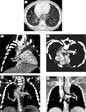
The chest radiograph demonstrated reduced volume of the right lung, with mild rightward shift of the heart (Fig. 1). The lungs showed abnormal fissures, and unusual orientations of the vascular markings. The tracheal column of air showed a segmental narrowing in the upper mediastinum.

Figure 1. The frontal chest radiograph shows hypoplasia of the right lung, with the heart displaced mildly to the right. Note the abnormally oriented vascular markings in the right lung. A vertical fissure is seen in the medial aspect of the right lung extending from the hilum to the diaphragm (arrows). A vertical fissure is also seen in the medial aspect of the left lower lung through the cardiac silhouette (arrow). A long segment of narrowing is seen in the trachea at the level of the thoracic inlet.
The contrast-enhanced computed tomographic images (Fig. 2) from the referring hospital confirmed hypoplasia of the right lung, and rightward shift of the heart. The cardiac apex, nonetheless, was orientated towards the left anterior chest wall. Both lungs possessed two lobes. The right and left bronchuses were of similar length and showed mild narrowing. The right lower lobe extended to the left side, forming a so-called “horseshoe lung”, or “cross-over lung”.6 The right pulmonary artery had an unusual downward meandering course in the medial aspect of the right lung. The left pulmonary artery arose distally from the pulmonary trunk, and coursed to the left, forming a pulmonary arterial sling around the distal trachea. Each lung was drained by a single pulmonary vein. The right pulmonary vein had an abnormal meandering course, accompanying initially the right pulmonary artery. It made a “U-loop” in the right lower lung, having a close spatial relationship with the inferior caval vein, but then achieved an unobstructed connection to the left atrium. There were double aortic arches, both patent, with the left arch being dominant. The trachea showed mild segmental narrowing. There were bilateral superior caval veins without a bridging vein. Bronchoscopy demonstrated the narrow distal trachea, with three complete cartilaginous rings.

Figure 2. Reformatted images of contrast-enhanced computerized tomography. An axial image at the level immediately above the diaphragm (a) shows the right pulmonary vein connecting to the scimitar vein (SV) near the inferior caval vein (ICV). Note an incomplete fissure in the right lower lung (arrows). The right lung medial to the fissure extends to the left thorax through the space between the heart and the esophagus (E) and descending aorta (DsAo), forming a horseshoe lung. The right and left lungs are in direct contact with an intervening fissure (arrow). The right anterior oblique view (b) shows the scimitar vein (SV) connecting to the left atrium (LA) via a vertical channel (asterisks), which courses along the anterior aspect of the right pulmonary artery (RPA). Note the close spatial relationship between the scimitar vein and inferior caval vein. The descending aorta (DsAo) is on the right side of the midline. The oblique axial view (c) shows the abnormal course of the pulmonary trunk, with a distal origin of the left pulmonary artery (LPA), which encircles the right side of the trachea (T) to course behind the trachea and in front of the descending aorta (DsAo) and esophagus (E). AsAo denotes the ascending aorta. The coronal view of the posterior chest (d) shows distal segments of the left (LAA) and right (asterisk) aortic arches. It also shows abnormal courses of right and left pulmonary arteries. The coronal view of the airway (e) shows segmental narrowing of the trachea where the left (LAA) and right (asterisk) aortic arches are located. The narrowing was considered to be a congenital tracheal stenosis rather than due to vascular compression. It was proved to be due to cartilaginous rings at operation. A small bronchus to the horseshoe lung segment (arrow) arises from the proximal right main bronchus.
The patient underwent a surgery with division of the right aortic arch, reimplantation of the left pulmonary artery to the pulmonary trunk, and tracheoplasty. She developed a stenosing web in the subglottic trachea, which was treated with bronchoscopic dilation. Echocardiography demonstrated mild stenosis of the left pulmonary artery with a pressure gradient of 20 millimetres of mercury. She was discharged at home in a good general condition.
Discussion
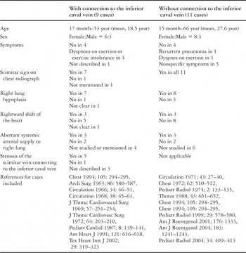
The essential part of the scimitar syndrome is the partially or totally anomalous pulmonary venous connection to the inferior caval right vein.1 There are rare cases in which the right pulmonary vein has an abnormal course that is similar to that of the classic scimitar vein, but has a normal connection to the left atrium, or dual connections to the inferior caval vein and left atrium.2–4 The cases having normal connections have been termed the “meandering pulmonary vein”,3 while the cases having dual connections are dubbed the “scimitar variant”.4 Already, however, the two terms have been used interchangeably.7 In addition, some of the cases reported as “anomalous unilateral single pulmonary vein” show similar features.8 It has been speculated that these rare anomalies are related pathogenetically to the classic scimitar syndrome.5 Our review of the literature (Table 1) shows that 7 of 9 cases with the right pulmonary vein having dual connections to the inferior caval vein and left atrium, and 8 of 11 cases with a scimitar-like vein connecting only to the left atrium, including the present case, were associated with hypoplasia of the right lung. Aberrant systemic arterial supply to the right lower lung was seen in 3 of 5 cases of each group in which the systemic arterial side was investigated. It is of particular interest that 5 cases with a scimitar vein having dual connections showed stenosis of the scimitar venous connection to the inferior caval vein. This tendency is highly suggestive of a common pathogenetic mechanism between these rare conditions and the classic scimitar syndrome. We speculate that, during early development, the scimitar vein has had dual connections to the inferior caval vein and left atrium. The connection to the left atrium is then obliterated in most cases. Rarely, its connection to the left atrium persists, with complete or partial obliteration of its connection to the inferior caval vein. Alternatively, the connection of the scimitar vein to the left atrium is secondary to obstruction of the scimitar venous connection to the inferior caval vein.
Table 1. Reported cases of the scimitar vein connecting to the left atrium with or without connection to the inferior caval vein.

In our patient, the systemic arterial supply to the right lower lung was normal. The patient did, however, show other features that are commonly or often seen in scimitar syndrome; horseshoe or crossover lung, abnormal lobation of the right lung, and abnormal bronchial and pulmonary arterial branching.1, 6 Our patient also had a pulmonary arterial sling. In this respect, she was similar to the patient reported by Filho et al.,9 in which scimitar syndrome was associated with horseshoe lung and pulmonary arterial sling. Hwang et al.,10 also reported a case in which horseshoe lung was associated with an abnormal course of the left pulmonary artery that was similar to an arterial sling, but without encircling the distal trachea. Their case was described as having normal pulmonary venous return. Although they did not state whether the right pulmonary vein was meandering, the right lung was hypoplastic. It is most likely, therefore, that the abnormal course of the left pulmonary artery in these cases, and in our own case, are simply variations of pulmonary arterial branching.
With all these common features among the cases of classic scimitar syndrome and the cases reported herein and by others, the classic scimitar syndrome, the so-called meandering pulmonary vein and scimitar variant can safely be considered to be closely related congenital anomalies with a common pathogenetic mechanism.
Acknowledgement
We acknowledge the contribution of Robert M. Freedom in the preparation of this case report. He sadly passed away before the completion of the manuscript. We also thank Hyun Woo Goo for his critical comments on our draft manuscript.