I. INTRODUCTION
Carbon nanotubes (CNTs) have attracted extensive attention as a part of the fundamental scientific study and investigation of nanoscale devices over the past decades due to their extraordinary physical and chemical properties.Reference Iijima1 The previous reports have shown that CNTs can be used as templates for confining and directing the growth of tubular or other one-dimensional nanostructures.Reference Ajayan, Stephan, Redlich and Colliex2–Reference Zhang, Fu, Shi, Fang and Li4 For example, Ajayan et al.Reference Ajayan, Stephan, Redlich and Colliex2 used CNTs as removable templates to synthesize V2O5 nanotubes by calcining mixtures of CNTs and V2O5 power at 750 °C, which provides a new synthesis route for the fabrication of nanostructures. Sun et al.Reference Sun, Yuan, Liu, Han and Zhang5 used CNT template to prepare α-Fe2O3 nanotubes by supercritical fluid at high temperature and pressure.
Double alkaline rare-earth tungstates with the formula of ARe(WO4)2 (A = Li+, Na+, K+, Rb+, Cs+; Re = trivalent rare earth ions) form a wide variety of inorganic compounds having tetragonal and monoclinic symmetries that have attracted great interest in recent years. Their structure diversity provides these crystals with numerous physical and chemical properties. Previous works about the ARe(WO4)2 family compounds were reported mostly on the single crystals to be used as laser crystal materials for a long period of time. Furthermore, ARe(WO4)2 powders were usually synthesized by the methods, such as solid state reaction,Reference Neeraj, Kijima and Cheetham6 flux growth,Reference Han and Wang7 Czochralski technique,Reference Li, Lin, Zhang and Zhang8–Reference Kuz’micheva, Lis, Subbotin, Rybakov and Zharikov11 and hydrothermal reaction.Reference Esteban-Betegoìn, Zaldo and Cascales12, Reference Yan, Lin, Wu and Lei13 However, the reports on the synthesis of double alkaline rare-earth tungstates nanotubes using CNTs as removable templates are scarce. As one compound of the family, NaLa(WO4)2 having a tetragonal symmetry with the structure of scheelite CaWO4 (W6+ is coordinated by four oxygen atoms in a tetrahedral site and La3+/Na+ site is eight coordinated) that has attracted great interest in recent years. To the best of our knowledge, up till now there are no reports on the synthesis of NaGd(WO4)2:Eu3+ nanotubes using CNTs as removable templates.
In this article, NaGd(WO4)2:Eu3+ nanotubes have been successfully synthesized by the hydrothermal method using CNTs as removable templates for the first time. Moreover, the structure, morphology, mechanism, and luminescence of the products calcined at various temperatures have been investigated in detail.
II. EXPERIMENTAL
All the chemicals were purchased from National Chemical Reagent Company (Shanghai, China). The CNTs used in the experiment were 40–60 nm in diameter. Other chemicals were analytical grade and directly used without further purification. Distilled water was used throughout the whole process. CNTs were first refluxed in a 65% nitric acid at 140 °C for 2 h, and then in 30% hydrogen peroxide for 24 h. The treated CNTs were dispersed in 10 mL of pyridine by magnetic stirrer at room temperature, at the same time mixed with 10 mL of Na2WO4·2H2O solution at a concentration of 15.5 mg·mL−1 to agitate for 1 h. Subsequently, 10 mL of the mixture of Gd(NO3)3 and Eu(NO3)3 solutions (Eu/Gd = 5 at.%) was dropped into the suspension solution and continued to stir for 24 h. The well-dispersed suspension solution was transferred into a 50-mL Teflon bottle held in a stainless steel autoclave, which was filled up to 60% capacity, sealed and maintained at 180 °C for 72 h. After the autoclave had been cooled down to room temperature naturally, the precipitate was collected by centrifugation and washed repeatedly with absolute ethanol, and then dried in an oven at 80 °C for 24 h. Finally, NaGd(WO4)2:Eu3+ nanotubes were obtained by calcining the samples in air at various temperatures.
The phase purity and crystallinity of the product were examined by x-ray powder diffraction (XRD) performed on a Rigaku D/max-3C x-ray diffractometer with Cu Kα radiation (1.54 Å), the operation voltage and current were 40 kV and 40 mA, respectively. The morphology and structure of the product were investigated by field-emission transmission electron microscopy (TEM; FEI Tecnai G20, Columbia, MD) equipped with an energy dispersive spectral (EDS) analyzer and selective area electron diffraction (SAED). Thermogravimetric and differential thermal analysis (TG-DTA) of the samples was carried out with a Perkin Elmer TGA-7 analyzer (Waltham, MA) at a heating rate of 10 °C·min−1 from room temperature to 800 °C. The photoluminescence (PL) excitation and emission spectra were performed on a JASCO FP-6500 spectrometer (Tokyo, Japan) equipped with a 150-W Xenon lamp as the excitation source at room temperature.
III. RESULTS AND DISCUSSION
A. Thermal analysis
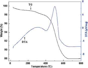
Figure 1 shows the TG-DTA curves of the precursor heated at a rate of 10 °C·min−1 in air. There are two main strong exothermic peaks near 312 and 518 °C in DTA curve. The peak near 312 °C is accompanied by the first weight loss step (2.7%) between 20 and 400 °C in TG curve, which is caused by the decomposition and oxidation of surfactant pyridine and the dehydration of combined water in the precursor, the strong exothermic peak near 518 °C is accompanied by the second weight loss (9.35%) between 400 and 650 °C in TG curve, which is attributed to the combustion of the CNTs and organic groups of surfactant. After 650 °C, there is no heat effect and the weight of the sample is stable, which indicates that the CNT is almost decomposed completely and NaGd(WO4)2:Eu3+ crystal has been formed.
Thermogravimetric and differential thermal analysis curves of the NaGd(WO4)2:Eu3+/ carbon nanotubes (CNTs) precursor.

B. Structural and morphology characterization
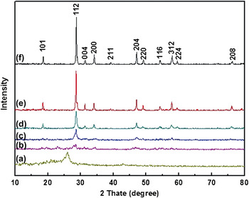
Figure 2 presents the XRD patterns of CNTs, the precursor NaGd(WO4)2:Eu3+ /CNTs, and the samples calcined from 500 to 800 °C. For the precursor, the diffraction peaks of CNTs were not obviously observed, and no resolved diffraction peaks belonging to NaGd(WO4)2:Eu3+ were detectable, indicating that NaGd(WO4)2:Eu3+ in the as-prepared composites (b) is mainly amorphous, and the diffraction peaks of CNTs were not obviously observed due to the dense and uniform coating layers.Reference Yang, Zhang, Shi and Fang14 After being calcined at (c) 500, (d) 600, (e) 700, and (f) 800 °C for 4 h, respectively, the samples crystallized. The obtained diffraction peaks of the product calcined at 500 °C can be readily indexed to the pure tetragonal phase of NaGd(WO4)2 with lattice parameters of a = 0.5243 nm and c = 1.1368 nm. Further increasing the annealing temperature from 600 to 800 °C, the peaks become even sharper and stronger, which is due to the increase of crystallinity and the elimination of carbon. All of the diffraction peaks are in good agreement with the reference data for NaGd(WO4)2 (JCPDS No. 25-0829). No additional peaks of other phases or impurities have been found, indicating that the doped Eu3+ ions has been effectively built into the crystal lattice of NaGd(WO4)2 . These results demonstrate that the well-crystallized tetragonal NaGd(WO4)2:Eu3+ are successfully synthesized by the hydrothermal method using CNTs as removable templates.
X-ray diffraction (XRD) patterns of (a) CNTs, (b) the as-prepared composites NaGd(WO4)2:Eu3+/CNTs and the samples calcined for 4 h at (c) 500, (d) 600, (e) 700, and (f) 800 °C.

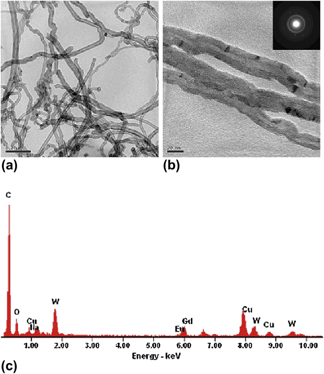
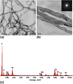
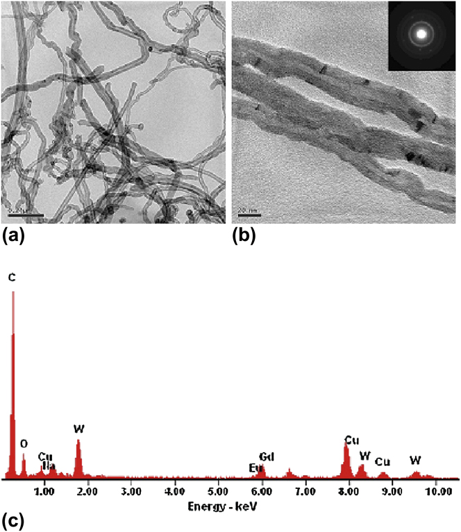
The as-prepared products were first examined by means of TEM. The TEM image [Fig. 3(a)] shows that the CNTs are uniformly sheathed with a dense layer of NaGd(WO4)2:Eu3+. Figure 3(b) shows a high-resolution TEM image of the morphology of the coating layer, and the inset shows the corresponding SAED pattern. The diffraction pattern displays diffuse rings, indicating that the coating layers are amorphous. The coating layer is about 7 nm thick in the as-prepared composites. According to the EDS analysis [Fig. 3(c)], which illustrates the presence of C, Na, Gd, Eu, W, and O, the atomic ratio of Na, Gd, Eu, W, and O is approximately 1:0.95:0.05:2:8. It suggests that the coating layer may be NaGd(WO4)2:Eu3+. In addition, the element Cu and part of element C come from the copper nets carrying carbon films, not the sample.
(a) Transmission electron microscopy (TEM) image of the as-prepared composites; (b) High-resolution TEM image of the as-prepared composites and corresponding selected area electron diffraction pattern (inset); (c) Energy dispersive spectral (EDS) analysis of the as-prepared composites.
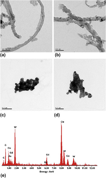
The images of the products calcined at various temperatures are shown in Figs. 4(a)–4(d). After calcination at 500 °C for 4 h, NaGd(WO4)2:Eu3+ nanotubes were obtained [Fig. 4(a)]. The obtained nanotubes have the outer diameter of 40 nm and the inner diameter of 13 nm. When the calcination temperature reaches 600 °C, NaGd(WO4)2:Eu3+ nanotubes are also obtained [Fig. 4(b)], but some nanoparticles appear on the side walls of nanotubes, which can be explained by the NaGd(WO4)2:Eu3+ nanoparticles tending to become more spherical to further minimize their surface energies at higher temperatures.Reference Shi, Lian, Wang, Liu, He, Dong, Wang and Ewing15 As the temperature gradually increased, nanoparticles continue to grow. As shown in Figs. 4(c) and 4(d), when the calcination temperature is increased to 700 and 800 °C, the NaGd(WO4)2:Eu3+ nanotubes collapse and convert into nanoparticles by the oriented agglomeration of small nanoparticles. When the NaGd(WO4)2:Eu3+ nanotubes are calcined at 700 °C, the EDS analysis [Fig. 4(e)] illustrates that C element basically does not exist, and there is almost no change in atomic ratios of the other elements. In addition, the elements Cu and few C are from the copper nets carrying carbon films, not the sample.
TEM images of the products calcined for 4 h at (a) 500, (b) 600, (c) 700, and (d) 800 °C; (e) EDS analysis of the NaGd(WO4)2:Eu3+ nanotubes calcined at 700 °C.

C. Possible formation mechanism of NaGd(WO4)2:Eu3+ nanotubes
In this work, the presence of pyridine plays a dominant role in the formation of NaGd(WO4)2:Eu3+/CNTs. It was reported that aniline grafted onto the CNTs could form a highly branched composite by flux.Reference Sun, Wilson and Schuster16 It is known that CNTs should be a good electron acceptor,Reference Riggs, Guo, Carroll and Sun17 and similar to aniline, pyridine is a good electron donor due to the presence of a pair of isolated nitrogen electrons that are delocalized over its entire aromatic ring,Reference Li, Sun, Kinloch, Zhi, Sirringhaus and Windle18 which indicates that the noncovalent bond may exist between pyridine and CNTs. Thus, we consider that pyridine may be grafted onto the surface of CNTs by means of noncovalent functionalization.
Li et alReference Li, Sun, Kinloch, Zhi, Sirringhaus and Windle18 have reported that the pyridine has a strong interaction with the CNTs, probably through conjugated π–π stacking. The interaction between the electron-rich CNTs and the aromatic pyridine may then make it much easier for nitrogen to accept a proton, which results in the concentration of WO42− around the CNTs being much higher than that in the bulk solvent. Owing to the relatively high concentration, the WO42− around the CNTs could react with Na+, Gd3+, and Eu3+ in the solvent, producing NaGd(WO4)2:Eu3+ nanoparticles which deposit on the surface of CNTs. According to the principle of Ostwald,Reference Talapin, Rogach, Shevchenko, Kornowski, Haase and Weller19 most of the nanoparticles that exist in the solvent do not disappear as time goes by. Under the solvothermal condition, the particles may self-assemble to make a continuous and uniform coating layer, and CNTs coated with dense layers of NaGd(WO4)2:Eu3+ were obtained at last. After calcining the as-prepared composites at 500 or 600 °C for 4 h, the NaGd(WO4)2:Eu3+ nanotubes were obtained. The possible mechanism of the formation of NaGd(WO4)2:Eu3+ nanotubes is shown in Fig. 5.
The possible formation mechanism of NaGd(WO4)2:Eu3+ nanotubes.

D. PL properties
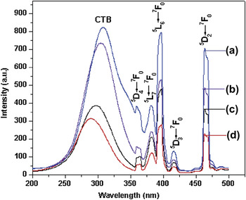
The room temperature excitation and emission spectra of NaGd(WO4)2:Eu3+ calcined at various temperatures are presented in Figs. 6 and 7. It can be clearly found that the excitation and emission spectra of samples are different in the intensity to some extent. The excitation spectra (Fig. 6) are obtained by monitoring the emission of the 5D0 → 7F2 transition of Eu3+ at 615nm. As shown in Fig. 6(a), the excitation spectrum that collected at the emission wavelength of 615 nm consists of a broad band ranging from 200 to 350 nm and some sharp lines from 350 to 500 nm in the longer wavelength region. The ultraviolet excitation spectrum with a strong and broad band from 200 to 350 nm can be attributed to charge transfer transition from O2− to W6+ in the WO42− group.Reference Lei, Yan, Chen and Zhao20 The strong charge transfer band (CTB) from host WO42− group is favorable for the effective energy transfer and luminescence of Eu3+ ions. In the longer wavelength region, the other bands were located at 363, 382, 396, 416, and 464 nm, corresponding to transitions 7F0 → 5D4, 7F0 → 5L7, 7F0 → 5L6, 7F0 → 5D3, and 7F0 → 5D2 of Eu3+ ions, respectively,Reference Wang, Endo, He and Wu21 and the strongest excitation peak is at 396 nm (7F0 → 5L6).
The excitation spectra of NaGd(WO4)2:Eu3+ calcined at (a) 500, (b) 600, (c) 700, and (d) 800 °C, respectively.

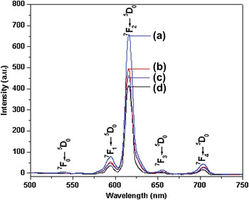
The emission spectra of NaGd(WO4)2:Eu3+ calcined at (a) 500, (b) 600, (c) 700, and (d) 800 °C, respectively.

Comparing the excitation spectrum of the samples in the range 200–350 nm, we can discover that, with decrease in annealing temperature, the maximum of CTB is red shifted gradually, which can be explained as follows. From the TG-DTA measurements, the CNTs were completely removed at 650 °C. When the calcination temperatures are lower than 650 °C, a few residual carbons were doped into the sample, and NaGd(WO4−x)2C2x:Eu3+ forms in the nanotubes. Tachikawa et al.Reference Tachikawa, Tojo, Kawai, Endo, Fujitsuka, Ohno, Nishijima, Miyamoto and Majima22 reported that carbon-doped TiO2 has a much narrower band gap as the energy of the C 2p orbit is higher than that of O 2p in the TiO2−xCx. Similarly, carbon doped into NaGd(WO4)2:Eu3+ could help to raise the valence band of the NaGd(WO4−x)2C2x:Eu3+. Therefore, NaGd(WO4−x)2C2x:Eu3+ has a narrower band gap in comparison with pure NaGd(WO4)2:Eu3+, which decreases the energy barrier that must be overcome to produce powerful radical species, leading to the red shift in the CTB.
The emission spectra of NaGd(WO4)2:Eu3+ phosphors excited under 396 nm are shown in Fig. 7. As can be seen, the emission spectrum exhibits five groups of emission lines ranging from 500 to 750 nm, which are ascribed to the 5D0 → 7FJ (J = 0, 1, 2, 3, 4) transitions of the Eu3+ ions. Luminescence originating from transitions between 4f levels is predominant due to electric dipole or magnetic dipole interactions.Reference Ignatovych, Holovey, Watterich, Vidóczy, Baranyai, Kelemen, Ogenko and Chuiko23 The intensity of electric dipole transitions depends strongly on the site symmetry in a host crystal. Magnetic dipole transitions are not affected much by the site symmetry because they are parity-allowed. The intensities of different 5D0 → 7FJ transitions and the splitting of these emission peaks depend on the local symmetry of the crystal field of Eu3+. If Eu3+ occupies an inversion symmetry site in the crystal lattice, magnetic dipole transition 5D0 → 7F1 (around 590 nm) is the dominant transition. On the contrary, if Eu3+ does not occupy the inversion symmetry site, the electric dipole transition 5D0 → 7F2 (around 600–630 nm) is the dominant transition.Reference Pizani, Leite, Pontes, Paris, Rangel, Lee, Longo, Delega and Varela24, Reference Chen, Liu and Li25 In Fig. 7, as can be seen that the most prominent red emission centered at 615 nm ascribed to 5D0 → 7F2 is the dominant transition, indicating that Eu3+ ions occupy the sites with no inversion center and have low crystal field symmetry.
Furthermore, it is interesting to note that the intensity of emission band at 615 nm of NaGd(WO4)2:Eu3+ nanotubes calcined at 500 °C is the strongest one in these samples, and the relative intensities of emission decreases with increasing calcination temperature from 500 to 800 °C, which is unexpected. In normal conditions, the PL intensity should increase with the increasing calcined temperature as the crystal grows well as the temperature increased. The unusual phenomenon could be explained as follows: (i) there are small amounts of residual carbon doped into the NaGd(WO4)2:Eu3+ nanotubes when the calcination temperature is 500 or 600 °C. Asahi et al.Reference Asahi, Morikawa, Ohwaki, Aoki and Taga26 have reported that TiO2 doped with N or C has a narrower band gap than pure TiO2, due to the results of a mixture of N 2p, C 2p, and O 2p states. Therefore, carbon-doped NaGd(WO4)2:Eu3+ could also have a narrower band gap in comparison with pure ones due to the result of a mixture of C 2p and O 2p states. Because the energy of the C 2p orbit is higher than that of O 2p, carbon doped into NaGd(WO4)2:Eu3+ could help to raise the valence band of the NaGd(WO4−x)2C2x:Eu3+. Therefore, the electronic transition from the valence band to the conduction band could be easier, which is beneficial for luminescence. (ii) Ajayan et al.Reference Ajayan, Stephan, Redlich and Colliex2 reported that when the CNTs used as templates are removed by calcination, some oxygen vacancies form in the nanotubes. Park et al.Reference Park, Kim and Bard27 reported that carbon-doped TiO2 (TiO2−xCx) nanotube arrays with a smaller band gap and more electron–hole pairs showed much higher photocurrent densities and more efficient water splitting under visible-light illumination than pure TiO2 nanotube arrays. Generally, defects such as oxygen vacancies are known to be the most common defects usually act as radiative centers in luminescence processes.Reference Gu, Wang, Lu, Zhou, Xu and Yuan28 Thus, it can clearly be found that the red emission will be enhanced effectively when the NaGd(WO4)2:Eu3+ nanotubes have a little residual carbon and more oxygen vacancies. When the calcination temperature reaches 700 or 800 °C, the CNTs have been completely removed. As there is no residual carbon doped into NaGd(WO4)2:Eu3+ nanotubes and the oxygen vacancies are modified, the intensities of luminescence decrease. In addition, the crystal shape of the NaGd(WO4)2:Eu3+ is more integrated with increasing calcination temperature.
IV. CONCLUSIONS
In summary, we have synthesized NaGd(WO4)2:Eu3+/CNTs composites using CNTs via a hydrothermal method at 180 °C. After calcining NaGd(WO4)2:Eu3+/CNTs composites at various temperatures, NaGd(WO4)2:Eu3+ nanotubes were obtained by removing the CNTs at 500 or 600 °C. The formation mechanism of NaGd(WO4)2:Eu3+ nanotubes have been investigated in detail. The PL studies indicated that the NaGd(WO4)2:Eu3+ nanotubes obtained have a characteristic red emission peak of Eu3+ ions at 615 nm, attributed to the 5D0 → 7F2 electric dipole transition, and the NaGd(WO4)2:Eu3+ nanotubes exhibit the strongest emission band when the calcination temperature is 500 °C. Furthermore, the emission intensity decreases with the increasing of annealing temperature, indicating that a few residual carbons doped into NaGd(WO4)2:Eu3+ nanotubes and many oxygen vacancies could promote the intensity of red emission peak of Eu3+ ions.
ACKNOWLEDGMENTS
This project was supported by the Hubei Province Nature Science Foundation (2010CDB04701) and the Hubei Province Education Office Key Research (D20101011) of China.