




No CrossRef data available.
Article contents
Histopathological follow-up after bailout stenting for early postoperative stenosis of a central aorto-pulmonary shunt
Published online by Cambridge University Press: 18 April 2005
Abstract
We treated a neonate with pulmonary atresia and a ventricular septal defect complicated by straddling of the atrioventricular valves by constructing a central aorto-pulmonary shunt. The postoperative course was complicated by obstruction of the shunt, which was treated by implantation of a coronary stent. Six months after the stenting, a Glenn anastomosis was created and the stented shunt removed. Analysis showed that the shunt was completely covered by a vascularized neointima. The stent had not produced injury to the shunt, with struts of the stent covered nicely by neoendothelium, with sparse inflammation surrounding the artificial implants.
Information
- Type
- Brief Report
- Information
- Copyright
- © 2003 Cambridge University Press
Implantation of stents has recently been reported for relief of acute postoperative obstruction of aorto-pulmonary shunts.1 Little information exists, however, on the biocompatibility of stents implanted into Dacron and polytetrafluoroethylene shunts. The aim of this study, therefore, was to assess the inflammatory response, and the extent to which the surface of the stent had been covered by neoendothelium, by means of cutting and grinding the specimen explanted at surgery.
Case report
A newborn patient was diagnosed with pulmonary atresia and ventricular septal defect. Despite a tripartite and moderately hypoplastic right ventricle, perforation of the atresia was not performed due to straddling of the atrioventricular valves. After initial stabilization of the patient by the administration of intravenous prostaglandin E1, a central aorto-pulmonary shunt, of 3.5 mm diameter, was constructed at 8 days of age. Ten days postoperatively, after extubation of the patient, the arterial saturations of oxygen declined to 62%, and the patient was diagnosed echocardiographically to have obstruction of the aorto-pulmonary shunt. Cardiac catheterization revealed a stenosis of the pulmonary insertion of the shunt. A coronary stent (AMG, Germany, length 9 mm) was crimped on a 3.5 mm Savvy (Cordis, Netherlands) coronary angioplasty catheter, and the stent was implanted into the distal shunt. After the intervention, arterial saturations of oxygen rose to levels above 80%, and the patient was discharged 8 days postinterventionally. At 6 months of age, elective cardiac catheterization demonstrated a patent stented shunt. The patient was scheduled for a Glenn operation, during which the stented shunt was removed and further histopathological work-up initiated. The postoperative course of the patient was uneventful.
Methods
The removed stented polytetrafluoroethylene shunt was documented photographically (Fig. 1) before the stented part was removed, rinsed with saline, and fixed in 4.5% formaldehyde. The unstented part of the shunt was embedded in paraffin wax for conventional histopathological work-up, being sectioned and stained with Hematoxylin and eosin, Elastic van Gieson, Azan, and Picrosirus-red stains. The stented part of the shunt was dehydrated in ascending alcohol concentrations before infiltration of the specimen was performed with methacrylate (Technovit 7200, Kulzer, Germany). The methacrylate-infiltrated specimen was then polymerized by blue and yellow light. Specimens of 150 μm thickness were sawn and ground, and polished to a final thickness of 25 μm. The specimens were then stained according to Richardson and colleagues.2

Figure 1. Macroscopic view (a) of the stented polytetrafluoroethylene shunt removed at surgery. The struts (b) are incorporated into loosely arranged and endothelialized neointima supported by the polyteterafluoroethylene shunt; Richardson stain, original magnification 5 ×. There is neointimal tissue (c) close to a strut with loosely connected collagenous fibers. The shunt itself, however, reveals no destructive changes. Richardson stain, original magnification 40 ×. The luminal surface of the shunt (d) is composed of loosely arranged vascularized (→) connective tissue, Richardson stain, original magnification 40 ×.
Results
Complete endothelialization of the polytetrafluoroethylene-shunt was demonstrated both macroscopically and histologically (Fig. 2). The struts were completely covered with, and incorporated in, a neointimal layer of fibroblastic cells, which was less than 50 μm in thickness. Close to the polytetrafluoroethylene shunt, there was poor vascularization of the neointima. The struts were surrounded by a single layer of fibroblasts, with little or no inflammatory reaction. In contrast, a mild to moderate granulomatous foreign body reaction, consisting of macrophages and polynuclear giant cells, was present along the luminal surface of the shunt attached to the neointima. Along the outer surface of the shunt, a mild granulomatous infiltration was present, comprising macrophages and single polynuclear giant cells.

Figure 2. Moderate granulomatous inflammation along the luminal surface of the shunt attached to the neointima (a) consists of macrophages and polynuclear giant cells (→); Hematoxylin–Eosin stain, original magnification 20 ×. The mild granulomatous infiltration along the outer surface of the shunt (b) consists of single macrophages and multinucleated giant cells (→). The polytetrafluoroethylene shunt itself displays irregular slit-like spaces. Hematoxylin–Eosin stain, original magnification 40 ×.
Conclusion
Stent implantation into polytetrafluoroethylene-shunts is not associated with marked inflammation. The surface of the stent is completely covered with “neointimal” and endothelial cells. Histopathological work-up of explanted cardiovascular implants by cutting and grinding should be routinely performed to evaluate medium- to long-term biocompatibility.
References
Peuster M, Fink C, Bertram H, Paul T, Hausdorf G. Transcatheter recanalization and subsequent stent implantation for the treatment of early postoperative thrombosis of modified Blalock-Taussig shunts in two children. Cathet Cardiovasc Diagn 1998; 45: 405–408.Google Scholar
Richardson KC, Jarett L, Finke EH. Embedding in epoxy resins for ultrathin sectionning in electron microscopy. Stain Technol 1960; 35: 313–325.Google Scholar

Macroscopic view (a) of the stented polytetrafluoroethylene shunt removed at surgery. The struts (b) are incorporated into loosely arranged and endothelialized neointima supported by the polyteterafluoroethylene shunt; Richardson stain, original magnification 5 ×. There is neointimal tissue (c) close to a strut with loosely connected collagenous fibers. The shunt itself, however, reveals no destructive changes. Richardson stain, original magnification 40 ×. The luminal surface of the shunt (d) is composed of loosely arranged vascularized (→) connective tissue, Richardson stain, original magnification 40 ×.
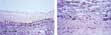
Moderate granulomatous inflammation along the luminal surface of the shunt attached to the neointima (a) consists of macrophages and polynuclear giant cells (→); Hematoxylin–Eosin stain, original magnification 20 ×. The mild granulomatous infiltration along the outer surface of the shunt (b) consists of single macrophages and multinucleated giant cells (→). The polytetrafluoroethylene shunt itself displays irregular slit-like spaces. Hematoxylin–Eosin stain, original magnification 40 ×.
 You have
Access