Case
In 2019, a married couple was referred to the Reproductive Medicine Department at Royale Hayat Hospital, Kuwait City, for treatment. They presented with a 5-year history of secondary infertility due to a male factor. The husband’s age was 44 years, and semen analysis showed oligoasthenoteratozooermia. His wife was 38 years old with regular ovulatory cycles and follicle-stimulating hormone levels of 8.6 mIU/ml. We counselled the couple regarding the need for intracytoplasmic sperm injection (ICSI) treatment, given the quality of sperm in the ejaculate. They underwent a full ICSI cycle at Royale Hayat Hospital, Kuwait City. The patient responded quite poorly to controlled ovarian stimulation. Briefly, 450 IU daily of recombinant human gonadotropin (Gonal-F, Merck, Rome, Italy) were administrated for 8 days (total gonadotrophin dose of 3150 IU). One dominant follicle was seen on the left ovary, whereas no follicles were seen on the right ovary. Oocyte maturation was triggered using a single injection of 5000 IU IVF-C (Human Chorionic Gonadotropin, Ciplamed) subcutaneously. Transvaginal ultrasound-guided oocyte retrieval was carried out 36 h after the trigger administration. Only one oocyte was retrieved during oocyte pick-up. At egg collection day, the sperm count was 7.8 × 10⁶/ml, total motility was 30%, and strict morphology 4%.
Oocyte retrieval, ICSI procedure, embryo culture and transfer
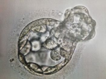
The cumulus–oocyte complex was isolated from follicular fluid and then rinsed in 1.0 ml G-MOPS™ plus medium (Vitrolife, Göteborg, Sweden). Following oocyte pick-up, the retrieved gamete was transferred to 1.0 ml equilibrated G-IVF™ medium (Vitrolife) at 37°C and 6% CO2, 5% O2, and nitrogen balance in a K-System incubator (K-System G210, Cooper Surgical, USA) until time of ICSI. Sperm used for the ICSI procedure was collected by masturbation and processed using a standard method as described by Bourne and colleagues (2004). Approximately 38–39 h after the trigger, the oocyte was treated with hyaluronidase (80 mIU/ml) for 45–60 s to remove the surrounding cumulus cells. At that time, the oocyte was immature at metaphase I (MI) and was placed back into culture in G-IVF™ medium. The oocyte was observed again a few hours later. At 43 h after the trigger, it was found to be at metaphase II (MII) with a clear extrusion of the first polar body. A single spermatozoon with normal morphology and progressive motility was selected under an inverted microscope (Nikon Eclipse Ti-S, Japan) and micro-injected with the use of electrohydraulic injectors (Narishige, Japan). The oocyte was kept still using a holding micropipette at the 9 o’clock position, and the polar body was oriented at the 12 o’clock position. The injecting pipette was then gently advanced through the zona pellucida and oolemma until the pipette was beyond the centre of the oocyte. Next, the sperm was gently deposited into the oocyte’s cytoplasm. The oocyte was examined for the presence of two pronuclei, and successful fertilization was confirmed 16–19 h after insemination. On day 2, the embryo was four-cell grade I, reaching eight cells on day 3. At this stage, an embryo biopsy was performed, as the couple was concerned about the risk of having a child with Down’s syndrome. The biopsy was performed with the use of a laser (Zilos-tk® Laser, Hamilton Thorne, USA) to make a hole on the zona pellucida. A single blastomere was aspirated with a fine biopsy pipette (Humagen, Cooper Surgical, USA) and sent to the genetics laboratory to assess three chromosomes (X, Y and 21) using fluorescence in situ hybridization (FISH) technology (Munné et al., Reference Munné, Lee, Rosenwaks, Grifo and Cohen1993). The embryo culture was completed adopting a sequential G-series medium system (Vitrolife, Göteborg, Sweden) as follows. The fertilized oocyte was initially placed into a 20-μl drop of G-1™ medium covered by mineral oil. On the morning of day 3, after the biopsy, the embryo was transferred from the G-1™ microdroplet to a 20-μl droplet of G-2™ medium and kept in culture to reach the blastocyst stage. On the morning of day 5, microscopic observation confirmed the formation of a grade 5Bb blastocyst (Fig. 1), which was transferred on the afternoon of the same day, after genetic report confirmation of an XX embryo with normal 21 chromosomes. The blastocyst was classified using Gardner’s score (Gardner and Schoolcraft, Reference Gardner and Schoolcraft1999) according to blastocyst expansion, the morphology of the inner cell mass (ICM) and trophectoderm (TE). The embryo transfer was performed under transabdominal ultrasound guidance using a soft transfer catheter (Wallace® Classic, Cooper Surgical, USA). Luteal phase support with vaginal progesterone gel twice a day (Crinone 8%, Merck) was started on the evening of the egg collection day and continued until the 9th gestation week.
Blastocyst transferred.

Pregnancy outcome
The embryo implanted and 6 weeks later the first scan was performed, which revealed the presence of two fetal heartbeat in a single amniotic sac. At the time of this report, gestation was at 18 weeks, and so far has been eventless and without any complications.
Discussion
Over 8 million in vitro fertilization (IVF) children have been born since 1978, when the first IVF baby was announced (Steptoe and Edwards, Reference Steptoe and Edwards1978). Worldwide, approximately 2.5 million medically assisted reproduction (MAR) cycles have been performed, resulting in over 500,000 deliveries annually (Adamson et al., Reference Adamson, de Mouzon, Chambers, Zegers-Hochschild, Mansour, Ishihara, Banker and Dyer2018). During the last 40 years, IVF techniques have improved steadily, therefore providing better pregnancy prospects for couples suffering from infertility. However, despite marked advances in MAR treatments, the development of human embryos in vitro is still suboptimal, and many good quality embryos fail to implant and generate a viable pregnancy (Hu et al., Reference Hu, Maxson, Hoffman, Ory, Eager, Dupre and Lu1998). IVF outcome is dependent on multiple factors, including oocyte and sperm quality, maternal age, infertility cause, lifestyle factors, as well as laboratory conditions for manipulation and embryo culture (Gardner, Reference Gardner2016; Sciorio and Smith, Reference Sciorio and Smith2019). Appropriate oocyte cytoplasmic and nuclear maturation is paramount to ensure an optimal embryonic developmental competence. While nuclear maturation is usually obtained by the time of oocyte retrieval, with the extrusion of the first polar body (Voronina and Wessel, Reference Voronina and Wessel2003), cytoplasmic maturation cannot be readily assessed and might be incomplete. The oocyte must be at the MII stage, which denotes its competence to be fertilized by a spermatozoon. Nonetheless, in the IVF laboratory, it is common to wait a few hours between oocyte collection and denudation in the assumption that the oocyte might need some time to recover after egg collection or in case the oocyte is found immature, at the MI stage. Several authors have reported that a waiting time of 2–4 h between egg retrieval and ICSI improves fertilization rate and embryo quality (Rienzi et al.,Reference Rienzi, Ubaldi, Anniballo, Cerulo and Greco1998; Isiklar et al., Reference Isiklar, Mercan, Balaban, Alatas, Aksoy and Urman2004). Dozortsev and colleagues (2004) found in human oocytes that the optimal insemination time to increase pregnancy outcomes was 37–41 h post-trigger. Conversely, excessive in vitro culture of human oocytes can affect their ultrastructural components. In the mice model, prolonged oocyte culture induces alterations in gene expression, histone modification, and increases spindle and chromosomes abnormalities (Trapphoff et al., Reference Trapphoff, Heiligentag, Dankert, Demond, Deutsch, Frohlich, Arnold, Grummer, Horsthemke and Eichenlaub-Ritter2016). In humans, in vitro ageing has been linked to changes in oocyte ultrastructure, alteration of the spindle and mitochondrial activities (Bianchi et al., Reference Bianchi, Macchiarelli, Micara, Linari, Boninsegna, Aragona, Rossi, Cecconi and Nottola2015), and also to pregnancy outcomes. Hence, it is advisable to limit the oocyte’s incubation time to improve pregnancy rate (Pujol et al., Reference Pujol, García, Obradors, Rodríguez and Vassena2018).
In this case report, a monoamniotic twin pregnancy occurred following the transfer of a single blastocyst; this is a rare event. An embryo can split into two or three and give rise to monozygotic twins (MZT) or triplets. The frequency of this event after MAR cycles varies from 0 to about 10% compared with 0.5% in live births achieved by natural conception (Hviid et al., Reference Hviid, Malchau, Pinborg and Nielsen2018). The time of embryo division will be responsible for the formation of the amniotic and chorionic sacs, which are the membranes surrounding the embryo in which the fetus develops (Fig. 2). If the embryo divides in the early stage after fertilization, the fetus will be dichorionic diamniotic, which occurs in 20–30% of monozygotic twins. If the division occurs around the blastocyst stage or soon after, the gestation will most likely become monochorionic diamniotic, which is observed in 70% or 75% of cases. Finally, if the division of the embryo is around the second week of development, the fetus will be a monochorionic monoamniotic twin, which is a sporadic occurrence observed in only 1–2% of the live births (Aston et al., Reference Aston, Peterson and Carrell2008). Little information is known about the mechanisms involved in early embryonic development that give rise to a twin pregnancy. Many investigators have hypothesized that the increase in the likelihood of MZT in MAR cycles might be associated with specific IVF procedures. It has been suggested that manipulation during or before embryo culture, including zona pellucida handling or insemination techniques (conventional IVF or ICSI), might affect the rate of MZT pregnancies (Alikani et al., Reference Alikani, Noyes, Cohen and Rosenwaks1994; Aston et al., Reference Aston, Peterson and Carrell2008). Several studies have examined the association between assisted hatching (AH) and MZT in MAR cycles. Results have shown a statistically significant association between AH and MZT pregnancies after IVF (Knopman et al., Reference Knopman, Krey, Oh, Lee, McCaffrey and Noyes2014; Nakasuji et al., Reference Nakasuji, Saito, Araki, Nakaza, Nakashima and Kuwahara2014; Kanter et al., Reference Kanter, Boulet, Kawwass, Jamieson and Kissin2015; Vaughan et al., Reference Vaughan, Ruthazer, Penzias, Norwitz and Sakkas2016). Blastocyst culture itself seems to be a risk factor for twin pregnancy. Moreover, extended culture might have an effect on the connection between cells of the ICM, which might induce embryo splitting (Abusheikha et al., Reference Abusheikha, Salha, Sharma and Brinsden2000). Indeed, several studies have shown that blastocyst transfer was associated with a statistically significant increase in MZT pregnancy risk when compared with cleavage-stage embryo transfer (Da Costa et al., Reference Da Costa, Abdelmassih, de Oliveira, Abdelmassih, Abdelmassih and Nagy2001; Jain et al., Reference Jain, Boostanfar, Slater, Francis and Paulson2004; Wright et al., Reference Wright, Schieve, Vahratian and Reynolds2004; Kawachiya et al., Reference Kawachiya, Bodri, Shimada, Kato, Takehara and Kato2011; Sotiroska et al., Reference Sotiroska, Petanovski, Dimitrov, Hadji-Lega, Shushleski and Saltirovski2015). Along these lines, embryo biopsy could also play a role and induce embryo splitting. It has been demonstrated that cleavage-stage biopsy significantly reduces human embryonic implantation potential, while blastocyst biopsy does not (Scott et al., Reference Scott, Upham, Forman, Zhao and Treff2013b). However, a study published by Verpoest et al. (Reference Verpoest, Van Landuyt, Desmyttere, Cremers, Devroey and Liebaers2009) evaluated the effect of embryo biopsy at the cleavage stage on MZT risk and found no association. Knopman and colleagues (2014) also examined the effect of ICSI and embryo biopsy at the cleavage stage and failed to demonstrate any association with MZT. In contrast, Vaughan et al. (Reference Vaughan, Ruthazer, Penzias, Norwitz and Sakkas2016) found an increased incidence of MZT among those pregnancies conceived after embryo biopsy performed on day 3 and extended embryo culture and transfer at the blastocyst stage. Several studies have shown that genetic factors, including ovarian stimulation, maternal age, and embryo quality might also play a role in embryo splitting (Derom et al., Reference Derom, Derom, Vlietinck, Maes and Van den Berghe1993; Alikani et al., Reference Alikani, Noyes, Cohen and Rosenwaks1994; Abusheikha et al., Reference Abusheikha, Salha, Sharma and Brinsden2000). However, the exact mechanisms explaining twinning after MAR treatments are not fully understood and, even if single embryo transfer (SET) is performed, multiple pregnancies, such as twin or more pregnancies, have been reported (Osianlis et al., Reference Osianlis, Rombauts, Gabbe, Motteram and Vollenhoven2014; Vega et al., Reference Vega, Zaghi, Buyuk and Jindal2018). Multiple pregnancies achieved with IVF/ICSI treatments experienced a significantly higher risk of obstetrical, peri-natal, and post-natal complications (Zheng et al., Reference Zheng, Chen, Yang, Yu, Wang and Qin2018). Therefore, patients should be always informed of a possible risk of monozygotic multiple pregnancies even after SET.
Different types of monozygotic twins are shown.

Another compelling aspect of this case report relates to the genetic test performed and its results showing a transferable embryo. It is well known that the likelihood of having a transferable embryo is very low after the biopsy of only one embryo, especially in women at advanced maternal age. Embryo aneuploidy is regarded as the primary reason for IVF failure, with higher maternal age associated with increased aneuploidy risk (Esteves et al., Reference Esteves, Carvalho, Martinhago, Melo, Bento, Humaidan and Alviggi2019). The application of preimplantation genetic testing in the human embryo, which was first successfully introduced in the late 1980s, helps to avoid the transmission of chromosomal abnormalities to the offspring. The process involves aspiration of one or more cells from an embryo generated through IVF that are subjected to genetic testing. The unaffected embryos are transferred to the uterus (Handyside et al., Reference Handyside, Kontogianni, Hardy and Winston1990).
In our case report, three chromosomes were analyzed (X, Y and 21) to primarily avoid the transfer of an embryo with trisomy of chromosome 21, and other syndromes associated with sex chromosomes. Klinefelter’s syndrome is an example of a disorder involving sex chromosomes, with a prevalence of one in 500 males, in which one extra X chromosome is included in the male karyotype, therefore invariably resulting in infertility if a viable offspring is born (Visootsak and Graham Jr, 2006; Beg et al., Reference Beg, Nieschlag, Abdel-Meguid, Alam, Abdelsalam, Haque, Mosli, Bajouh, Abuzenadah and Al-Qahtani2019). Another example is Turner’s syndrome, which is due to a total or partial loss of one of the X chromosomes and is associated with short stature and primary ovarian insufficiency and infertility. The latter occurs in one every 2500 female and is one of the most common chromosomal abnormalities in women (Bouet et al., Reference Bouet, Godbout, El Hachem, Lefebvre, Bérubé, Dionne, Kamga-Ngande and Lapensée2016). Furthermore, trisomy of chromosome 21, which might result in the delivery of an offspring with Down’s syndrome, is the most common non-lethal trisomy in humans, usually associated with advanced maternal age (Glasson et al., Reference Glasson, Hussain, Petterson, Montgomery and Bittles2002). Down’s syndrome is also associated with cognitive impairment, congenital heart defects, and is the only autosomal trisomy compatible with survival beyond puberty (Bovicelli et al., Reference Bovicelli, Orsini, Rizzo, Montacuti and Bacchetta1982). In addition, a genetic rearrangement (translocation) involving chromosome 21 is associated with an increased risk of acute myeloid leukaemia (Ustun et al., Reference Ustun, Morgan, Moodie, Pullarkat, Yeung and Broesby-Olsen2018). The couple’s concern about the health of the future child, at least related to the sex chromosomes and Down’s syndrome justified the shared decision to perform embryo biopsy and genetic testing. Although other methods exist to identify those abnormalities post-implantation, including non-invasive diagnostic modalities, they would likely lead to pregnancy termination, which is not allowed in Kuwait. Therefore, embryo preimplantation genetic assessment was deemed considered as a valid approach to obtain information of the embryo genetic content. In addition, we acknowledge that day 3 embryo biopsy and FISH analysis do not provide a comprehensive genetic assessment of the whole chromosomal content (Esteves et al., 2020). Other methods, such as whole-genome amplification and next-generation sequencing, are best suited for this purpose, but are not routinely available in our settings.
Finally, although live birth would have been the ultimate endpoint for such report, we do believe the ongoing pregnancy at 18 weeks to be relevant to stimulate discussion on the unusual aspects of our case, including the immaturity of the oocyte and its prolonged culture, the delayed insemination, the extended in vitro embryo culture and the blastomere biopsy, all of which may have contributed to the outcome.
Conclusion
Besides in vitro ageing of the oocyte, which could not be avoided, the abnormal embryo separation might also be related to extended culture at the blastocyst stage and blastomere biopsy for preimplantation genetic screening. Our case report stimulates discussion on the above aspects, and emphasizes that, despite pregnancy being achieved following sperm injection of a single oocyte, which was immature at retrieval, embryos cultured in vitro should not be exposed to non-necessary risk of damage. Considering that the pregnancy is not yet at term and that the actual consequences are still unknown, clinicians and embryologists should exercise caution to routinely recommend extended embryo culture and preimplantation genetic testing.
Financial Support
This research received no specific grant from any funding agency, commercial or not-for-profit sectors.
Conflict of Interest
SCE has received research grants from Merck, and lecture fees from Merck and Gedeon-Richter outside the submitted work. No potential conflict of interest was reported by RS, EA, NA and MA.
Ethical Standards
Not applicable.