





Introduction
Cor triatriatum sinister represents approximately 0.1% of CHDs.Reference Nassar and Hamdan1 It involves a fibromuscular membrane dividing the left atrium into proximal and distal chambers. The clinical course is determined by the size of the membrane’s fenestration. According to the Loeffler classification, restrictive cor triatriatum sinister is characterised by a small fenestration and usually manifests in early infancy with severe pulmonary venous obstruction or heart failure.Reference Loeffler6 Survival into the second decade with such a restrictive orifice is exceedingly rare, often masked by a long history of presumed respiratory disease, because pulmonary venous obstruction may mimic chronic pulmonary disease or asthma.Reference Karthik, Kelpis, Abela and Weerasena4,Reference Powell, Hanke, Tweddell and Madsen5 We present a unique adolescent case of restrictive, calcified cor triatriatum sinister managed successfully with surgery.
Case presentation
A 14-year-old male (173 cm, 62 kg) was referred to our clinic following an episode of sustained palpitations. His history was notable for chronic exercise-induced dyspnoea and wheezing, leading to a long-standing diagnosis of refractory asthma. Despite various inhaled bronchodilator therapies, his symptoms persisted (NYHA Class II).
Physical examination revealed a loud P2 and a right ventricular heave.
Transthoracic echocardiography demonstrated preserved left ventricular systolic function with an ejection fraction of 60%. The aortic root measured 3.2 cm, and the left atrium was enlarged, measuring 4.6 cm. The right-sided chambers were markedly enlarged. The left ventricle had a D-shaped configuration due to systolic septal flattening, indicating significant right ventricular pressure overload. Mitral regurgitation and tricuspid regurgitation were both moderate. The estimated pulmonary artery pressure was 85 mmHg. The aortic valve maximum velocity was 1.9 m/s. The interatrial septum was subsequently confirmed to be intact by transesophageal echocardiography, and the left atrial membrane with a restrictive 2–3 mm fenestration was clearly delineated. A fibromuscular membrane was clearly visualised dividing the left atrium (Video 1), featuring a highly restrictive 2–3 mm fenestration with high-velocity turbulence.
Transesophageal echocardiography was performed in midesophageal 0, 30, 40, 50, and 60-degree four-chamber views and 90–120-degree bicaval views. The interatrial septum was intact. A left atrial membrane causing obstruction to pulmonary venous return was visualised, confirming cor triatriatum sinister. To further delineate the membrane’s morphology prior to surgery, transesophageal echocardiography revealed a tiny, 2–3 mm fenestration located on the aspect of the membrane facing the interatrial septum (Video 2).
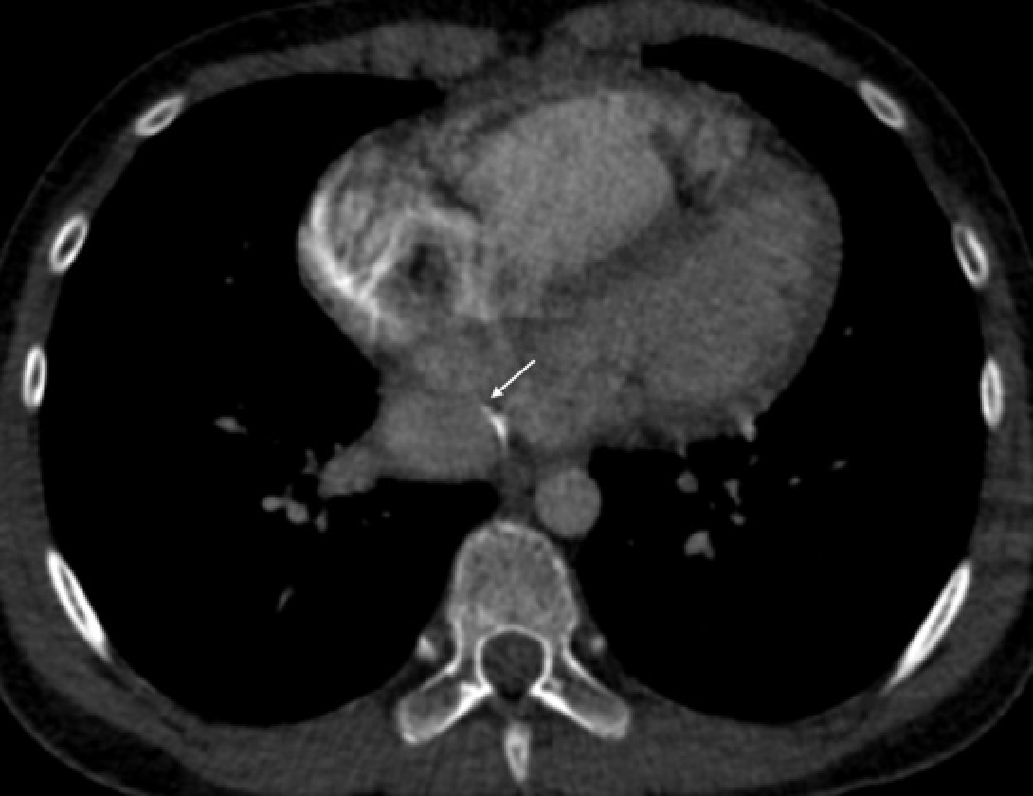
Cardiac CT confirmed the diagnosis, identifying a calcified linear structure within the left atrium (Figure 1).
Cardiac CT scan demonstrating the calcified linear membrane within the left atrium, indicating long-standing obstruction.

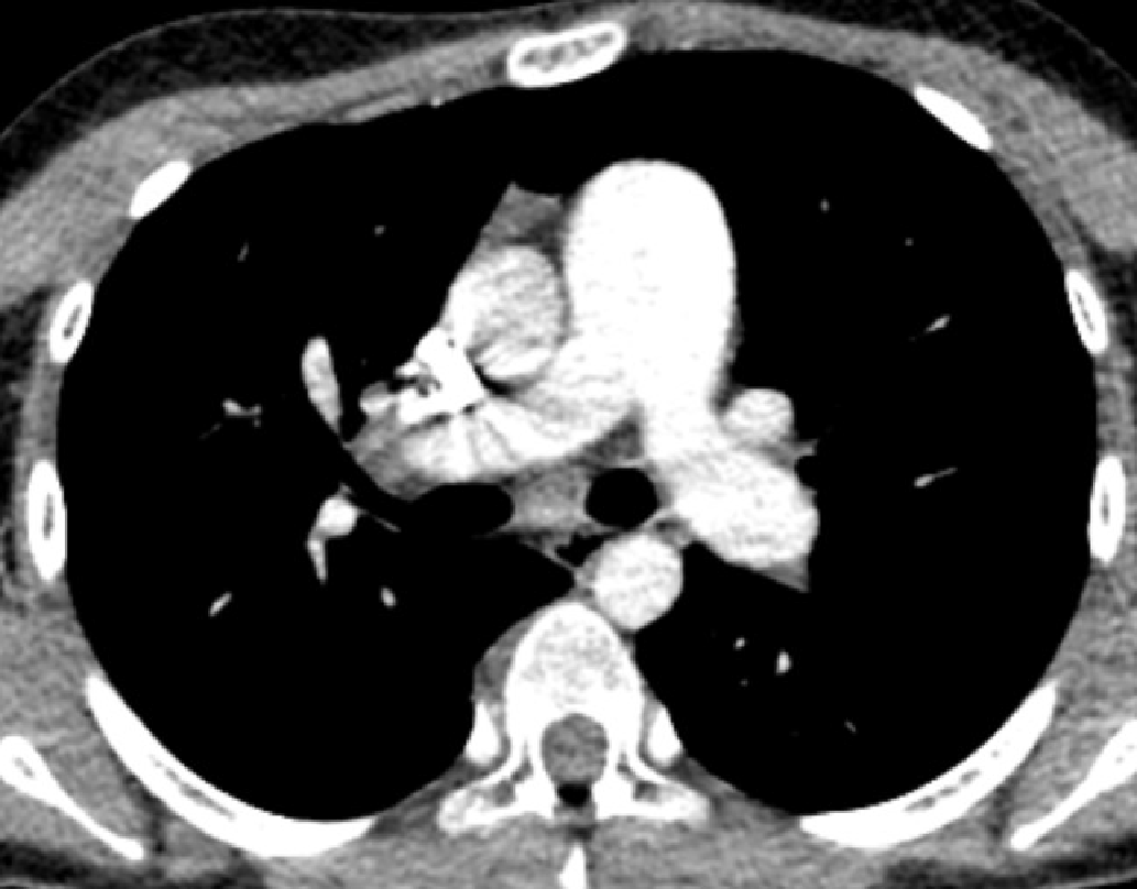
The main pulmonary artery (36 mm) was severely dilated, alongside significant enlargement of the right-sided chambers (Figure 2).
Axial CT imaging highlighting the significantly dilated main pulmonary artery and enlarged right heart chambers due to chronic pressure overload.

Diagnostic cardiac catheterisation and angiography were performed to define the haemodynamic severity and assess operability. Pressures were as follows: RA 5 mmHg, PA 50/30 mmHg (mean 39 mmHg), RPA 51/30 mmHg (mean 37 mmHg), pulmonary capillary wedge pressure 28 mmHg, aorta 79/61 mmHg (mean 68 mmHg), and LV end-diastolic pressure 4 mmHg. Oxygen saturations were 80.4% in the superior vena cava, 84.1% in the pulmonary artery, and 99% in the aorta. Calculated Qp was 2.00 L/min, and Qs was 1.75 L/min, with Qp/Qs of 1.14. Pulmonary vascular resistance was 4.50 Wood units, and indexed PVR was 7.43 Wood units·m2; systemic vascular resistance was 37.72 Wood units, and indexed SVR was 62.26 Wood units·m2. The PVR/SVR ratio was 0.119, supporting preserved pulmonary vascular reversibility despite severe post-capillary pulmonary hypertension. Angiography demonstrated normal coronary and aortic anatomy. Pulmonary venous return filled the proximal left atrial chamber first, followed by the distal left atrial chamber beneath the membrane, and subsequently the left ventricle and aorta. These findings confirmed that the pulmonary hypertension was predominantly post-capillary and secondary to obstruction caused by the restrictive cor triatriatum sinister membrane. The preoperative echocardiographic and cardiac catheterization findings are summarized in Table 1.
Preoperative echocardiographic and cardiac catheterisation findings

Table 1. Long description
A table comparing preoperative echocardiographic and cardiac catheterization findings. The table has 22 rows and 2 columns. Column headers are Parameter and Finding. Row 1: Parameter, Echocardiographic diagnosis; Finding, Cor triatriatum sinister. Row 2: Parameter, Left atrial membrane; Finding, Fibromuscular membrane dividing the left atrium into proximal and distal chambers. Row 3: Parameter, Fenestration size; Finding, 2-3 mm. Row 4: Parameter, Interatrial septum; Finding, Intact on TEE. Row 5: Parameter, LV systolic function; Finding, Preserved; EF 60 percent. Row 6: Parameter, LV morphology; Finding, D-shaped LV due to septal flattening. Row 7: Parameter, Aortic root; Finding, 3.2 centimeters. Row 8: Parameter, Left atrium; Finding, 4.6 centimeters, enlarged. Row 9: Parameter, Right-sided chambers; Finding, Markedly enlarged. Row 10: Parameter, Mitral regurgitation; Finding, Moderate. Row 11: Parameter, Tricuspid regurgitation; Finding, Moderate. Row 12: Parameter, Estimated pulmonary artery pressure by TTE; Finding, 85 millimeters of mercury. Row 13: Parameter, Aortic valve Vmax; Finding, 1.9 meters per second. Row 14: Parameter, Main pulmonary artery diameter on CT; Finding, 36 millimeters. Row 15: Parameter, RA pressure; Finding, 5 millimeters of mercury. Row 16: Parameter, PA pressure; Finding, 50/30 millimeters of mercury; mean 39 millimeters of mercury. Row 17: Parameter, RPA pressure; Finding, 51/30 millimeters of mercury; mean 37 millimeters of mercury. Row 18: Parameter, Pulmonary capillary wedge pressure; Finding, 28 millimeters of mercury. Row 19: Parameter, Aortic pressure; Finding, 79/61 millimeters of mercury; mean 68 millimeters of mercury. Row 20: Parameter, LV end-diastolic pressure; Finding, 4 millimeters of mercury. Row 21: Parameter, Oxygen saturations; Finding, SVC 80.4 percent; PA 84.1 percent; aorta 99 percent. Row 22: Parameter, Qp/Qs/Qp:Qs; Finding, 2.00 liters per minute/1.75 liters per minute/1.14.
The patient underwent surgical repair via a right anterolateral thoracotomy. Cardiopulmonary bypass was established via femoral and superior vena cava cannulation. Following systemic hypothermia and antegrade blood cardioplegia (1500 cc), a left atriotomy was performed. Intraoperative findings revealed a thick, partially calcified membrane obstructing the mitral inflow, with a tiny central orifice. Adhesions on the anterior mitral leaflet were released, and the membrane was completely excised. Total cardiopulmonary bypass time was 49 minutes; cross-clamp time was 27 minutes.
The patient was successfully extubated within 24 hours. Post-operative transthoracic echocardiography showed a significant reduction in right ventricular pressures. He was discharged on day 7 on enalapril and metoprolol to manage systemic pressures and support right ventricular remodelling.
Discussion
Survival to age 14 with a 2–3 mm cor triatriatum sinister fenestration is a rare clinical phenomenon. Most obstructive cases are diagnosed during infancy or childhood, whereas adult or late-presenting cases are usually less obstructive or have atypical clinical presentations.Reference Nassar and Hamdan1–Reference Ozyuksel, Yildirim and Avsar3,Reference Niwa, Tanaka and Tateno7–Reference Hamdan, Mirochnik, Celermajer, Nassar and Iserin9
Late-presenting paediatric or adolescent cor triatriatum sinister is therefore exceptional. A previously reported 14-year-old patient presented with respiratory distress mimicking pulmonary disease,Reference Powell, Hanke, Tweddell and Madsen5 while larger surgical series show a wide age range but generally include a heterogeneous anatomic spectrum, often with associated lesions.Reference Alphonso, Nørgaard, Newcomb, d’Udekem, Brizard and Cochrane2,Reference Ozyuksel, Yildirim and Avsar3,Reference Kazanci, Emani and McElhinney10,Reference Saxena, Burkhart, Schaff, Daly, Joyce and Dearani11 Compared with these reports, our case is notable for isolated, severely restrictive (2–3 mm) cor triatriatum sinister, a high left atrial membrane gradient of 25 mmHg, calcification of the membrane, and absence of an atrial septal decompression pathway.
The correlation between echocardiographic and catheterisation findings was central to clinical decision-making in this case. Echocardiography demonstrated severe mechanical obstruction at the left atrial membrane, marked right-sided chamber enlargement, septal flattening, and elevated estimated pulmonary pressure, while catheterisation confirmed elevated pulmonary venous pressure with PCWP of 28 mmHg and a low PVR/SVR ratio of 0.119. This combination supported passive, post-capillary pulmonary hypertension rather than irreversible pulmonary vascular obstructive disease, thereby justifying surgical correction.Reference Stout, Daniels and Aboulhosn12 The low PVR/SVR ratio of 0.11 provided crucial evidence that the pulmonary hypertension was passive and reversible.
Despite 14 years of exposure to high left atrial pressures, the pulmonary arterial bed maintained its elasticity and did not transition into an irreversible obstructive state. This preserved vascular integrity explains the patient’s rapid clinical improvement; once the mechanical barrier (the membrane) was surgically removed, the compliant pulmonary vessels were immediately able to accommodate normal blood flow without resistance.
Following the successful surgical relief of the mechanical obstruction, postoperative medical management was individualised to facilitate right ventricular reverse remodelling and manage systemic haemodynamics. The patient was started on enalapril and metoprolol. While the primary lesion was excised, the right ventricular had undergone significant compensatory hypertrophy over 14 years. ACE inhibitors such as enalapril may help reduce systemic afterload and mitigate neurohormonal pathways that drive adverse cardiac remodelling, while beta-blockers such as metoprolol may help control postoperative sympathetic activation and optimise right ventricular filling time.
This dual pharmacological approach, combined with the removal of the restrictive membrane, ensured a stable recovery and addressed the long-standing secondary effects of chronic pressure overload.
This case highlights the danger of premature closure in clinical reasoning. The chronic pulmonary venous congestion mimicked asthma for years, a recognised diagnostic trap in late-presenting cor triatriatum sinister and other obstructive pulmonary venous lesions.Reference Karthik, Kelpis, Abela and Weerasena4,Reference Powell, Hanke, Tweddell and Madsen5 The onset of palpitations and the discovery of a calcified membrane suggest that even congenital structures may be dynamic, with calcification likely worsening the obstruction over time.
Supplementary material
The supplementary material for this article can be found at https://doi.org/10.1017/S1047951126124032.


 Open access

