INTRODUCTION
Dusky shark Carcharhinus obscurus occurs in tropical and warm temperate continental oceans worldwide (Compagno, Reference Compagno1984). It is a placental viviparous species, and the young are born at 70–100 cm total length (TL) (Last & Stevens, Reference Last and Stevens1994).
The dusky shark is caught occasionally in fisheries in the Gulf of California (Instituto Nacional de Pesca, 2000) and there is little biological information for this species in this area. Embryonic abnormalities have not been recorded for this species.
Several types of abnormality have been reported in elasmobranchs, mostly involving cases of albinism (Schwartz, Reference Schwartz1959; Joseph, Reference Joseph1961; McKenzie, Reference McKenzie1970; Cohen, Reference Cohen1973; Nakaya, Reference Nakaya1973; Talent, Reference Talent1973; Follett, Reference Follett1976; Schwartz & Safrit, Reference Schwartz and Safrit1977; McKay & Beinssen, Reference McKay and Beinssen1987/1988; Taniuchi & Yanagisawa, Reference Taniuchi and Yanagisawa1987; De Jesus, Reference De Jesus-Roldan1990; Smale & Heemstra, Reference Smale and Heemstra1997; Brahim et al., Reference Brahim, Secka and Capapé1998; Castro, Reference Castro2000; Ishihara et al., Reference Ishihara, Homma and Nakamura2001; Clark, Reference Clark2002; Teixeira & Góes de Araújo, Reference Teixeira and Góes de Araújo2002; Deynat, Reference Deynat2003; Saïdi et al., Reference Saïdi, Bradaï, Marouani, Guélorget and Capapé2006; Sandoval-Castillo et al., Reference Sandoval-Castillo, Mariano-Meléndez and Villavicencio-Garayzar2006; Bottaro et al., Reference Bottaro, Ferrando, Gallus, Girosi and Vacchi2008a, Reference Bottaro, Consalvo, Ferrando, Gallus, Girolsi, Psomadakis, Atkinson and Vacchib; Reum et al., Reference Reum, Paulsen, Pietsch and Parker-Stetter2008; Veena et al., Reference Veena, Thomas, Raje and Durgekar2011) and vertebral deformities (Officer et al., Reference Officer, Clement and Rowler1995; Heupel et al., Reference Heupel, Simpfendorfer and Bennet1999).
However, there is no previous report of cyclopia in elasmobranchs. Cyclopia is a rare congenital malformation, resulting from the division of the embryonic brain that leads to fusion of the eyes to form a single, central eye. It is always associated with an extreme degree of alobar holoprosencephaly which is a malformation in which the brain does not separate and is usually associated with severe facial anomalies (Gaffield & Keeler, Reference Gaffield and Keeler1996).
MATERIALS AND METHODS
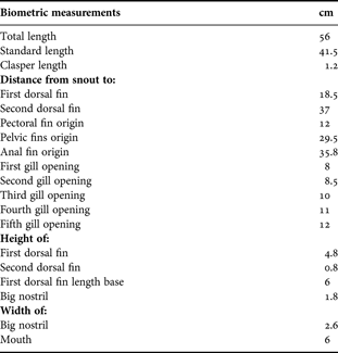
A pregnant female dusky shark was caught around Cerralvo Island (24°22′24.04″N 109°55′53.44″W) in the Gulf of California, Mexico. The specimen was caught at the north point of the island on 16 February 2011 with a simplera (a small, bottom longline with only a few (~30) hooks). The female was 2.5 m TL and weighed approximately 300 kg. The female had nine embryos in the uteri, one of which was abnormal. Embryos were removed, and the embryo with malformations was measured and photographed. The specimen was preserved in ethanol and is in custody of the fishermen who found the shark. Biometric measurements are presented in Table 1.
Morphometric measurements (expressed in centimetres, cm) of abnormal male embryo of dusky shark Carcharhinus obscurus, in the Gulf of California.

The shark was identified using the taxonomic key of Compagno et al. (Reference Compagno, Krupp, Schneider, Fischer, Krupp, Schneider, Sommer, Carpenter and Niem1995). A shark tomography was taken to describe the skull malformation, where the skull and the spine were the reference points in the tomography. The scanner used was a Toshiba, Aquilon, TSX 101A 1F Multislice CT Scan System.
RESULTS
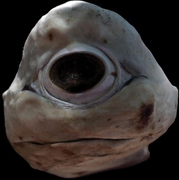
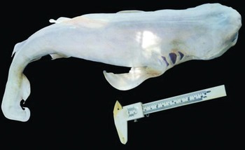
The malformed male embryo was 56 cm TL, and the main characteristic was the presence of a single, large (2.6 cm wide) eye (cyclop) located in the middle of the snout. The absence of nostrils was also evident (Figure 1). The embryo was entirely albino (Figure 2), and the head of the embryo had a pronounced bump and short (flat) snout. The gill slits were normal and well formed. A ventral mouth with small teeth in both jaws was present.
Abnormal male embryo of the dusky shark, Carcharhinus obscurus showing the big eye (cyclop).

Lateral view of an abnormal dusky shark, Carcharhinus obscurus with albinism and showing some malformations in caudal tail and head.

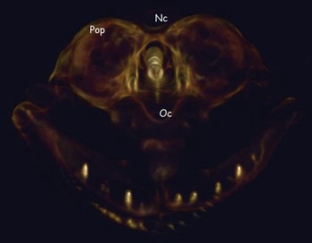
The tomography of the head showed that post-orbital processes were formed, although a single nasal capsule was developed under the single orbit capsule (Figure 3). Comparing the structure of the skull of this shark with a normal one, the skull was formed only to post-orbital processes. The medial rostral cartilage was not developed, which explained the flat snout. The anterior portions of the skull and its components were not well developed, including: nasal capsule; ectethmoid condyle; and medial rostral cartilage. According to the features described, it is confirmed that the shark had alobar holoprosencephaly.
Tomography of the dusky shark, Carcharhinus obscurus head showing post-orbital processes (pop), a single nasal capsule (nc) that was developed under the single orbit capsule (oc).

There was a slight deformity in the vertebral column and a curvature pronounced to the caudal fin. The tomography shows the evidence of fusion of about four vertebrae, confirming this deformity. The rest of the body was apparently normal, with well formed dorsal, pectoral, pelvic and anal fins. Table 1 shows other measurements from this specimen.
DISCUSSION
There have been several reports of abnormalities in sharks and rays from the Pacific waters of Mexico, including skeletal deformities, malformations in the cephalic region, bicephaly and albinism (e.g. Castro-Aguirre & Torres-Villegas, Reference Castro-Aguirre and Torres-Villegas1979; De Jesus-Roldán, Reference De Jesus-Roldan1990; Castro, Reference Castro2000; Sandoval-Castillo et al., Reference Sandoval-Castillo, Mariano-Meléndez and Villavicencio-Garayzar2006; Escobar-Sánchez et al., Reference Escobar-Sanchez, Galvan-Magaña, Downton-Hoffman, Carrera-Fernandez and Alatorre-Ramirez2009; Bejarano-Álvarez et al., Reference Bejarano-Alvarez, Galván-Magaña and Ochoa-Báez2011; Galván-Magaña et al., Reference Galván-Magaña, Escobar-Sánchez and Carrera-Fernández2011).
Bonfil (Reference Bonfil1989) reported a malformed reef shark, Carcharhinus perezi from Yucatán, Mexico, where the shark had prenatal developmental abnormalities in the cephalic region with a reduced snout and a ventral positioning of the eyes. He reported that abnormalities occurred simultaneously, with two eyes positioned in front of the buccal aperture, and with fusion of eyelids and lack of nostrils.
Also there has been a case of a Carcharhinus plumbeus embryo (Saïdi et al., 2006) with abnormalities in the nostrils, and this specimen did not have a fully developed gill opening.
However, there is no published information about abnormal dusky sharks, and our report is the first of cyclopia in an elasmobranch of this species. Cyclopia is a rare congenital deformity, both in human and other animals, and is associated with an extreme degree of alobar holoprosencephaly, which is the most common structural anomaly of the developing forebrain, resulting from incomplete midline cleavage of the prosencephalon and associated with neurological impairment and dysmorphism of the brain and face (Gaffield & Keeler, Reference Gaffield and Keeler1996).
Albinism is a genetic anomaly that produces a reduction in biosynthesis of the pigment melanin during embryonic development. Albinism has been reported occasionally in various elasmobranchs (e.g. Gopalan, Reference Gopalan1971; Castro, Reference Castro2000; Sandoval-Castillo et al., Reference Sandoval-Castillo, Mariano-Meléndez and Villavicencio-Garayzar2006) but the present report is considered a first record of albinism in C. obscurus. The abnormal embryo in our study showed total albinism, and the lack of pigmentation would be congenital. The mother and the other embryos were normally pigmented.
In relation to albinism, it is more frequent in elasmobranchs. According to Sandoval-Castillo et al. (Reference Sandoval-Castillo, Mariano-Meléndez and Villavicencio-Garayzar2006), albinism is a genetic anomaly that produces a reduction in melanin pigment biosynthesis. The abnormal embryo in our study presented total albinism, which is the lack of pigmentation, and can be congenital. The mother was normally pigmented, indicating a mutation for the recessive albino allele. There are other reported cases of albinism in elasmobranchs (Schwartz, Reference Schwartz1959; Joseph, Reference Joseph1961; McKenzie, Reference McKenzie1970; Cohen, Reference Cohen1973; Nakaya, Reference Nakaya1973; Talent, Reference Talent1973; Follett, Reference Follett1976; Schwartz & Safrit, Reference Schwartz and Safrit1977; McKay & Beinssen, Reference McKay and Beinssen1987/1988; Taniuchi & Yanagisawa, Reference Taniuchi and Yanagisawa1987; De Jesus, Reference De Jesus-Roldan1990; Smale & Heemstra, Reference Smale and Heemstra1997; Brahim et al., Reference Brahim, Secka and Capapé1998; Castro, Reference Castro2000; Ishihara et al., Reference Ishihara, Homma and Nakamura2001; Clark, Reference Clark2002; Teixeira & Góes de Araújo, Reference Teixeira and Góes de Araújo2002; Deynat, Reference Deynat2003; Saïdi et al., 2006; Sandoval-Castillo et al., 2006; Bottaro et al., Reference Bottaro, Ferrando, Gallus, Girosi and Vacchi2008a, Reference Bottaro, Consalvo, Ferrando, Gallus, Girolsi, Psomadakis, Atkinson and Vacchib; Reum et al., Reference Reum, Paulsen, Pietsch and Parker-Stetter2008; Veena et al., Reference Veena, Thomas, Raje and Durgekar2011), but there are no reports on this type of anomaly in the dusky shark Carcharhinus obscurus, so our report is considered a first record of albinism in this shark species in Mexico and probably in the world.
The abnormality in the vertebral column near the caudal fin is a consequence of a spinal deformity. Although there are no previous reports of such an abnormality in C. obscurus, there are reports of skeletal malformations in other carcharhinid sharks, including bull shark Carcharhinus leucas, sandbar shark C. plumbeus and lemon shark Negaprion brevirostris, and also the sand tiger shark Odontaspis taurus (Hoening & Walsh, Reference Hoenig and Walsh1983). Cases of spinal malformations may be caused by parasites, arthritis, injuries, tumours or poor nutrition (Sadowsky, Reference Sadowsky1971; Schwartz, Reference Schwartz1973; Heupel et al., 1999). Several studies in teleost fish suggest that spinal deformities can be produced by a high water temperature during embryonic development (Hubbs, Reference Hubbs1959) or vitamin C deficiency, inducing lordosis (curvature of the spine, especially towards the end of the vertebrae column) (Wimberger, Reference Wimberger1993).
ACKNOWLEDGEMENTS
We thank Jesus Enrique Lucero León, Manuel Lucero León and Jose Luis León Lucero, fishermen of Cerralvo Island (BCS, México), who allowed us to study the specimen. The first author thanks CONACYT and PIFI. F.G.M. thanks the Instituto Politécnico Nacional for fellowships (COFAA and EDI). We thank Angela M. Cheek and Lucía Briseño Haro for editing the English-language text, Dr Carlos Rodríguez and Dr Manuel Martínez Marín for the analysis and discussion with tomography and X-rays, and Rubén García Gómez and Juan Pablo Hernández Sánchez for editing the images. We also thank two anonymous referees whose comments improved the manuscript.