

MS is the most common chronic inflammatory disease of the nervous system in young adults. Two subtypes of MS onset have been described: relapsing-remitting (RRMS; 85%) and primary progressive (15%). MS is a major cause of physical disability in young adults (Pugliatti et al., Reference Pugliatti, Rosati, Carton, Riise, Drulovic, Vécsei and Milanov2006; Tullman, Reference Tullman2013). Cognitive dysfunctions and mood disorders occur in a large proportion of MS patients (Chiaravalloti & DeLuca, Reference Chiaravalloti and DeLuca2008; Montreuil & Pelletier, Reference Montreuil, Pelletier, Defer, Brochet and Pelletier2011; Rao, Leo, Bernardin, & Unverzagt, Reference Rao, Leo, Bernardin and Unverzagt1991). Cognitive deficits mainly affected include executive functions, memory, attention and information processing speed. The most frequently described mood disorders are depression and anxiety (José Sá, Reference José Sá2008; Kinsinger, Lattie & Mohr, Reference Kinsinger, Lattie and Mohr2010). However, some behavioural changes presented by MS patients, such as relationship problems, irritability, inexplicable states of agitation and also disinhibition, have received little attention (Chiaravalloti & DeLuca, Reference Chiaravalloti and DeLuca2003; Lima et al., Reference Lima, Simioni, Bruggimann, Ruffieux, Dudlerc, Felley and Schluep2007). While these behavioural changes might represent reactions to lifestyle changes induced by the disease, they could also be the result of changes in social cognition.
Social cognition is the ability to analyse and interpret relevant social stimuli in order to adapt behavioural responses in a way that is consistent with the context (Adenzato, Cavallo, & Enrici, Reference Adenzato, Cavallo and Enrici2010; Adolphs, Reference Adolphs2009; Brüne & Brüne-Cohrs, Reference Brüne and Brüne-Cohrs2006). In additional to cortical areas (e.g., the prefrontal cortex), social cognitive processing is subsumed by activation of a broad subcortical neuronal network (Adolphs, Reference Adolphs2009). MS leads to lesions in white matter that may disrupt this network, thereby impairing the process. Researchers have only recently started to assess social cognition in MS and investigations have primarily focused on the recognition of basic emotional facial expressions and the assessment of Theory of Mind abilities (ToM, the capacity to attribute affective and cognitive mental states to others). Among these studies, some that have specifically explored the ability to attribute mental states to others have, despite methodological differences, found impairments in MS patients compared to controls in both verbal (Banati et al., Reference Banati, Sandor, Mike, Illes, Bors, Feldmann and Illes2010) and non-verbal tasks (Banati et al., Reference Banati, Sandor, Mike, Illes, Bors, Feldmann and Illes2010; Henry et al., Reference Henry, Phillips, Beatty, McDonald, Longley, Joscelyne and Rendell2009; Ouellet et al., Reference Ouellet, Scherzer, Rouleau, Métras, Bertrand-Gauvin, Djerroud and Duquette2010). The same results have also been found using more naturalistic tasks, such as the video-based instrument, the Movie for the Assessment of Social Cognition, which is designed to approximate the demands of everyday social interaction (Kraemer et al., Reference Kraemer, Herold, Uekermann, Kis, Wiltfang, Daum and Abdel-Hamid2013; Pöttgen et al., Reference Pöttgen, Dziobek, Reh, Heesen and Gold2013). Moreover, impaired facial emotion recognition has been reported in MS (Phillips et al., Reference Phillips, Henry, Scott, Summers, Whyte and Cook2011; Prochnow et al., Reference Prochnow, Donell, Schäfer, Jörgens, Hartung, Franz and Seitz2011) in connection with the emotions of both anger and fear (Henry et al., Reference Henry, Phillips, Beatty, McDonald, Longley, Joscelyne and Rendell2009; Henry et al., Reference Henry, Tourbah, Chaunu, Rumbach, Montreuil and Bakchine2011; Krause et al., Reference Krause, Wendt, Dressel, Berneiser, Kessler, Hamm and Lotze2009). Sadness recognition is also affected, albeit to a lesser extent (Krause et al., Reference Krause, Wendt, Dressel, Berneiser, Kessler, Hamm and Lotze2009). Using functional Magnetic Resonance Imagery (MRI), these latter authors observed a decrease of cerebral activation in the insula and ventro-lateral prefrontal cortex among patients who exhibited a deficit in the recognition of emotions compared to unimpaired patients and controls.
The factors responsible for these impaired social cognition abilities are not still fully understood. The links between social cognition and other cognitive functions, in particular executive functions, remain controversial (Eslinger et al., Reference Eslinger, Moore, Troiani, Antani, Cross, Kwok and Grossman2007; Lough et al., Reference Lough, Kipps, Treise, Watson, Blair and Hodges2006). In MS, some authors have found a correlation between ToM performance and executive functions (Banati et al., Reference Banati, Sandor, Mike, Illes, Bors, Feldmann and Illes2010; Ouellet et al., Reference Ouellet, Scherzer, Rouleau, Métras, Bertrand-Gauvin, Djerroud and Duquette2010), such as, for example, verbal fluency (Henry et al., Reference Henry, Phillips, Beatty, McDonald, Longley, Joscelyne and Rendell2009), while others have not (Di Bitonto et al., Reference Di Bitonto, Longato, Jung, Fleury, Marcel and Collongues2011; Henry et al., Reference Henry, Tourbah, Chaunu, Rumbach, Montreuil and Bakchine2011). Differences in the employed methodologies, heterogeneity of the population studied and the small sample sizes used in certain studies may explain some of this divergence (Kraemer et al., Reference Kraemer, Herold, Uekermann, Kis, Wiltfang, Daum and Abdel-Hamid2013; Ouellet et al., Reference Ouellet, Scherzer, Rouleau, Métras, Bertrand-Gauvin, Djerroud and Duquette2010; Prochnow et al., Reference Prochnow, Donell, Schäfer, Jörgens, Hartung, Franz and Seitz2011). Moreover, the progression of the disease, as reflected by disease duration and physical disability, may increase social cognition impairment. Indeed, even though numerous studies have identified cognitive impairments as of the onset of the disease (e.g., Deloire et al., Reference Deloire, Salort, Bonnet, Arimone, Boudineau and Amieva2005), the prevalence of cognitive impairments is higher during its later stages (progressive forms, Huijbregts et al., Reference Huijbregts, Kalkers, de Sonneville, de Groot and Polman2006) and appears to increase with disease duration (Chiaravalloti & DeLuca, Reference Chiaravalloti and DeLuca2008).
We hypothesize that social cognition changes as a function of the progress of the disease, as indicated by the study conducted by Banati et al. (Reference Banati, Sandor, Mike, Illes, Bors, Feldmann and Illes2010), which showed that the progression of clinical symptoms influences the ability to attribute mental states. The aim of Banati et al. study was not to specifically analyse social cognition abilities (including facial emotion recognition assessment) of patients with RRMS. For these reasons, Banati et al. (Reference Banati, Sandor, Mike, Illes, Bors, Feldmann and Illes2010) state that no definitive conclusions can be made about patients with RRMS. In RRMS, disease duration, level of disability and cognitive impairment are variable. Moreover, clinical features like disease duration or level of disability are only moderately correlated. Exploring the relationship between social cognition and other cognitive functions in MS, taking into account the clinical features of the disease seems necessary to deepen our knowledge of social cognition deficits in RRMS.
The aims of this study were to evaluate social cognition in a large cohort of RRMS patients, and more specifically with reference to the recognition of basic emotional facial expressions and ToM stories tasks, and to assess the link with other cognitive functions and disease progression. To do so, we used a rapid experimental procedure that can be readily adopted for clinical use. We expected that MS patients would perform worse than healthy participants on social cognition measures. Moreover, we hypothesized that patients with cognitive deficits, a higher degree of neurological disability and a longer disease duration would have more severe deficits in social cognition measures.
Method
Participants
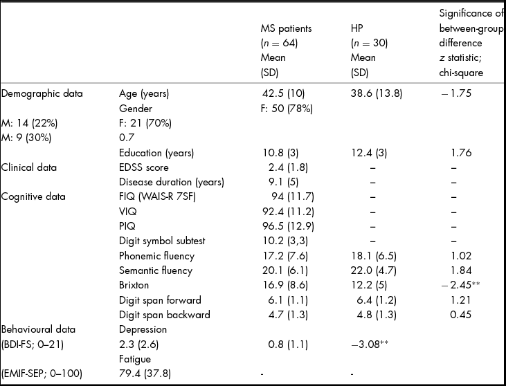
The patients of the study met the following inclusion criteria: (i) RRMS defined according to the revised McDonald criteria (Polman et al., Reference Polman, Reingold, Edan, Filippi, Hartung, Kappos and Wolinsky2005); (ii) a baseline physical disability score (Expanded Disability Severity Scale, EDSS) between 0 and 5.5 (Kurtzke, Reference Kurtzke1983); (iii) Schooling in France. The non-inclusion criteria were: (a) a previous history of other neurological disease (e.g., severe head trauma, encephalitis etc.); (b) visual impairments that made it impossible to perform the tests; (c) a history of major psychiatric illness excepted major depressive disorders and anxiety disorders controlled by medication (according to the Axis I criteria of the Diagnostic and Statistical Manual of Mental Disorders – Fourth Edition); (d) alcohol or drug abuse; (e) recent treatment with corticoids (less than four weeks prior to the evaluation), in order to avoid any effect of treatment on emotional state. The patients for the study were recruited from the departments of Neurology of Reims and Besancon in France. The patients' performances on social cognition tasks and executive measures were compared with those of healthy participants matched for age, gender and education level. The characteristics of the population are presented in Table 1.
Demographic, Clinical, Cognitive and Behavioural Characteristics of Patients with Multiple Sclerosis (MS) and Healthy Participants

Note: FIQ: Full Intelligence Scale; VIQ: Verbal Intelligence Scale; PIQ: Performance Intelligence Scale; BDI-FS: Beck Depression Inventory – Fast Screen; EMIF-SEP: Échelle modifiée d'impact de la Fatigue – Sclérose en Plaques; HP: Healthy Participants. SD: Standard deviation.
**p < .01.
Measures
Functional Measure
This scale is the most widely used to quantify disability in MS. The EDSS scale ranges from 0 to 10 in 0.5 unit increments (except from level 0 to level 1). Level 0 corresponds to the absence of clinical symptoms. Levels 1.0 to 4.5 refer to MS patients who are able to walk without an aid and is based on measures of impairment using 8 functional systems: (1) Pyramidal - weakness or difficulty moving limbs, (2) Cerebellar – ataxia (unsteadiness of gait), loss of coordination or tremor, (3) Brainstem - problems with speech, swallowing, eye movements (double vision or jerky eye movements), (4) Sensory - numbness or loss of sensations, (5) Bowel, bladder and sexual function, (6) Visual function – reduction or loss of vision or a blind spot (scotoma), (7) Cerebral (or mental) functions and fatigue (8) other. EDSS steps 5.0 to 9.5 are defined by the impairment to ambulation and usual equivalents in functional systems scores are provided.
General Cognitive and Psychological Assessment
Wechsler adult intelligence scale-revised (WAIS-R)
The abbreviated seven subtests form of the WAIS-R (WAIS-R 7SF; Ward, Reference Ward1990; Wechsler, Reference Wechsler1981) was used in order to estimate verbal (VIQ), performance (PIQ) and full intelligence quotients (FIQ). The seven WAIS-R subtests used in this short form were Information, Digit Span, Arithmetic, Similarities, Picture Completion, Block Design and Digit Symbol. The Digit Span subtest is divided into Digit Span Forward and Digit Span Backward. Digit Span Backward provides a measure of working memory and Digit Symbol a measure of processing speed, while Similarities and Picture Completion assess abstract reasoning. Given the good standardization of the WAIS-R, it was administered to MS patients only. A subtest score in at least the 9th percentile below the mean normative values was defined as impaired. Given the WAIS-R is an older version of the WAIS test, a less conservative cut-off value of 9% was used to minimise the likelihood of patients with cognitive impairment being misclassified as non-cognitively impaired (such a scenario might occur if a more conservative cut-off of 2 or 5% was used) due to known Flynn Effects (average IQ increases three points per decade; Flynn, Reference Flynn2009).
Executive functions
For the evaluation of executive functions, all the participants completed the Brixton Spatial Anticipation test (Burgess & Shallice, Reference Burgess and Shallice1997) together with measures of phonemic and semantic fluency (using R and Fruits respectively, Cardebat, Doyon, Puel, Goulet, & Joanette, Reference Cardebat, Doyon, Puel, Goulet and Joanette1990).
Brixton spatial anticipation test
This task provides a measure of rule deduction and mental flexibility (i.e., the capacity to detect a rule, to follow it and to change it in order to adopt a new rule), which is often impaired in MS (Drew, Tippett, Starkey, & Isler, Reference Drew, Tippett, Starkey and Isler2008). Participants were presented with a 56-page stimulus booklet. Each page contained an array of 10 circles (two rows of five circles) which were each numbered from 1 to 10. On each page, one circle was filled in with a black colour. The position of this filled circle changed from one page to the next, according to a series of simple rules that varied without warning. On each page, participants were asked to point to where they thought the filled circle would be on the next page based on the rule inferred from the previous pages. Responses were considered correct if they followed the present rule, and on trials where the rule changed, a response was correct if it followed where the black dot would have next moved if the rule had not changed. This test did not require a complex verbal or motor response by the subject or any visual evaluation of colours and was not subject to any time constraints. The psychometric properties of the Brixton test are reported as adequate (Cronbach's alpha =.71; Burgess & Shallice, Reference Burgess and Shallice1997). The total number of errors across 55 trials was used as the outcome measure.
Fluency test
Phonemic and semantic fluency testing require the activation of the process involved in initiating and monitoring executive functions (Rosser & Hodges, Reference Rosser and Hodges1994). Fluency tests are one of the most sensitive measures of executive dysfunction in MS (Wishart and Sharpe, Reference Wishart and Sharpe1997; Zakzanis et al., Reference Zakzanis2000). The validity and reliability of verbal fluency are high (respectively around .70 and .74). For each condition (phonemic and semantic), the participants were given two minutes to produce as many exemplars as possible (using the probes: r and fruits, respectively). The patients' performances were then compared with those of the healthy participants.
Beck depression inventory-fast screen (BDI-FS)
From the 13-item short form Beck Depression Inventory (BDI-II; Beck, Steer, & Brown, Reference Beck, Steer and Brown1996, French translation; Pichot & Lempérière, Reference Pichot and Lempérière1964), we derived the 7-item short form BDI-FS (Beck, Steer, & Brown, Reference Beck, Steer and Brown2000) to evaluate depressive symptoms. The BDI-FS is a short version of the BDI-II with only seven items. Scoring is similar to the BDI-II. The BDI-FS was developed specifically for evaluating depression in patients whose behavioural and somatic symptoms are attributable to medical that may confound the diagnosis of depression. Because neurological manifestations of MS (fatigue, poor concentration) overlap with those of depression, BDI-FS score has been preferred. The BDI-FS shows a good concurrent and discriminative validity in MS (Benedict, Fishman, McClellan, Bakshi & Weinstock-Guttman, Reference Benedict, Fishman, McClellan, Bakshi and Weinstock-Guttman2003). The score varies between 0 and 21. The patients' scores were compared with those of the healthy participants.
Échelle modifiée d'Impact de la fatigue – sclérose en plaques (EMIF-SEP)
Fatigue was measured using the French version of the Modified Fatigue Impact Scale (Debouverie, Pittion-Vouyovitch, Louis, & Guillemin, Reference Debouverie, Pittion-Vouyovitch, Louis and Guillemin2007). The EMIF-SEP is a self-report questionnaire assessing the perceived impact of fatigue on four dimensions (cognitive, physical, social role and psychological) that was developed exclusively for use in MS. EMIF-SEP is composed of 40 items. Each item is rated on a five-point scale ranging from 0 to 4 (3 = it's always true, 2 = it's sometimes true, 1 = it's sometimes wrong, 0 = it's always wrong). The fatigue score can vary from 0 (no fatigue) through to 123 (high degree of fatigue). The score is converted into a percentage. Each dimension has a high internal consistency (Cronbach's alpha > .80). The test-retest reliability is satisfactory (all intra-class correlation coefficients are above .70; Debouverie et al., Reference Debouverie, Pittion-Vouyovitch, Louis and Guillemin2007).
Social Cognitive Testing
Faux pas tasks
These tasks are similar to the one described by Baron-Cohen, O'Riordan, Stone, Jones, and Plaisted (Reference Baron-Cohen, O'Riordan, Stone, Jones and Plaisted1999). These tests assess subtle ToM abilities and can be administered successfully at the age of around 9–11 years. The subject must detect whether someone unintentionally says something which will hurt the feelings of another person. To understand that a faux pas has occurred, one has to represent two mental states: a cognitive mental state and an affective mental state. In order to attribute a cognitive mental state, it is necessary to understand that the person making the faux pas does not know that they should not speak as they do. In order to attribute an affective mental state it is necessary to understand that the person hearing the faux pas would feel insulted or hurt.
In this study, two scenarios involving a faux pas were presented. These scenarios are extracted from a French battery of social cognition assessment (Ehrlé, Henry, Pesa & Bakchine, Reference Ehrlé, Henry, Pesa and Bakchine2011). Normative values were collected from 150 healthy participants between 20 and 69 years old (data not published). To verify the reliability, we computed Cronbach's alpha for the faux-pas items included in this study (not including the comprehension item). Cronbach's alpha was excellent (α = .90). Each story was followed by a faux pas detection question and three other questions: (i) the faux pas detection question evaluated whether the subject correctly detected the faux pas and identified who made it. Correct answers indicated that the subject correctly detected and identified the faux pas; (ii) the false belief question checked that the subject had understood that the faux pas was a consequence of the speaker's false belief; (iii) there was an intention-related question to assess the subject' understanding that the faux pas was unintentional; (iv) finally, there was a question relating to story comprehension (comprehension question). Correct answers were rated 1, incorrect answers were rated 0. The maximum score a subject could achieve in each task was 4. The maximum score for the two stories was 8.
Facial emotion recognition task
This task was derived from the Facial Expressions of Emotion: Stimuli and Tests (Young, Perrett, Calder, Sprengelmeyer, & Ekman, Reference Young, Perrett, Calder, Sprengelmeyer and Ekman2002). The stimuli consisted of a series of 60 black-and-white photographs of the same female face displaying the six primary emotions (anger, disgust, fear, surprise, sadness, and happiness) as described by Ekman and Friesen (Reference Ekman and Friesen1976). The pictures were presented on a computer screen in a 4×3-inch format. Each face was presented once, for 5 s, and was followed by a screen displaying six boxes bearing the names of the six basic emotions. The participants were asked to point to the label that best described the facial expression shown on the screen. The testing phase was preceded by a short training phase. During the testing phase, the 60 faces were presented in a pseudo-random order in which the same emotion was never presented three times in succession. The participants were allowed to take as much time as they needed to make their decision but received no feedback. One point was attributed for each correct answer. The maximum score was 10 for each emotion, and 60 for the whole task.
Procedure
Patients were invited to participate during the course of their regular appointments in the neurology department at the university medical centre. The healthy participants were recruited from hospital staff or by means of an announcement. All subjects gave written informed consent and the study was conducted in accordance with the Helsinki Declaration. All the participants completed several cognitive measures, a self-report measure of depression and fatigue, as well as a measure of facial emotion recognition and ToM. The duration of the protocol was 1h30 for the patients and 30 minutes for the healthy participants.
Statistical Analysis
All statistical analyses were performed using JMP software (SAS Institute Inc. Copyright©2010, Version 9.0.0). Non-parametric statistical tests were used due to the nonhomogeneity of variances and non-normality of the distributions. An alpha level of .05 was selected for the two pairwise comparisons. A Mann–Whitney U test was performed to compare the two groups in terms of demographics, cognitive measures and performance on the social cognition tests. The chi-square test was used for categorical variables. The relationship between age, sex, years of education, disease duration, EDSS, executive function and social cognition was determined using Spearman correlations.
In order to examine the impact of cognitive abilities, we formed two patient groups. We used the same type of procedure as Rao et al. (Reference Rao, Leo, Bernardin and Unverzagt1991). More specifically, a participant was deemed to have failed a test if his or her score was at least the 9th percentile below the mean normative values (see Measures). Subjects who failed two tests or more were defined as cognitively impaired (CI-MS) and those who failed less than two tests were considered to be cognitively preserved (CP-MS). For the analysis, we evaluated both the number of subjects fulfilling our criterion for cognitive impairment and the number of tests failed by each subject.
Severity (disability) level was assigned to one of two groups based on EDSS: moderate (EDSS 0–3) vs. more severe (EDSS 3.5–5.5) disability. This corresponds to the idea of a two-stage physiopathology for the disease, which comprises an inflammatory stage and a degenerative stage (Leray et al., Reference Leray, Yaouanq, Le Page, Coustans, Laplaud, Oger and Edan2010). Short-term (0–7 years) and long-term (8 or more years) MS subgroups were created on the basis of the median values of the whole MS group. MS subgroups and healthy participants were compared using the Kruskall–Wallis test. p values for the Steel–Dwass–Chritchlow–Fligner post-hoc test were calculated. Applying Bonferonni corrections to the three pairwise comparisons yielded an adjusted critical p value of .01.
Results
Sixty-four patients with a diagnosis of RRMS according to the revised McDonald criteria (Polman et al., Reference Polman, Reingold, Edan, Filippi, Hartung, Kappos and Wolinsky2005) and an EDSS score ≤ 5.5 and 30 healthy participants participated in this study. The healthy participants had no history of neurological disease, psychiatric illness or drug abuse. The MS and control groups did not differ significantly with respect to gender distribution, age and years of education (Table 1). Descriptive data and comparison statistics for performance in standard neuropsychological tests and self-report questionnaires scores are summarised in Table 1. Mean IQ scores were in the average range for MS patients (standard score = 90–109; 25–75th percentile), as defined by Wechsler (Reference Wechsler1981). No patient exhibited an impaired performance level (> 9th percentile) on the full scale IQ estimate. Moreover, mean digit symbol subtest scores were in the average range for MS group (Table 1). On the measures of executive functions, MS patients performed significantly worse than healthy participants on the Brixton test (p < .01). No difference was observed between these two groups for phonetic and semantic fluency, or for verbal working memory (measured by backward digit span) performances. In addition, the MS patients scored significantly higher than the healthy participants on the BDI-FS (p <.01). However, the scores were low (mean = 2.3) and point to a minimal level of depressive symptoms among the patients. These results are summarized in Table 1.
Faux Pas Task
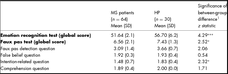
As opposed to the healthy participants, analyses using Mann–Whitney U found that MS patients performed significantly worse in the Faux pas tests (p <.02) (see Table 2). The MS group achieved significantly lower scores than the control group on the intention-related question (p =.02), whereas the same participants were able to respond correctly to the faux pas detection question (p =.09), as shown in Table 2. The absence of a significant difference on the comprehension question suggests that the difficulties encountered by the MS patients in the Faux Pas tasks were not related to language or working memory difficulties.
Performances of MS Patients and Healthy Participants On the Emotion Recognition Test and the Faux Pas Test
Note: HP: Healthy Participants; SD: Standard deviation.
1 Mann–Whitney U-test; ***p < .001, *p < .05.
Facial Emotion Recognition Task
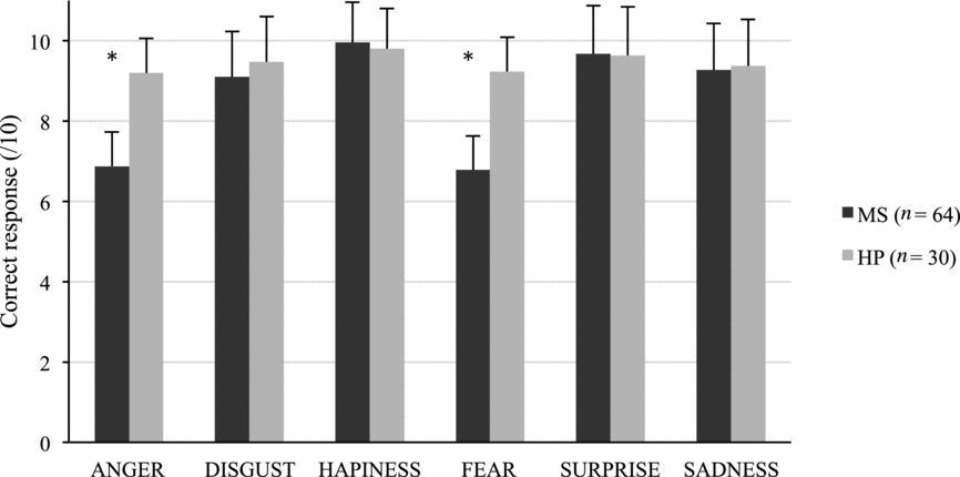
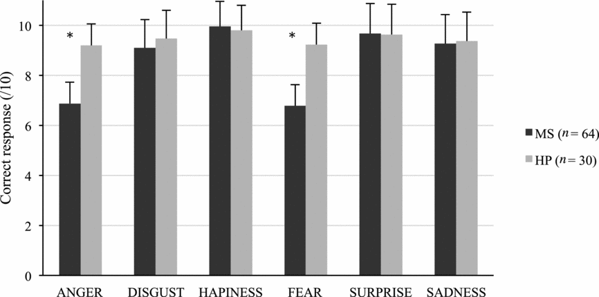
The results of the Mann–Whitney U test revealed that patients with MS performed significantly poorer than the healthy participants on the emotion facial recognition task (p <.001) (see Figure 1). The recognition of fear (p <.001) and anger (p <.001) were impaired in the MS patients. No differences between MS patients and healthy participants were observed with regard to the recognition of disgust (p =.47), happiness (p =.58) and surprise (p =.15).
Performances of MS patients and healthy participants (HP) on facial emotion recognition task.
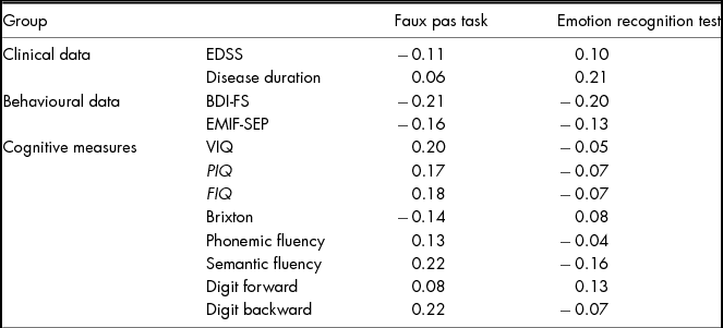
Spearman's correlations were calculated to test the association: (i) between Faux Pas tasks and facial emotion recognition performances, in order to examine if these two processes are related; (ii) between the measures of social cognition (facial emotion recognition and Faux Pas tasks) and clinical features of the disease (EDSS, disease duration, EMIF-SEP, or BDI-FS), in order to explore the role of disability, disease duration and symptoms of depression in social cognition and (iii) between social cognition performances and demographic factors (age, sex, education level). Only two correlations were observed: a positive correlation between fear recognition and performance in the Faux Pas tasks (rho = .26; p <.03) and between anger recognition and performance in the Faux Pas tasks (rho = −.21; p <.03). No other significant correlation was observed. We also examined the link between more general cognitive functions (executive functioning, speed of information processing) and social cognition. No significant correlation was observed in the MS or the control group. As no significant correlation between social cognition, clinical features of the disease (EDSS, disease duration) or cognitive performances was observed, no regression analysis has been performed. The correlations for MS group are summarized in Table 3.
Spearman's Correlations Between Social Cognition Test with Other Clinical, Behavioural and Cognitive Measures for MS Groups

Note: FIQ: Full Intelligence Scale; VIQ: Verbal Intelligence Scale; PIQ: Performance Intelligence Scale; BDI-FS: Beck Depression Inventory – Fast Screen; EMIF-SEP: Échelle modifiée d'impact de la Fatigue – Sclérose en Plaques.
Effect of Cognitive Impairment on Social Cognition
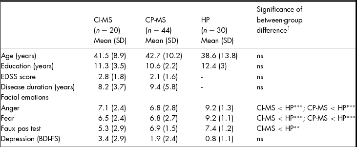
To assess whether performance on either of the social cognition measures was related to more general cognitive functioning, the MS group was split into two groups based on cognitive abilities. As a result, CI-MS (performances below the 9th percentile on at least two tests) and CP-MS patients were compared. Their performances were also compared to those of healthy participants (Table 4). No difference in terms of age, education level, EDSS and disease duration was observed between the subgroups of MS and healthy participants (See Table 4). The performances of the CI-MS patients in the Faux Pas tasks were significantly lower than those of the control group. By contrast, the CP-MS patients achieved performances that were statistically similar to those of the control group. No difference was detected between CI-MS patients and CP-MS patients in social cognition measures.
Effect of Cognitive Abilities on Social Cognition and Depression
Note: CI: Cognitively Impaired; CP: Cognitively Preserved, HP: Healthy Participants; BDI-FS: Beck Depression Inventory-Fast Screen; SD: Standard Deviation; ns = not significant. 1Kruskall–Wallis test; ***p < .001, **p < .01.
Effect of Physical Disability
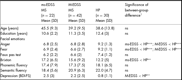
The MS group was also subdivided in terms of physical disability: moderately disabled (moderate-EDSS: 0–3) and more severely disabled patients (more severe-EDSS: 3.5–5.5). Analyses using post-hoc Kruskall–Wallis tests found no differences in terms of age and education were observed between the subgroups of MS and healthy participants (See Table 5). No significant difference was observed for educational level or gender between the ‘moderate-EDSS’ and ‘more severe-EDSS’ MS groups. However, the ‘more severe-EDSS’ MS group was older and had experienced a longer period of illness than the ‘moderate-EDSS’ MS group.
Effect of Disability (EDSS) on Social Cognition, Executive Functions and Depression
Note: ms-EDSS: More severe-EDSS; M-EDSS: Moderate-EDSS; HP: Healthy Participants; ns = not significant; BDI-FS: Beck Depression Inventory-Fast Screen; SD: Standard deviation.
1 Kruskall–Wallis test; ***p <.001, **p <.01.
No difference was observed between the ‘moderate-EDSS’ and ‘more severe-EDSS’ MS groups regarding the social cognition tests (Faux Pas tasks and fear and anger recognition), the Brixton test or depression (BDI-FS). As opposed to the healthy participants, the patients in the MS subgroups performed worse in the facial emotion recognition test (Table 5). Only a statistical trend was observed for the ‘more severe-EDSS’ group compared to healthy participants in Faux Pas tasks (p =.02). These results seem to indicate that the MS subjects encountered facial emotion recognition difficulties regardless of their disability. In the cognitive tasks, no difference was detected in terms of overall degree of intelligence (FIQ) between the two MS groups (p =.32). The ‘more severe-EDSS’ MS participants performed worse than the healthy participants in the Brixton test. The performances of the ‘moderate-EDSS’ group were similar to those of the control group. These results are summarized in Table 5.
Effect of Disease Duration
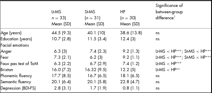
The MS group was divided into two subgroups on the basis of disease duration (0–7 years vs. 8 years and greater). Post-hoc Kruskall–Wallis test revealed that age, education level and gender were comparable between the two groups of MS and healthy participants (Table 6). No significant difference was observed between the ‘short disease duration’ and the ‘long disease duration’ MS groups in terms of the overall scores for the facial recognition of fear and anger, the Faux Pas test score, the Brixton executive test score, the BDI-FS or IQ (FIQ, p =.81).
Effect of Disease Duration on Social Cognition, Executive Functions and Depression
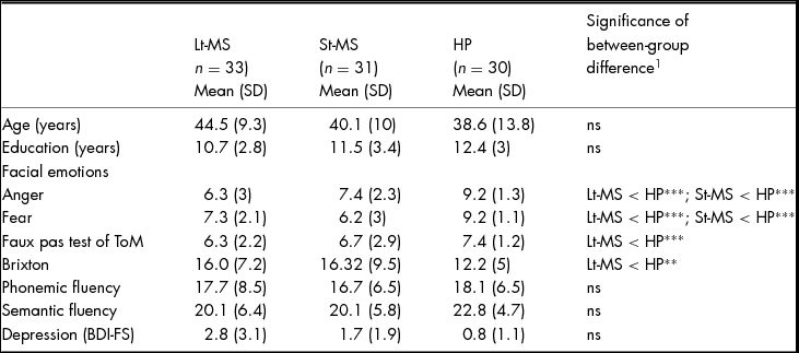
Note: Lt-MS: Long-term EDSS; St-MS: Short-term EDSS; HP: Healthy Participants; ns = not significant; BDI-FS: Beck Depression Inventory-Fast Screen; SD: Standard deviation.
1 Kruskall–Wallis test; ***p < .001, **p < .01.
The ‘short disease duration’ and ‘long disease duration’ groups achieved lower scores than the control subjects on the facial emotion recognition test and the BDI-FS. Only the ‘long disease duration’ group performed worse than the control group on the Faux Pas tasks and executive tasks. These results are presented in Table 6.
Discussion
The aim of our study was to examine socio-cognitive abilities in RRMS by means of a short examination suitable for use in clinical practice. We also examined the effect of the characteristics of the disease (physical disability and disease duration) on socio-cognitive abilities. RRMS patients were significantly less able to recognize facial emotions than the controls, irrespective of the level of cognitive or physical disability. However, MS patients with longer disease duration and those with cognitive impairments seem to have more difficulties to interpret the faux pas.
The difficulties in facial recognition, particularly for fear and anger, irrespective of the severity of their disability, disease duration or cognitive impairments, are consistent with previous that also reported specific impairments of fear and anger recognition (Henry et al., Reference Henry, Phillips, Beatty, McDonald, Longley, Joscelyne and Rendell2009; Henry et al., Reference Henry, Tourbah, Chaunu, Rumbach, Montreuil and Bakchine2011; Prochnow et al., Reference Prochnow, Donell, Schäfer, Jörgens, Hartung, Franz and Seitz2011). Prochnow et al. (Reference Prochnow, Donell, Schäfer, Jörgens, Hartung, Franz and Seitz2011) found similar difficulties, which also applied to the recognition of surprise and sadness. Other studies have not found facial emotion recognition impairments in MS patients (Di Bitonto et al., Reference Di Bitonto, Longato, Jung, Fleury, Marcel and Collongues2011; Jehna et al., Reference Jehna, Neuper, Petrovic, Wallner-Blazek, Schmidt and Fuchs2010; Pinto et al., Reference Pinto, Gomes, Moreira, Rosa, Santos and Silva2012). The fact that these studies used different patient selection criteria and different methodologies could explain the apparently contradictory results (small cohort, positive and negative valence judgments, participants with very recently diagnosed isolated clinical syndrome). Deficits in the recognition of anger and fear in MS seem to be consistent between studies. These data may reflect the specific involvement of a neural network that contributes to the decoding of these particular emotional facial expressions. However, the localization and extent of MS lesions are highly variable. We would therefore expect different profiles of emotion recognition impairments. Unfortunately, our study could not explore this issue since MRI data were not available.
Only a small number of MRI studies have investigated neuronal activation patterns in emotion processing tasks in MS patients (Jehna et al., Reference Jehna, Langkammer, Wallner-Blazek, Neuper, Loitfelder and Ropele2011; Krause et al., Reference Krause, Wendt, Dressel, Berneiser, Kessler, Hamm and Lotze2009; Passamonti et al., Reference Passamonti, Cerasa, Liguori, Gioia, Valentino and Nisticò2009). These studies report the involvement of ventrolateral prefrontal cortex in MS patients. This region is also associated with more general cognitive processes such as working memory, maintaining attention or planning (Passingham, Toni, & Rushworth, Reference Passingham, Toni and Rushworth2000). MS patients are often impaired on these cognitive processes (selective attention, working memory). Consequently, impairments in the recognition of facial emotions in MS may not be specific to emotional information processing. They may depend on task complexity and the integration of information. In our study, we assessed these processes and, in particular, working memory and processing speed, and did not find any correlation between these processes and facial emotion recognition performances. This suggests that emotion decoding impairments were not the result of a reduced information processing speed or reduced working memory capacity in our sample. Our study confirms the presence of these deficits at an early stage of the disease even in the absence of cognitive impairment.
In our study, we found that the patients performed significantly worse only on the intention-related question asked following Faux Pas stories. This indicates that the MS patients correctly detected the faux pas, but erroneously interpreted it as an intentional utterance. These data are consistent with previous findings (Banati et al., Reference Banati, Sandor, Mike, Illes, Bors, Feldmann and Illes2010) indicating that MS patients achieve lower global scores in Faux Pas tasks. However, most other studies have only analysed the global score and the authors have concluded that MS patients have impaired faux pas detection (Banati et al., Reference Banati, Sandor, Mike, Illes, Bors, Feldmann and Illes2010). In our study, we analysed the scores of each question and our results highlight the subtle deficits that can be revealed by a detailed analysis of the answers. This deficit could lead to misinterpretations of real-life social situations and impair the quality of social interactions. Unlike in the case of emotion recognition, we found an effect of disability and executive impairments on performance in Faux Pas tasks.
In our study, we investigated the impact of the characteristics of the disease (physical disability, disease duration and cognitive impairment) on social cognition abilities. Our sample, which was larger than that used in other studies, enabled us to form groups based on illness duration and physical disability. Even though, in the MS literature, the relationship between disability and cognitive decline is unclear, the results of our study reveal differences in faux pas performances. Cognitive impairment and disease duration seem to be associated with the performance of patients in the Faux Pas test. MS patients with longer disease duration and those with cognitive impairments both performed worse than patients with shorter disease duration and without cognitive impairment. This deficit could not be explained by other clinical characteristics such as physical disability, intensity of fatigue or depression. However, this results should be interpreted with caution in our study. Indeed, to have an assessment applicable in clinical practice, we evaluated patients on two Faux Pas stories only. Our results provide some indication that faux pas interpretation may be impaired in MS patients. Other studies with more faux pas items are needed to confirm these results.
By contrast, the impaired recognition of fear and anger could be detected even during the very early stages of the disease, regardless of cognitive abilities, disease duration and physical disability. Faux Pas tasks might be more dependent on the integrity of executive processes than facial emotion recognition. Several studies (e.g., Banati et al., Reference Banati, Sandor, Mike, Illes, Bors, Feldmann and Illes2010) have shown a link between executive functions (working memory, mental flexibility,. . .) and performance in Faux Pas tasks. The correlations between executive functions and facial emotion recognition tasks are less often reported (Henry et al., Reference Henry, Phillips, Beatty, McDonald, Longley, Joscelyne and Rendell2009). Recent research used more ecological ToM tasks with real life conversation vignettes and compare performance of these ToM tasks under higher executive demands to those with traumatic brain injury and lower executive demand. These studies showed that ToM impairments in everyday communication may be due to working memory demands (Honan, McDonald, Gowland, Fisher, & Randall, Reference Honan, McDonald, Gowland, Fisher and Randall2015; McDonald et al., Reference McDonald, Gowland, Randall, Fisher, Osborne-Crowley and Honan2014).
Facial emotion recognition and Faux Pas tasks are two different but overlapping aspects of social cognition. Facial emotion recognition requires the ability to infer the emotional states of others from facial expressions. Faux Pas tasks require the ability to ascribe both cognitive and affective mental states. It is possible that our results may be due to the fact that the attribution of affective mental states may be disrupted in the early stages of the disease, while it is only later that the disruption of cognitive mental states occurs. Our study cannot answer this question because we did not perform separate analyses of the attribution of affective and cognitive mental states in our Faux Pas tasks. However, Roca et al. (Reference Roca, Manes, Gleichgerrcht, Ibáñez, González de Toledo, Marenco and Sinay2014) did conduct such an analysis and revealed a disruption in the attribution of cognitive mental states in the absence of any disruption to the attribution of affective mental states in a small cohort with very low disability. These results suggest that the underlying cognitive ToM network might tend to be disrupted in MS. However, this assumption is not consistent with the impaired recognition of fear and anger observed in our own and in other studies (Henry et al., Reference Henry, Phillips, Beatty, McDonald, Longley, Joscelyne and Rendell2009; Prochnow et al., Reference Prochnow, Donell, Schäfer, Jörgens, Hartung, Franz and Seitz2011). Recognizing facial emotions requires the attribution of affective mental states. If these attributions are impaired in MS patients, we should find deficits in affective ToM as assessed in the Faux Pas test. It is possible that the affective ToM process involved in Faux Pas tasks is different from that involved in facial emotion recognition tasks. The heterogeneity of MS, even when studying one and the same form of the disease, makes comparisons between samples difficult. These differences require further studies exploring social cognitive abilities based on the use of tests designed to distinguish between cognitive and affective mental states, such as the Yoni task (Shamay-Tsoory, Simone, & Aharon-Peretz, Reference Shamay-Tsoory and Aharon-Peretz2007).
This study has several limitations. The control and patient samples could be increased in order to permit better comparisons between subgroups (e.g., duration of the disease, EDSS, cognitive functioning). Larger samples might also reveal additional profiles. In our sample, the ‘more severe-EDSS’ group was older than the ‘moderate-EDSS’ group, thereby limiting the interpretation of the results. The relationship between socio-cognitive abilities, neuropathology and disease progression remains to be established. Our sample only contained patients with RRMS. To avoid the risk of misclassifying patients and include patients with secondary progressive MS, EDSS was limited to a maximal value of 5.5. This inclusion criteria could be too restrictive. It would be interesting to replicate this research without this limit and extent it to other clinical forms of the disease since disability disrupted some aspects of social cognition in our study. The use of an older version of the WAIS-R is also a limitation, despite the use of a less conservative cut-off value. In addition, we used only two Faux Pas stories to provide social cognition assessment applicable in clinical practice but this approach is unusual. Our results provide some indication that ToM may be impaired in RRMS but it needs to be verified in future studies with more faux pas items. In addition to these behavioural data, it seems important to include a more systematic neuroimaging investigation in order to explore anatomo-functional correlations. Another limitation of our study lies in the lack of measures to assess the functional impact of these disorders during the daily life of MS patients. We consider this important because social cognition disorders can impair everyday social functioning and thus thereby impairing the effectiveness of rehabilitation activities undertaken during the course of the disease (Wolwer & Frommann, Reference Wolwer and Frommann2011). Such measures would also help assess the need to develop specific approaches to rehabilitation with regard to the socio-cognitive aspects. More studies are needed to better define the social cognition difficulties in this population before starting to consider rehabilitation-related aspects.
Social cognition impairments are associated with disturbances to interpersonal functioning such as poor communication and inappropriate social behaviour. Although this deficit is not the only one to affect MS patients it still needs to be better defined since all studies assessing the attribution of mental states in MS have found impairments (Banati et al., Reference Banati, Sandor, Mike, Illes, Bors, Feldmann and Illes2010; Henry et al., Reference Henry, Phillips, Beatty, McDonald, Longley, Joscelyne and Rendell2009; Henry et al., Reference Henry, Tourbah, Chaunu, Rumbach, Montreuil and Bakchine2011; Ouellet et al., Reference Ouellet, Scherzer, Rouleau, Métras, Bertrand-Gauvin, Djerroud and Duquette2010). However, this area remains unexplored and further studies are needed. If cognitive disorders are a major factor limiting employment status and social functioning, disorders affecting social cognition could be even more critical. Indeed, many studies of other neurological and psychiatric diseases have highlighted the significant impact of these disorders on individuals' social functioning. Thus, it is important to integrate the assessment of social cognition in clinical evaluation. The methodology used in this study permits a relatively rapid assessment of ToM abilities (< 20 minutes) compared to the experimental tests administered in other studies. However, 20 minutes of assessment during routine clinical practice is still too long.
Although the SDMT is an effective brief screening operation for detecting cognitive impairment in MS (Deloire et al., Reference Deloire, Bonnet, Salort, Arimone, Boudineau, Petry and Brochet2006), we do not have any screening test for behavioural disturbances comparable to the Neuropsychiatric Inventory in the case of neurodenegenerative diseases. The Neuropsychiatric Inventory was developed to provide a means of assessing neuropsychiatric symptoms and psychopathology of patients with Alzheimer's disease and other brain disorders. This scale may be useful in the assessment of behavioural changes in several areas (Delusions, Hallucinations, Agitation/Aggression, Elation/Euphoria, Apathy/Indifference, Disinhibition, Irritability/Lability,. . .).
A tool such as Barrow Neurological Institute Screen for Higher Cerebral Functions (BNIS, Prigatano, Reference Prigatano1991) could be used in MS. In addition to assessing cognitive functions (language, orientation, concentration, visuospatial function, memory), the BNIS provides an assessment of affect (affective expression, spontaneous emotion, emotion perception and mood control). The BNIS is easy and fast to administer (13.8 minutes for neurological and psychiatric populations and 11.8 minutes for healthy participants, Prigatano et al., Reference Prigatano, Tonini, Truelle and Montreuil2013). Furthermore, several studies (Boosman, Visser-Meily, Post, Duits, and van Heugten, Reference Boosman, Visser-Meily, Post, Duits and van Heugten2013; Prigatano et al., Reference Prigatano, Montreuil, Chapple, Tonini, Toron, Paquet and Truelle2014; Redfors et al., Reference Redfors, Hofgren, Eriksson, Holmegaard, Samuelsson and Jood2014) have shown that the BNIS is more sensitive than the MMSE in various neurological diseases (brain injury, stroke) that share characteristics with MS (e.g., slowing of processing speed). This screening tool might be of value in MS for the detection of both cognitive and behavioural disturbances and in helping to select patients for testing on the basis of a more complete cognitive and affective battery.
In summary, our results are the first to show, in a RRMS sample, a heterogeneity of social cognition impairments based on the progression of the disease and cognitive impairment. Indeed, facial affect recognition is impaired regardless of severity of physical disability, disease duration or cognitive impairments, thus suggesting that there is an affective ToM deficit at an early stage of MS. At the same time, performance on Faux Pas tests seems to be impaired in patients with a longer disease duration or patients with cognitive deficits, thus suggesting that cognitive ToM deficits occur later. Banati et al. (Reference Banati, Sandor, Mike, Illes, Bors, Feldmann and Illes2010) have already shown that patients with a shorter disease duration score higher on affective ToM. However, these authors did not investigate the cognitive components of ToM. In order to corroborate our results, further studies are required to investigate social cognition abilities in different forms of the disease and with different degrees of severity. Impairments to facial affect recognition and ToM may exacerbate the difficulties some patients experience in maintaining effective interpersonal relationships. It therefore seems crucial for clinicians to investigate these aspects and to take them into account in the patient's care. Before initiating social cognition rehabilitation in MS, it is necessary to perform additional investigations in order to find out more precisely the nature of social cognition impairments as well as their functional outcomes.
Financial Support
This research received no specific grant from any funding agency, commercial or not for profit sectors.
Conflict of Interest
None.
Ethical Standards
The authors assert that all procedures contributing to this work comply with the ethical standards of the relevant national and institutional committees on human experimentation and with the Helsinki Declaration of 1975, as revised in 2008.