INTRODUCTION
Extracellular purine (adenosine-5′-triphosphate (ATP), adenosine-5′-diphosphate (ADP)) and pyrimidine (uridine-5′-triphosphate (UTP), uridine-5′-diphosphate (UDP) and UDP-sugars) nucleotides are widespread signaling molecules involved in a variety of physiological and pathological processes, including pain transmission (Burnstock, Reference Burnstock1996, Reference Burnstock2006; Liu and Salter, Reference Liu and Salter2005). By activating the ligand-gated cationic channels P2X receptors (P2X1−7) (Jacobson et al., Reference Jacobson, Jarvis and Williams2002; North, Reference North2002) and the metabotropic G-protein coupled P2Y receptors (P2Y1,2,4,6,11,12,13,14) (Abbracchio et al., Reference Abbracchio, Burnstock, Boeynaems, Barnard, Boyer and Kennedy2006), extracellular nucleotides act as co-transmitters and neuromodulators in sensory systems (Chizh and Illes, Reference Chizh and Illes2001). The neuronal homomultimer P2X3 and the heteromultimer P2X2/3 receptors have been recognized as the predominant P2 receptors involved in ATP-evoked painful signals (Jarvis, Reference Jarvis2003; Burnstock, Reference Burnstock2006), but it is now increasingly clear that P2Y receptors can as well affect the processing of noxious sensory inputs (Svichar et al., Reference Svichar, Shmigol, Verkhratsky and Kostyuk1997; Gerevich and Illes, Reference Gerevich and Illes2004; Nakayama et al., Reference Nakayama, Yamashita, Konishi, Kazama and Kokubun2004; Jarvis, Reference Jarvis2010). Moreover, emerging evidence indicates that other purinergic-related molecules (e.g. adenine) may also contribute to nociception (Burnstock, Reference Burnstock2009a; the present data), suggesting that this system can modulate nociceptive sensitivity at multiple and different levels.
The situation is further complicated by the recent demonstration that, in a similar way to glial cells in the central nervous system (CNS), also satellite glial cells (SGCs), which wrap around neurons in sensory ganglia, are active participants in the generation and maintenance of pain following inflammation or damage to peripheral tissues (Ohara et al., Reference Ohara, Vit, Bhargava, Romero, Sundberg and Charles2009; Takeda et al., Reference Takeda, Takahashi and Matsumoto2009). Upon activation, SGCs release cytokines and probably other bioactive molecules that potentiate neuronal transmission of pain. In addition, like other glial cells, SGCs maintain ionic homeostasis (Vit et al., Reference Vit, Ohara, Bhargava, Kelley and Jasmin2008) and express receptors for various neurotransmitters, including some P2 receptors (Weick et al., Reference Weick, Cherkas, Hartig, Pannicke, Uckermann and Bringmann2003; Gerevich and Illes, Reference Gerevich and Illes2004; Ceruti et al., Reference Ceruti, Fumagalli, Villa, Verderio and Abbracchio2008), which enable them to respond to neuronally released ATP (Zhang et al., Reference Zhang, Chen, Wang and Huang2007). Thus, it is becoming increasingly clear that activation of SGCs purinoceptors by extracellular nucleotides can participate in the modulation of sensory transmission (Gerevich and Illes, Reference Gerevich and Illes2004; Takeda et al., Reference Takeda, Tanimoto, Kadoi, Nasu, Takahashi and Kitagawa2007, Reference Takeda, Takahashi and Matsumoto2009; Zhang et al., Reference Zhang, Chen, Wang and Huang2007; Ceruti et al., Reference Ceruti, Fumagalli, Villa, Verderio and Abbracchio2008; Chen et al., Reference Chen, Zhang, Wang, Li, Gu and Huang2008). This suggests that the pharmacological manipulation of the purinergic system on glial cells might represent a new strategy for treating painful situations that still deserve an effective cure, such as migraine and neuropathic pain.
P2X receptors in SGCs from sensory ganglia
Since the publication of early works demonstrating ATP involvement in pain (Bleehen and Keele, Reference Bleehen and Keele1977; Krishtal et al., Reference Krishtal, Marchenko and Pidoplichko1983), the expression and function of P2X receptors in sensory neurons, including those from dorsal root (DRG), trigeminal (TG), nodose and petrosal ganglia have been characterized (for reviews see Dunn et al., Reference Dunn, Zhong and Burnstock2001; Burnstock, Reference Burnstock2009b). All P2X receptor subtypes, with the exception of the P2X7 subtype, were found in sensory neurons (Burnstock, Reference Burnstock2001), with the P2X3 receptor showing the highest level of expression (Kim et al., Reference Kim, Paik, Cho, Shin, Bae and Moritani2008).
Unlike sensory neurons, SGCs show a lower expression of P2X receptors. In fact, some conflicting reports on the expression of P2X2 receptors by satellite glia (Vulchanova et al., Reference Vulchanova, Riedl, Shuster, Buell, Surprenant and North1997; Petruska et al., Reference Petruska, Cooper, Gu, Rau and Johnson2000), and a single report demonstrating the presence of P2X4 mRNA on both neuronal and glial cells in DRG (Kobayashi et al., Reference Kobayashi, Fukuoka, Yamanaka, Dai, Obata and Tokunaga2005) have been published. Interestingly, the P2X7 subunit has been demonstrated to be clearly and selectively expressed by SGCs (Chessell et al., Reference Chessell, Hatcher, Bountra, Michel, Hughes and Green2005; Kobayashi et al., Reference Kobayashi, Fukuoka, Yamanaka, Dai, Obata and Tokunaga2005; Zhang et al., Reference Zhang, Han, Faltynek, Jarvis and Shieh2005; Chen et al., Reference Chen, Zhang, Wang, Li, Gu and Huang2008). From a pharmacological point of view, in DRG-derived cultures the non-selective P2X3/P2X7 agonist 3′-O-(4-benzoyl)benzoyladenosine 5′-triphosphate (BzATP) evoked a concentration-dependent increase in inward Na+/Ca2+ currents in both neurons and non-neuronal cells. However, BzATP currents were significantly reduced by the P2X3 selective antagonist A-317491 in neurons, but not in non-neuronal cells, indicating that in the latter they were not mediated by the activation of P2X3 receptors (Zhang et al., Reference Zhang, Han, Faltynek, Jarvis and Shieh2005). Pharmacological characterization of these responses in non-neuronal cells showed a rank order of potency for known P2X receptor agonists, as follows: BzATP > ATP > α,β-meATP, and the inhibition of 100 µM BzATP-evoked currents by pyridoxal-phosphate-6-azophenyl-2′,4′-disulfonate (PPADS) and Mg2+. This pharmacological profile was consistent with the involvement of P2X7 receptors (Bianchi et al., Reference Bianchi, Lynch, Touma, Niforatos, Burgard and Alexander1999). Moreover, confocal microscopy analysis revealed a positive P2X7 immunoreactivity on the plasma membrane of almost all non-neuronal cells, while no immunostaining could be observed in neurons. Of note, P2X7 receptors in non-neuronal cells were not associated to formation of large pores after prolonged exposure to BzATP (Zhang et al., Reference Zhang, Han, Faltynek, Jarvis and Shieh2005).
In 2005, Chessell and colleagues have shown that the P2X7 knock out mouse did not develop either mechanical allodynia or thermal hyperalgesia after inflammatory and neuropathic injury, while normal nociceptive processing was preserved (Chessell et al., Reference Chessell, Hatcher, Bountra, Michel, Hughes and Green2005). Interestingly, this receptor was found upregulated in DRGs and injured nerves from chronic neuropathic pain patients (Chessell et al., Reference Chessell, Hatcher, Bountra, Michel, Hughes and Green2005).
Given that P2X7 stimulation is known to induce interleukin (IL)-1β release from macrophages/microglia and reactive astrocytes (Bianco et al., Reference Bianco, Pravettoni, Colombo, Schenk, Möller and Matteoli2005b, Reference Bianco, Colombo, Saglietti, Lecca, Abbracchio and Matteoli2009; Sanz et al., Reference Sanz, Chiozzi, Ferrari, Colaianna, Idzko and Falzoni2009) and IL-1β levels were also strongly upregulated in the inflamed paws following Complete Freund's Adjuvant (CFA) intraplantar injection (Chessell et al., Reference Chessell, Hatcher, Bountra, Michel, Hughes and Green2005), it has been suggested that the P2X7 receptor subtype might play a pivotal role in the molecular cascade of events leading to inflammatory and neuropathic pain. In this context, growing evidence supports a role for P2X7 receptors as a ‘sensor of danger’ that, by monitoring ATP extracellular levels, may participate to the modulation of IL-1β processing and release (Bianco et al., Reference Bianco, Pravettoni, Colombo, Schenk, Möller and Matteoli2005b, Reference Bianco, Colombo, Saglietti, Lecca, Abbracchio and Matteoli2009; Ferrari et al., Reference Ferrari, Pizzirani, Adinolfi, Lemoli, Curti and Idzko2006).
To further confirm this issue, systemic administration of selective P2X7 receptor antagonists, such as A-740003 (Honore et al., Reference Honore, Donnelly-Roberts, Namovic, Hsieh, Zhu and Mikusa2006) and A-438079 (McGaraughty et al., Reference McGaraughty, Chu, Namovic, Donnelly-Roberts, Harris and Zhang2007) was associated with a significant dose-dependent reduction of both allodynia and hyperalgesia in several animal models of neuropathic and inflammatory pain. The antinociceptive actions of A-438079 were correlated to its ability to reduce both the evoked and the spontaneous firing of different classes of spinal neurons in neuropathic animals, and to inhibit the release of IL-1β (McGaraughty et al., Reference McGaraughty, Chu, Namovic, Donnelly-Roberts, Harris and Zhang2007).
Thus, these results would suggest a specific role for glial P2X7 receptors in neuronal–glial cell interaction associated with ongoing pain (Zhang et al., Reference Zhang, Han, Faltynek, Jarvis and Shieh2005; Jarvis, Reference Jarvis2010). However, this view has been recently challenged by a study demonstrating that activation of P2X7 receptors expressed by SGCs, tonically inhibits neuronal P2X3 receptor expression (Chen et al., Reference Chen, Zhang, Wang, Li, Gu and Huang2008). This effect was mediated by ATP released from SGCs upon P2X7 activation, and by the subsequent activation of P2Y1 receptors on neurons. Consistent with this finding, application of the P2Y1 receptor agonist 2-methylthio adenosine 5′-diphosphate (2MeSADP) inhibited the expression of P2X3 receptors on neurons, whereas the P2Y1 receptor antagonist MRS2179 significantly increased P2X3 receptor levels (Chen et al., Reference Chen, Zhang, Wang, Li, Gu and Huang2008). Moreover, an in vivo analysis after silencing of P2X7 receptors by siRNA showed an exaggerated nociceptive behavior in response to the injection of the P2X3 agonist α,β-methylene adenosine 5′-triphosphate (α,β-meATP) into the hind paws. In line with these findings, activation of P2X7 receptors with its agonist BzATP prevented the development of allodynia in a CFA-model of inflammation (Chen et al., Reference Chen, Zhang, Wang, Li, Gu and Huang2008). It was therefore proposed that the direct targeting of SGCs-expressed P2X7 receptors might not be the best pharmacological strategy for the treatment of chronic pain conditions (Chen et al., Reference Chen, Zhang, Wang, Li, Gu and Huang2008). Collectively, these conflicting results suggest that the role of glial P2X7 receptor in pain transmission need to be further investigated.
Finally, a crucial role for the P2X4 receptor subtype in the induction and maintenance of neuropathic pain has been recently demonstrated. This receptor has been found upregulated in spinal cord microglia following nerve injury (Tsuda et al., Reference Tsuda, Kuboyama, Inoue, Nagata, Tozaki-Saitoh and Inoue2009), and mice treated with a P2X4 antisense oligonucleotide or P2X4 KO mice show attenuated tactile allodynia after nerve injury (Tsuda et al., Reference Tsuda, Tozaki-Saitoh and Inoue2010). To date, no role in pain transmission or modulation for this receptor subtype has been demonstrated in resident glial cells in sensory ganglia.
P2Y receptors in SGCs from sensory ganglia
Despite their widespread expression in sensory ganglia, the exact role of metabotropic P2Y receptors in pain transmission has not been fully understood. At variance from the selective expression of specific P2X subtypes in neurons and SGCs (see above), P2Y receptors were found widely expressed in both cell types.
In sensory neurons, P2Y receptors are located not only at the cell body, but also at the peripheral and central terminals (Gerevich and Illes, Reference Gerevich and Illes2004; Burnstock, Reference Burnstock2009a). Here, their activity is integrated within the complex molecular network associated to the transmission of nociceptive signals to the CNS. For example, activation of the P2Y1 subtype inhibits P2X3 receptor function (Gerevich et al., Reference Gerevich, Muller and Illes2005; Chen et al., Reference Chen, Zhang, Wang, Li, Gu and Huang2008), possibly leading to analgesia, whereas P2Y2 receptor activation can contribute to nociception by modulating the extracellular concentrations of the calcitonin gene-related peptide (CGRP) in DRGs (Sanada et al., Reference Sanada, Yasuda, Omatsu-Kanbe, Sango, Isono and Matsuura2002; Zimmermann et al., Reference Zimmermann, Reeh and Averbeck2002; Huang et al., Reference Huang, Wu, Nicol, Meller and Vasko2003).
Very few data are available on the role of the P2Y receptor family in SGCs. In DRG, in situ hybridization histochemistry studies detected mRNA expression on these cells only for the P2Y12 and P2Y14 receptor subtypes (Kobayashi et al., Reference Kobayashi, Fukuoka, Yamanaka, Dai, Obata and Tokunaga2006). However, immunofluorescence studies showed staining for P2Y1 on both neurons and SGCs (Chen et al., Reference Chen, Zhang, Wang, Li, Gu and Huang2008). Unfortunately no functional studies, which could help clarifying this issue, are available for DRG-derived SGCs.
RT-PCR experiments revealed mRNA expression for all the P2Y receptors in SGCs from mouse TG (see below). Immunofluorescence staining confirmed the selective expression of P2Y4 on SGCs from both mouse and rat TG (Weick et al., Reference Weick, Cherkas, Hartig, Pannicke, Uckermann and Bringmann2003; Vit et al., Reference Vit, Jasmin, Bhargava and Ohara2006). It should be noted, however, that these data are in disagreement with a previous work indicating a selective expression of the P2Y4 receptor subtype in sensory neurons (Ruan and Burnstock, Reference Ruan and Burnstock2003). This discrepancy could be explained by the later demonstrated lack of specificity of anti-P2Y4 commercially available antibodies (Tung et al., Reference Tung, Choi, Siow, Jiang, Ling and Simon2004), or by the differences in the staining protocols utilized by the two research groups. Calcium imaging experiments on intact TG revealed that application of ATP and UTP caused functional raises in [Ca2+]i in almost all SGCs (Weick et al., Reference Weick, Cherkas, Hartig, Pannicke, Uckermann and Bringmann2003). This effect persisted in nominally Ca2+-free extracellular solution, but was blocked by cyclopiazonic acid, an inhibitor of intracellular Ca2+ release from the endoplasmic reticulum. These experiments confirmed that ATP/UTP responses on SGCs were due to functional P2Y receptors, likely the P2Y2/P2Y4 receptor subtypes (Weick et al., Reference Weick, Cherkas, Hartig, Pannicke, Uckermann and Bringmann2003). Moreover, [Ca2+]i increases were observed in SGCs after application of adenosine 5′-[β-thio]-diphosphate (ADPβS) and 2MeSADP, two P2Y1 receptor selective agonists (Abbracchio et al., Reference Abbracchio, Burnstock, Boeynaems, Barnard, Boyer and Kennedy2006), therefore suggesting that P2Y1 receptors are also expressed and functional in these cells (Weick et al., Reference Weick, Cherkas, Hartig, Pannicke, Uckermann and Bringmann2003). Single-cell calcium imaging analysis in primary mouse trigeminal cultures demonstrated that exposure to ADP resulted in calcium increases in about 80% of SGCs (Ceruti et al., Reference Ceruti, Fumagalli, Villa, Verderio and Abbracchio2008). Experiments with MRS2179 (P2Y1 receptor antagonist) and Cangrelor (P2Y12/P2Y13 receptor antagonist) showed that, in glial cells, response to ADP mainly involved P2Y1 receptors, with a smaller contribution of the P2Y12/P2Y13 receptor subtypes (Ceruti et al., Reference Ceruti, Fumagalli, Villa, Verderio and Abbracchio2008). In the same study, UTP-induced calcium transients on SGCs were partially inhibited by pretreatment with both Suramin and Reactive blue 2, thus supporting the involvement of functional P2Y2/P2Y4 receptors on SGCs. Only minor functional responses to UDP were found, indicating a low P2Y6 receptor expression, and, finally, no SGCs showed responses to the P2Y14 agonist UDP-glucose despite its expression at the mRNA level (Ceruti et al., Reference Ceruti, Fumagalli, Villa, Verderio and Abbracchio2008). Overall, these results show that, in line with previous reports indicating a wide expression of P2Y receptors in CNS glial cells (Abbracchio and Verderio, Reference Abbracchio and Verderio2006; Inoue, Reference Inoue2008; Verkhratsky et al., Reference Verkhratsky, Krishtal and Burnstock2009), also SGCs from TG bear a wide panel of functional P2Y receptors, thus suggesting a primary role for nucleotides in modulating cell-to-cell communication in sensory ganglia.
In sensory ganglia, P2Y receptors undergo significant changes of their expression after neuropathic or inflammatory conditions. For example, it has been shown that P2Y1 mRNA is upregulated in rat DRGs after peripheral axotomy of the sciatic nerve (Xiao et al., Reference Xiao, Huang, Zhang, Bao, Lu and Guo2002). Additionally, P2Y2 mRNA levels were strongly upregulated after CFA injection in the hindpaw, whereas P2Y1,4,6 RNAs were downregulated (Malin et al., Reference Malin, Davis, Koerber, Reynolds, Albers and Molliver2008). Moreover, at difference from wild-type animals, P2Y2−/− mice failed to develop thermal hyperalgesia after CFA-induced inflammation. On the contrary, no differences in the extent of mechanical allodynia between wild type and P2Y2−/− mice were observed (Malin et al., Reference Malin, Davis, Koerber, Reynolds, Albers and Molliver2008). These results demonstrate that neuronal P2Y2 receptor subtype is specifically involved in thermal nociceptive transmission (Moriyama et al., Reference Moriyama, Iida, Kobayashi, Higashi, Fukuoka and Tsumura2003; Malin et al., Reference Malin, Davis, Koerber, Reynolds, Albers and Molliver2008). It has been also demonstrated that receptor subtypes that are expressed but not functional upon basal conditions (e.g. P2Y14) are upregulated under specific chronic pathological painful situations, such as after neuropathic nerve injury (Vega-Avelaira et al., Reference Vega-Avelaira, Géranton and Fitzgerald2009). These receptors could therefore represent innovative drug targets with an ideal therapeutic profile, since their pharmacological manipulation should not interfere with physiological transmission.
P2Y receptor levels are modulated during inflammatory conditions in SGCs as well. In fact, a chronic exposure of trigeminal neuron-satellite glia cultures to the well-known pro-inflammatory agent bradykinin (BK) resulted in highly significant increases in the mean calcium amplitudes observed in SGCs after exposure to ATP, ADP and UTP. Application of UDP did not produce statistically different increases in [Ca2+]i, but an enhancement in the percentage of responding SGCs was detected. Interestingly, only very few SGCs (about 3%) responded with increase in [Ca2+]i to BK exposure compared to a higher percentage of responding neurons (about 35%). Therefore, the observed changes in P2Y receptors functionality on SGCs might be mediated by a yet-to-be identified factor released from neurons upon BK stimulation, which contributes to a possible potentiation of neuron-to-glia signaling in TG during painful inflammatory conditions (Ceruti et al., Reference Ceruti, Fumagalli, Villa, Verderio and Abbracchio2008).
SGCs are a distinct type of glial cells, but they share many characteristics with astrocytes (Hanani, Reference Hanani2005). One typical feature of CNS astrocytes is their ability to generate intercellular calcium waves (ICWs). Any pharmacological, mechanical or electrical stimulation of a single astrocyte is associated to increases of cytosolic [Ca2+], which is subsequently spread out to adjacent astrocytes, leading to the generation of ICWs involving the entire astrocytic network (Giaume and Venance, Reference Giaume and Venance1998). ICWs are propagated by both cell-to-cell communication through gap junctions, and by extracellularly released ATP acting on P2Y1 and P2Y2 receptors on the surface of neighboring cells (Haydon, Reference Haydon2001; Scemes and Giaume, Reference Scemes and Giaume2006). In a recent paper, it has been shown that a similar mechanism takes place also for SGCs in sensory ganglia (Suadicani et al., Reference Suadicani, Cherkas, Zuckerman, Smith, Spray and Hanani2009). Under basal conditions, focal electrical or mechanical stimulation of a single neuron in primary TG cultures evoked a raise in cytosolic calcium not only in the stimulated cells, but also among adjacent glial cells, spreading as a typical ICW. Propagation of ICWs could be evoked also by the stimulation of SGCs (Suadicani et al., Reference Suadicani, Cherkas, Zuckerman, Smith, Spray and Hanani2009). The pharmacological characterization made in this study does not allow to clearly identify the specific P2 receptors involved in spreading the calcium signals, but considering that: (i) ICWs are mediated by P2Y receptors in spinal cord astrocytes (see above), and (ii) trigeminal SGCs bear functional P2Y receptors (Weick et al., Reference Weick, Cherkas, Hartig, Pannicke, Uckermann and Bringmann2003; Ceruti et al., Reference Ceruti, Fumagalli, Villa, Verderio and Abbracchio2008), it can be anticipated that also trigeminal ICWs are mediated by this receptor family.
It is noteworthy that after peripheral inflammation by submandibular CFA injections, the bidirectional Ca2+ signaling between neurons and SGCs, and among SGCs was found to be significantly altered (Suadicani et al., Reference Suadicani, Cherkas, Zuckerman, Smith, Spray and Hanani2009). Indeed, the amplitude of the Ca2+ signal generated in adjacent SGCs and neurons in response to a single SGCs activation was increased by two-fold in cultures obtained from CFA-treated mice. This finding correlates with recent studies indicating increases in SGCs cell coupling through gap junctions following inflammatory or chronic neuropathic conditions (Hanani et al., Reference Hanani, Huang, Cherkas, Ledda and Pannese2002; Ohara et al., Reference Ohara, Vit, Bhargava and Jasmin2008; Huang et al., Reference Huang, Belzer and Hanani2009; Ledda et al., Reference Ledda, Blum, De Palo and Hanani2009). The exact significance of this increase in SGCs signaling needs to be better elucidated, but it could be associated to the spreading of ‘nociceptive signals’ among sensory ganglia, and could therefore be implicated in the development of hyperalgesia and allodynia.
Other purinoceptors involved in pain transmission
Besides the predominant role played by nucleotides in pain signaling, extracellular adenosine (Ado), deriving from the metabolic degradation of ATP, can further modulate nociception, by the activation of its P1 G-protein-coupled receptors (the A1, A2A, A2B, A3 receptors) (Fredholm et al., Reference Fredholm, IJzerman, Jacobson, Klotz and Linden2001). In general, activation of A1 receptors decreases nociception, inflammation and cellular excitability, whereas A2A receptors exert excitatory effects on primary sensory afferents (Liu and Salter, Reference Liu and Salter2005). Very little is known on the presence and functionality of P1 receptors in sensory ganglia (Sawynok, Reference Sawynok1998; Burnstock, Reference Burnstock2009a). Immunocytochemical studies and western blot analysis revealed the presence of A1 receptors in a subpopulation of trigeminal sensory neurons (Carruthers et al., Reference Carruthers, Sellers, Jenkins, Jarvie, Feniuk and Humphrey2001), and recently, the selective A1 receptor agonist 2-chloro-N6-cyclopentyladenosine has been demonstrated to significantly decrease the basal and evoked pro-nociceptive ATP release in DRGs from neuropathic rats, probably by acting on neuronal A1 receptors, whose expression was previously highlighted in these cells (Dolphin et al., Reference Dolphin, Forda and Scott1986; Matsuka et al., Reference Matsuka, Ono, Iwase, Mitrirattanakul, Omoto and Cho2008). The importance of the antinociceptive action of Ado is further supported by clinical findings indicating that its intravenous infusion reduces some features of neuropathic and postoperative pain (Sawynok, Reference Sawynok2007). Although the mechanisms are still not clear, these studies support a possible role for Ado in the management of neuropathic pain.
Interestingly, adenine, the nucleobase moiety of ATP, ADP and Ado, might represent a new regulator of painful sensory information. By using a receptor activation bioassay, adenine has been identified as the endogenous ligand of a previously orphan G-protein coupled receptor (oGPCR) sequence related to a family of oGPCRs (Mrgs), which is specifically expressed in sensory neurons (Dong et al., Reference Dong, Han, Zylka, Simon and Anderson2001; Bender et al., Reference Bender, Buist, Jurzak, Langlois, Baggerman and Verhasselt2002). This new receptor was originally cloned from rodent tissue, and mRNA distribution studies demonstrated moderate levels in lung, peripheral blood leukocytes and ovaries, whereas significant receptor expression was found in the nervous system, with moderate levels in cortex and hypothalamus. Interestingly, the highest receptor levels were found in a subset of small DRG neurons, suggesting a possible role in nociception. Recently, the adenine receptor was found to be present also at the protein level in membrane preparations from both native tissues and cell lines, including rat brain cortex, striatum and the mouse neuroblastoma NG108-15 (Gorzalka et al., Reference Gorzalka, Vittori, Volpini, Cristalli, von Kugelgen and Muller2005).
Finally, it has been recently hypothesized that also the guanine nucleoside guanosine (Guo) may be involved in pain transmission by participating in the neuron-to-SGC communication in sensory ganglia (Vit et al., Reference Vit, Jasmin, Bhargava and Ohara2006). In the CNS, Guo has been reported to increase the astroglial expression of an inward rectifier K+ channel (Kir4.1) (Benfenati et al., Reference Benfenati, Caprini, Nobile, Rapisarda and Ferroni2006), suggesting that the postulated neuroprotective role of Guo (Rathbone et al., Reference Rathbone, Middlemiss, Andrew, Caciagli, Ciccarelli and Di Iorio1998) may also reside in its ability to potentiate astroglial K+ buffering. Interestingly, it has been anticipated that also SGCs express the Kir4.1 channel (Vit et al., Reference Vit, Ohara, Bhargava, Kelley and Jasmin2008). In particular, in TG, this glial K+ channel participate to the crucial role of SGCs in maintaining a low extracellular K+ concentration, which is fundamental for controlling neuronal hyperexcitability, a typical sign of some pathological pain states, such as migraine (Neusch et al., Reference Neusch, Weishaupt and Bahr2003; Vit et al., Reference Vit, Jasmin, Bhargava and Ohara2006, Reference Vit, Ohara, Bhargava, Kelley and Jasmin2008). In this respect, by modulating the activity of glial K+ channels Guo might be of therapeutic interest for controlling the alterations of extracellular K+ concentrations that occur in some pathological pain conditions. A key and still open question deals with the mechanism through which Guo exerts its actions, since to date no clear evidence for the existence of a specific Guo receptor has been reported. Binding data have demonstrated the presence of specific Guo-binding sites in rat brain (Traversa et al., Reference Traversa, Bombi, Di Iorio, Ciccarelli, Werstiuk and Rathbone2002) and there is also evidence that in brain slices the administration of Guo determines an increase of intracellular cyclic AMP (cAMP) (Traversa et al., Reference Traversa, Bombi, Camaioni, Macchiarulo, Costantino and Palmieri2003). However, further studies are needed to clarify this issue, and the real structure of the Guo-responding receptor is still unknown.
OBJECTIVE
In the Introduction section, we have tried to compose an updated picture on what is currently known on the expression and function of purinergic receptors in sensory ganglia involved in nociception (i.e. DRG and TG; see above). New data will be now provided to suggest that other purine compounds, namely adenine and Guo, may also participate to pain transmission. Special attention will be devoted to highlight the functional contribution of the P2 receptors expressed by SGCs in modulating sensory transmission. Finally, an in vitro model of purified SGCs from TG will be presented as a simple model to evaluate receptor function and plasticity upon different culturing conditions.
METHODS
Cell cultures
Primary mixed neuron–SGCs cultures were prepared from TG of P11 C57-Black/6 mice (Charles River Lab., Calco, Italy) as previously described (Ceruti et al., Reference Ceruti, Fumagalli, Villa, Verderio and Abbracchio2008). Experiments were performed 2 days after TG dissociation. For immunocytochemical and calcium imaging experiments, cells were plated on poly-l-lysine coated 24 mm diameter glass coverslips.
SGCs purified cultures were prepared from mixed neuron–SGCs cultures 5 days after TG dissociation. Culture medium (Ham's F-12 with 10% fetal calf serum and 1% penicillin/streptomycin) was replaced at days 1 and 3. At day 5, cells were detached from the Petri dish by a 5-min treatment with 0.5% trypsin/0.2% EDTA (Sigma-Aldrich, Milan, Italy) at 37°C, resuspended in fresh culture medium, and replated on uncoated wells or 24 mm diameter coverslips. This procedure completely removed all neurons without affecting SGCs adhesion and growth (England et al., Reference England, Heblich, James, Robbins and Docherty2001; Capuano et al., Reference Capuano, De Corato, Lisi, Tringali, Navarra and Dello Russo2009). Culture medium was replaced 24 hours after replating of cells and experiments were performed 2 and 4 days after SGCs purification.
Immunocytochemistry
Primary mixed cultures and SGCs purified cultures were fixed at room temperature (RT) for 25 min with 4% paraformaldehyde in 0.1 M phosphate-buffered saline (PBS; Euroclone, Milan) containing 0.12 M sucrose. Cells were subsequently incubated for 20 min at RT in goat serum dilution buffer (GSDB; 450 mM NaCl and 20 mM sodium phosphate buffer, pH 7.4, 15% goat serum, and 0.3% Triton X-100), and then transferred in the solution containing the mouse anti-NeuN antibody (Millipore, Vimodrone, Italy) diluted 1:500 in GSDB. After an overnight incubation at 4°C, cells were rinsed three times for 10 min in a high salt buffer solution (500 mM NaCl and 20 mM sodium phosphate buffer, pH 7.4), and then incubated for 1 h at RT with the secondary goat anti-mouse antibody conjugated to AlexaFluor® 488 (1:600 in GSDB; Molecular Probes, Invitrogen, Milan). After this incubation, nuclear labeling with the fluorescent dye Hoechst-33258 (1:10,000 in PBS, Molecular Probes, Invitrogen) was performed. Cells were rinsed three times in high salt buffer, once in PBS, and finally once in 5 mM sodium phosphate buffer, pH 7.4. Coverslips were mounted with fluorescent mounting medium (Dako, Milan), and analyzed by an inverted fluorescence microscope (200 M; Zeiss, Jena, Germany) equipped with a CCD camera (AxioCam HRm; Zeiss), connected to a PC computer equipped with the software Axiovision (Zeiss). Non-specific staining was evaluated on sections where the primary antibody adding was omitted from the staining procedure.
Intracellular calcium measurements
Intracellular calcium imaging experiments were performed on both primary mixed cultures and SGCs purified cultures. Cells were loaded for 45 min at 37°C with 2 µM Fura-2 pentacetoxy methylester in Krebs-Ringer solution buffered with HEPES (KRH; 125 mM NaCl, 5 mM KCl, 1.2 mM MgSO4, 2 mM CaCl2, 10 mM glucose and 25 mM HEPES/NaOH, pH 7.4), as previously described (Bianco et al., Reference Bianco, Fumagalli, Pravettoni, D'Ambrosi, Volonte and Matteoli2005a), and transferred to the recording chamber of an inverted microscope (Axiovert 100TV; Zeiss) equipped with a calcium imaging unit. Polychrome IV (TILL Photonics, Germany) was used as light source. Fura-2 fluorescence images were collected with a CCD camera Imago-QE (Till Photonics), and analyzed with the Tillvision 4.0.1 software. Images were acquired at 1 ratio/s. Neurons were distinguished from glial cells by their responsiveness to 50 mM KCl. The total number of cells analyzed for any given condition is indicated as ‘n’.
For calcium measurements, the following pharmacological agents were tested: ATP (100 µM), ADP (100 µM), UTP (10 and 100 µM), adenine (100 µM), Guo (100 µM). All reagents were obtained from Sigma-Aldrich (Milan, Italy).
Total RNA isolation and PCR analysis
Total RNA was extracted from primary trigeminal cultures and SGCs purified cultures, using the TRIZOL® reagent (Invitrogen). Manufacturer's instructions were followed. DNAse digestion, retrotranscription to cDNA and PCR reactions were carried out as previously described (Bianco et al., Reference Bianco, Fumagalli, Pravettoni, D'Ambrosi, Volonte and Matteoli2005a). For a complete list of the P2X3 and P2Y1,2,4,6,12,13,14 receptor primers utilized, see Bianco et al. (Reference Bianco, Fumagalli, Pravettoni, D'Ambrosi, Volonte and Matteoli2005a) and Ceruti et al. (Reference Ceruti, Fumagalli, Villa, Verderio and Abbracchio2008).
Statistical analysis
All results are expressed as mean ± s.e.m. of at least three independent experiments. Statistical significance between groups was derived from one-way ANOVA with the SPSS software for MacIntosh.
RESULTS
P2Y receptor expression and functionality in purified SGCs cultures from TG
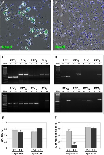
As previously mentioned, the purinergic cross-talk between sensory neurons and SGCs represents a key step in the transmission and integration of painful stimuli within sensory ganglia (see Introduction). In vitro culture systems are invaluable tools to evaluate the role of the various receptor subtypes in cell-to-cell communication, but the influence of culturing conditions on receptor expression should be always taken into account. We have previously demonstrated the presence of several functional P2Y receptor subtypes on both neurons and SGCs in mixed primary culture from mouse TG (see Introduction; Ceruti et al., Reference Ceruti, Fumagalli, Villa, Verderio and Abbracchio2008). Thus, in order to verify whether the expression and function of P2Y receptors on SGCs could be influenced by the presence or absence of neurons, we set up an in vitro model of purified SGCs from mouse TG. Five days after preparation of primary mixed neuron–SGCs cultures (Fig. 1A; Ceruti et al., Reference Ceruti, Fumagalli, Villa, Verderio and Abbracchio2008), SGCs approached confluence and were replated on uncoated wells. This procedure completely removed all neurons, as demonstrated by the lack of staining of the neuronal marker NeuN (Fig. 1B). RT-PCR analysis from SGCs cultures revealed that all the P2Y receptors were expressed after 2 days in culture, with no significant differences when compared to mixed neuron–SGCs cultures (Fig. 1C). Conversely, 4 days after SGCs purification, only P2Y1 and P2Y14 receptor mRNAs were detected (Fig. 1D), indicating that the time in culture without neurons can substantially influence P2Y receptor expression (see above). This hypothesis is further supported by the observation that, although RT-PCR is a semi-quantitative analysis, a trend to reduction in the expression of some P2Y receptor subtypes can be also observed in mixed neuron–glia cultures (cf. Fig. 1C,D). It is well known that TG neurons undergo spontaneous degeneration and death with time in culture (Simonetti et al., Reference Simonetti, Fabbro, D'Arco, Zweyer, Nistri and Giniatullin2006). The progressive loss of neuronal population might therefore influence P2Y receptor expression by SGCs, as more markedly shown in purified cultures. P2X3 receptor expression was never detected in purified cultures, thus confirming its selective neuronal localization (Chen et al., Reference Chen, Akopian, Sivilotti, Colquhoun, Burnstock and Wood1995).
Changes in P2Y receptor expression in purified trigeminal SGCs as a function of time in culture. (A) Representative brightfield of primary neuron-satellite glia mixed cultures 48 h after TG dissociation and cell plating. NeuN-positive neurons are shown in green. (B) Representative brightfield showing the typical morphology of purified SGCs, 2 days after replating of the mixed cultures on uncoated wells. This procedure completely removed all neurons, as demonstrated by the lack of NeuN staining. Nuclei were stained with the Hoechst-33258 dye (blue in A and B). Scale bars: 60 μm. (C, D) Expression of the various P2Y receptor subtypes obtained by RT-PCR analysis following RNA isolation from either mixed or purified cultures. Specific primers for each rodent P2Y receptor subtype, and for the P2X3 receptor subtype, were used. C and D show the comparison between mixed cultures (M), and purified SGCs (P) at 2 or 4 days after their preparation, respectively (see Methods for details). P2X3 receptor expression was never detected in purified cultures, confirming its neuronal-specific expression. (E, F) Histograms showing the evoked mean calcium increases (E), and the percentage of responding cells (F), obtained by the application of either 100 µM UTP or 1 µM ADP to purified SGCs at day 2 (2 d) and day 4 (4 d) in culture. A significant reduction in the percentage of UTP-responding cells was detected after 4 d in culture, in accordance with the downregulation of P2Y2 and P2Y4 receptor expression (see D). ***P < 0.001 with respect to the percentage of UTP-responding cells in 2 d cultures; one-way ANOVA.

To test for the functional outcome of this observation, at either 2 or 4 days in culture, SGCs were loaded with the calcium indicator Fura-2 and subjected to calcium imaging experiments. After 2 days in culture, a significant percentage of SGCs showed increases in [Ca2+]i after application of both 100 µM UTP (53.0 ± 5.3%, n = 200) and 1 µM ADP (66.1 ± 6.0%, n = 299; Fig. 1F). The mean calcium responses, evaluated as the increase of the 340/380 fluorescence ratio (ΔF340/380), were 0.56 ± 0.10 and 0.51 ± 0.06 for UTP and ADP, respectively (Fig. 1E). In agreement with the maintenance of P2Y1 receptor expression, 4 days after purification, SGCs responded to 1 µM ADP with no significant differences with respect to 2 days old cultures (% of responding cells: 62.0 ± 4.18; ΔF340/380: 0.63 ± 0.07; n = 153; Fig. 1E,F). On the contrary, application of 100 µM UTP induced changes in the [Ca2+]i in a significantly lower percentage of cells when compared with 2 days old cultures (% of responding cells: 10.0 ± 3.42; ΔF340/380: 0.49 ± 0.06; n = 120, P < 0.001; Fig. 1E,F), thus confirming the lower expression of the P2Y2 and P2Y4 receptor subtypes in this cell population. These results validate this purified culture model as a helpful tool for studying the role of P2Y receptors selectively in SGCs. However, to avoid possible drawbacks, the time in culture should be carefully taken into account, to avoid possible artifacts due to the lack of neurons.
A possible role for other purinoceptors in pain signaling within TG
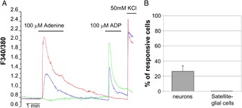
As mentioned in the Introduction, two additional and ‘atypical’ purine-based molecules, namely adenine and Guo, could contribute to the generation and integration of painful signals. However no information is currently available on their role in TG and, in particular, on their actions on SGCs. On this basis, we investigated the effect of adenine application to mouse trigeminal mixed primary cultures (Ceruti et al., Reference Ceruti, Fumagalli, Villa, Verderio and Abbracchio2008). By utilizing single-cell calcium imaging, we demonstrated that adenine (100 µM) was able to induce transient increases of calcium in a subset (26.4 ± 7.2%, n = 58) of trigeminal neurons (Fig. 2), with a mean calcium response of 0.59 ± 0.15. Interestingly, about 40% of adenine-responding neurons were also responsive to ADP, thus suggesting that a sub-population of neurons co-expresses both P2Y and adenine receptors. Moreover, the expression of functional adenine receptors seems to be restricted to neurons, since no calcium responses were found in glial cells (n = 110) (Fig. 2, see also discussion).
Adenine induced [Ca2+]i increases only in primary neurons, but not in SGCs from mouse TG. (A) Representative temporal plot of the [Ca2+]i changes recorded from three cells upon stimulation with 100 µM adenine, followed by 100 µM ADP. 50 mM KCl was utilized as a depolarizing agent to identify neurons (blue and red traces). Although no changes of [Ca2+]i were recorded in SGCs exposed to adenine (green trace), a significant calcium response to ADP was detected in these cells, as expected (Ceruti et al., Reference Ceruti, Fumagalli, Villa, Verderio and Abbracchio2008). (B) Histograms showing the percentage of adenine-responding cells (from a total of 58 neurons and 110 SGCs).

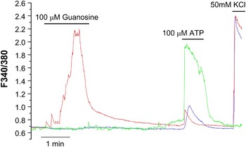
Concerning Guo, we observed that Guo application to our neuron–glia trigeminal cultures was able to increase calcium levels in a fraction of the neuronal population (22.5 ± 1.3%; n = 9) with a mean calcium response of 1.02 ± 0.48 (Fig. 3), while no calcium increases were observed in SGCs (n = 15). Thus, our data suggest that, if expressed by trigeminal SGCs, Guo receptor is not coupled to increases in [Ca2+]i but to a different yet-to-be identified second messenger system (see below). Interestingly, we have found that all the Guo responding neurons also showed calcium increases after application of ATP, indicating that Guo might influence pain transmission by modulating the endogenous ATP-mediated purinergic signaling.
Guo-evoked [Ca2+]i responses in primary cultures from TG. Representative temporal analysis of [Ca2+]i increases recorded from three cells, upon stimulation with 100 µM Guo, followed by 100 µM ATP. Neurons were identified by their response to 50 mM KCl (blue and red traces). No Guo-evoked [Ca2+]i changes were detected in SGCs (green trace) that responded to the stimulation by ATP.

CONCLUSIONS
• The purinergic system plays a crucial role in the modulation of neurons-to-SGCs communication within sensory ganglia, and consequently in the transmission and integration of pain sensations.
• In sensory ganglia, neuronal P2X3 and glial P2X7 are the most important ATP-activated ionotropic receptors involved in nociception.
• The expression and function of P2Y metabotropic receptors by SGCs can be significantly influenced in vitro by the presence or absence of neuronal cells.
• Other purine-based extracellular molecules, such as adenine and Guo, might be involved in the transmission of nociceptive signals within sensory ganglia.
DISCUSSION
ATP is one of the key mediators in nociception, and emerging evidence indicates that other extracellular nucleotides (ADP, UTP and UDP) and nucleosides (Ado) are involved in modulation of pain signaling as well (Liu and Salter, Reference Liu and Salter2005; Burnstock, Reference Burnstock2009a; Jarvis, Reference Jarvis2010). One of the most peculiar characteristics of purinergic transmission is the widespread expression of P1 and P2 receptors in the nervous system (Illes and Ribeiro, Reference Illes and Ribeiro2004; Burnstock, Reference Burnstock2008). Indeed, these receptors have been found to be expressed by both neurons and glial cells involved in nociceptive transmission (Donnelly-Roberts et al., Reference Donnelly-Roberts, McGaraughty, Shieh, Honore and Jarvis2008; Burnstock, Reference Burnstock2009a). Thus, it is becoming increasingly clear that the effects of purines on nociception are extremely complex and related to the specific receptor subtypes involved and/or their cellular localization. For instance, both P2X (e.g. P2X4) and P2Y (e.g. P2Y6,12) purinergic receptors are expressed by activated microglia following nerve injury, and any pharmacological or biotechnological manipulation has been demonstrated to critically modulate the development and maintenance of neuropathic pain (Tsuda et al., Reference Tsuda, Kuboyama, Inoue, Nagata, Tozaki-Saitoh and Inoue2009, Reference Tsuda, Tozaki-Saitoh and Inoue2010).
In sensory ganglia, P2 receptors were found on neurons (Gerevich and Illes, Reference Gerevich and Illes2004; Burnstock, Reference Burnstock2009a), Schwann cells (Fields and Stevens, Reference Fields and Stevens2000) and SGCs (Weick et al., Reference Weick, Cherkas, Hartig, Pannicke, Uckermann and Bringmann2003; Zhang et al., Reference Zhang, Chen, Wang and Huang2007; Ceruti et al., Reference Ceruti, Fumagalli, Villa, Verderio and Abbracchio2008), whereas the localization of Ado P1 receptors, at least in SGCs, is still largely unknown (Sawynok, Reference Sawynok2007). Although the role of P2 receptors in sensory neurons and terminals has been extensively studied (Burnstock, Reference Burnstock2009a, Reference Burnstockb), only little information concerning their role in the modulation of SGCs functions is available.
As already mentioned, SGCs are peculiar glial cells which envelop the cell bodies of primary sensory neurons in sensory ganglia, with the formation of a morphological and functional unit (Hanani, Reference Hanani2005). Recent evidence suggests that, within this functional unit, a non-synaptic cross communication between neurons and SGCs can increase the excitability of both primary afferents and centrally projecting neurons, leading to the development of hyperalgesia and allodynia (Takeda et al., Reference Takeda, Takahashi and Matsumoto2009). Under physiopathological conditions, the somata of sensory neurons release chemical mediators, such as CGRP, substance P and ATP, which in turn activate their receptors expressed on other neurons or surrounding SGCs. Activated SGCs release in turn cytokines, such as IL-1β and tumor necrosis factor α (TNFα), which may further potentiate neuronal excitability, leading to an auto-amplifying loop of neuronal sensitization (Takeda et al., Reference Takeda, Tanimoto, Kadoi, Nasu, Takahashi and Kitagawa2007, Reference Takeda, Takahashi and Matsumoto2009; Thalakoti et al., Reference Thalakoti, Patil, Damodaram, Vause, Langford and Freeman2007; Zhang et al., Reference Zhang, Chen, Wang and Huang2007; Li et al., Reference Li, Vause and Durham2008). The P2 purinergic receptors expressed by SGCs participate primarily to this bidirectional neuron–glial communication inside the sensory ganglia (Zhang et al., Reference Zhang, Chen, Wang and Huang2007; Suadicani et al., Reference Suadicani, Cherkas, Zuckerman, Smith, Spray and Hanani2009). Indeed, a seminal work by Zhang and colleagues (Zhang et al., Reference Zhang, Chen, Wang and Huang2007) reported that the electrical stimulation of DRG neurons elicited robust vesicular ATP release from their somata. The rate of ATP release was dependent on the frequency of nerve stimulation, and on the entry of external Ca2+ into the neuron. In addition, released ATP could activate P2X7 receptors on SGCs, leading to the release of TNFα, which in turn potentiated P2X3-mediated ATP currents in neurons (Zhang et al., Reference Zhang, Chen, Wang and Huang2007).
Interestingly, it has been hypothesized that other purinergic ligands (i.e. adenine and Guo) may also be involved in pain transmission (Bender et al., Reference Bender, Buist, Jurzak, Langlois, Baggerman and Verhasselt2002). The adenine receptor was found predominantly expressed in small DRG neurons; however, information on the presence and function of this receptor in other sensory ganglia is still lacking (Bender et al., Reference Bender, Buist, Jurzak, Langlois, Baggerman and Verhasselt2002). In a similar way, Guo has been recently reported to exert antinociceptive effects after systemic administration in a chronic rat pain model (Schmidt et al., Reference Schmidt, Paniz, Schallenberger, Bohmer, Wofchuk and Elisabetsky2010). It has been postulated that its antinociceptive activity might be related to the activation of a yet-to-be identified-specific receptor (Traversa et al., Reference Traversa, Bombi, Di Iorio, Ciccarelli, Werstiuk and Rathbone2002, Reference Traversa, Bombi, Camaioni, Macchiarulo, Costantino and Palmieri2003), leading to the stimulation of trophic factors release, and to the modulation of glutamatergic activity or of other signaling pathways, such as NO, GABA and noradrenergic pathways, known to be associated to the development of neuropathic pain (Willis, Reference Willis2001; Schmidt et al., Reference Schmidt, Paniz, Schallenberger, Bohmer, Wofchuk and Elisabetsky2010). However, no definitive data are currently available on both the identity of this putative Guo membrane receptor and on its second messenger coupling in native systems.
In the present study, we exploited our in vitro model of primary cultures from dissociated mouse TG to assess the possible involvement of adenine and Guo in calcium-dependent neuron–glia communication. We report for the first time that a significant percentage of trigeminal sensory neurons respond with calcium increases to the application of both adenine and Guo. These data highlight the potential importance of these mediators in neuron–glia calcium homeostasis and also suggest that these receptors might be involved in the generation of enhanced calcium currents that are typical of some pathological pain conditions (van den Maagdenberg et al., Reference van den Maagdenberg, Pietrobon, Pizzorusso, Kaja, Broos and Cesetti2004; Ceruti et al., Reference Ceruti, Fumagalli, Villa, Verderio and Abbracchio2008). Our calcium imaging analysis indicates a lack of functional adenine and Guo receptors linked to the modulation of [Ca2+]i on SGCs. To date, our data may simply signify that, under the experimental conditions utilized, SGC do not express the receptors for adenine and Guo. An alternative explanation is that the two putative receptors are indeed expressed in SGCs, but they are linked to a different transduction mechanism. For example several GPCRs can be coupled to Gi/Gs proteins which in turn lead to the modulation of cytosolic cAMP concentrations, as previously observed in brain slices for Guo and in NG108-15 cells for the adenine receptor (Traversa et al., Reference Traversa, Bombi, Camaioni, Macchiarulo, Costantino and Palmieri2003; Gorzalka et al., Reference Gorzalka, Vittori, Volpini, Cristalli, von Kugelgen and Muller2005). If this proves true, it can be envisaged that adenine and Guo, might participate to neuron–glia communication in sensory ganglia by acting on SGCs and possibly modulating glial K+ channels (see above), thus contributing to the modulation of chronic pain sensations (Vit et al., Reference Vit, Jasmin, Bhargava and Ohara2006).
Overall, evidence is accumulating depicting a scenario where SGCs in sensory ganglia play a fundamental role in pain transmission not only in normal conditions, but also under chronic pathological situations characterized by intense and untreatable pain sensations. The signaling pathways activated in SGCs (particularly the purinergic system) might therefore represent interesting pharmacological targets to be exploited for the development of new analgesic strategies.
A significant percentage of the data summarized in the present paper have been obtained by using in vitro models of primary cultures from sensory ganglia (see above). These models have been developed in order to get better insights into both the molecular mechanisms leading to SGCs sensitization upon painful conditions and in the signaling pathways involved in cell-to-cell communication. Although mixed neuron–SGCs cultures from sensory ganglia are generally used, thanks to their better preservation of the relationships observed in vivo between the two cell types, purified neuronal or SGCs cultures might help studying the signaling pathways at the molecular level in a single-cell population. For example, an in vitro model of purified SGCs cultures has been recently set up and utilized to demonstrate that IL-1β and NO treatment elicited the extracellular release of prostaglandin PGE2, and that the application of the SGCs-conditioned medium to trigeminal neurons was able to augment their capsaicin-evoked release of CGRP (Capuano et al., Reference Capuano, De Corato, Lisi, Tringali, Navarra and Dello Russo2009). To test whether the absence of neurons might influence the expression of purinergic receptors in SGCs, we purified these cells from trigeminal neurons (see Methods). As mentioned in the Results section, significant differences were detected depending upon the time in culture, since the expression and function of P2Y receptors was still comparable to both intact TG and mixed primary cultures after 2 days but not after 4 days in culture. Currently, we do not have a definitive explanation for these changes, but it can be envisaged that they could be due to the absence of the neuronal signals that normally maintain SGCs in their ‘physiological’ functional status. Thus, our results clearly show that one should be aware of the intrinsic limitations of TG culture models, which should be utilized when still reflecting the conditions observed in vivo (e.g. not later than 2 days in culture for P2Y receptors). On the other hand, it is clear that the purified SGCs culture model represents a useful tool to study SGCs sensitization at the molecular level after chronic algogenic conditions, thus providing useful information to be tested and confirmed in additional in vitro and in vivo models.
ACKNOWLEDGEMENTS
Authors wish to thank Dr. Matteo Zanardelli for his help in preparing purified trigeminal SGCs cultures. This work was supported by the grant #GGP07032A on: ‘Genetic and micro-environmental factors regulate the role of ATP as transmitter of pain in a migraine model’ from The Italian Comitato Telethon Fondazione Onlus to MPA.
Statement of interest
None.