Sub-pulmonary membrane is a rare disease and was described in relation to supracristal ventricular septal defects with or without aortic insufficiency, Reference Duggal, Bajaj and Bansal1,Reference Raff, Gaynor, Weinberg, Spray and Gleason2 with valvular pulmonary stenosis, Reference Tefera, Bermudez-Canete and Rubio3 and as an isolated form causing critical neonatal sub-pulmonic stenosis. Reference Tomar, Radhakrishnan and Sharma4 Van Praagh believes that the fibrous membrane is a developmental condition related to the semilunar valve rather than the infundibulum, whereas the muscular membrane arises from the pulmonary infundibulum. Anderson states that this is an acquired condition produced by fibrous growth on top of a sub-pulmonary infundibular narrowing, also described in cases with Fallot’s tetralogy. Reference Anderson and Jacobs5 Cases of sub-pulmonary membrane have also been described following correction of transposition of the great arteries and respond to percutaneous balloon pulmonary valvuloplasty. Reference Vasec and Goetz6
The conal septum and semilunar valves form embryologically from the conal septal cushions that fuse: the septal cushions become muscularised, while the valvular cushions remain fibrous. Reference Spicer, Bridgeman, Brown, Mohun and Anderson7 Fibrous sub-aortic stenosis is well known to cause 8%–20% of left ventricular outflow tract obstructions. Reference Foker8 Yet, to the best of our knowledge, the late development of sub-pulmonary membrane following successful intervention to relieve pulmonary atresia in cases with intact interventricular septum, atrio-ventricular and ventriculo-arterial concordance has not yet been described.
Description of cases
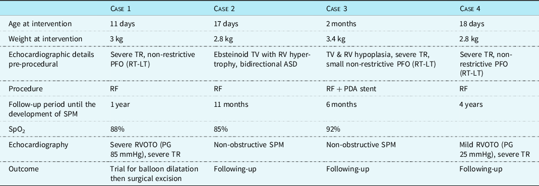
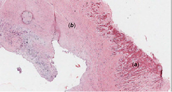
Out of our series of 79 cases of pulmonary atresia with intact interventricular septum that were treated during the period from 2009 to 2019 by radiofrequency perforation and balloon dilatation of the pulmonary valve with or without ductal stenting, we observed the development of a sub-pulmonary membrane in 4 cases on the long-term follow-up. Patients with pulmonary atresia with intact interventricular septum were attempted at radiofrequency perforation + balloon pulmonary valvuloplasty if the atresia was membranous with a preserved stem of main pulmonary artery, with a tripartite or bipartite right ventricle having a tricuspid annular Z-score not smaller than −4, and no evident coronary sinusoids indicating an right ventricle-dependent coronary circulation. In addition, Patent ductus arteriosus (PDA) stenting was attempted in cases with small right ventricles that were felt not enough to support the cardiac output. The degree of Tricuspid regurge (TR) or tricuspid dysplasia did not contraindicate the procedure. Table 1 provides a summary of the pre-operative findings in the cases, operative technique and outcome. In the first case, sub-pulmonary membrane caused significant obstruction that was resistant to balloon dilatation (Fig 1) and was sent for surgical excision: revealing a fibrous membrane on pathological examination (Fig 2). Another case had a right ventricular outflow tract gradient of 25 mmHg by echocardiography and is still following-up; the other 2 cases only demonstrate the sub-pulmonary membrane on echocardiography without significant gradient across the right ventricular outflow tract till the writing of this paper (Figs 3 and 4).
Summary of cases

PDA = Patent ductus arteriosus; RF = radiofrequency; SPM = sub-pulmonary membrane; TR = Tricuspid regurge.
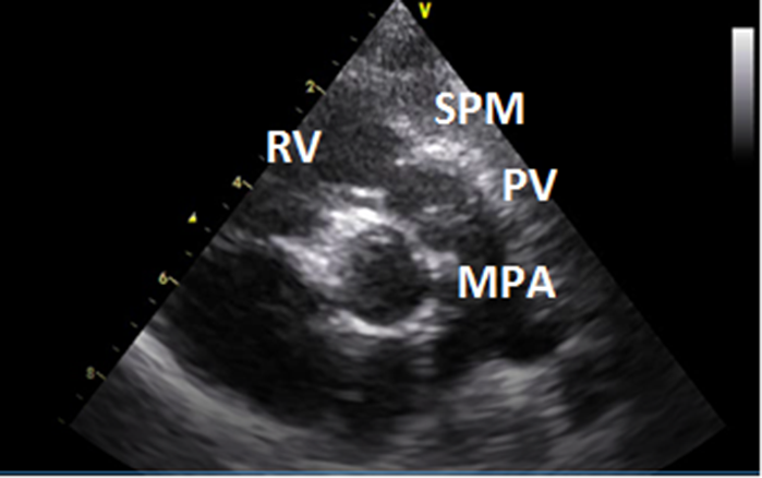
PS view showing the SPM. MPA = main pulmonary artery; PV = pulmonary valve; RV = right ventricle; SPM = sub-pulmonary membrane.

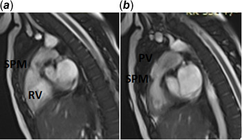
Sagittal cardiac MRI view showing the RVOT in (a) diastole and (b) systole. PV = pulmonary valve; RV = right ventricle; SPM = sub-pulmonary membrane.

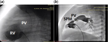
Lateral angiograms (a) prior to the perforation of pulmonary valve showing a blind RVOT; (b) after 1 year showing the SPM. PV = pulmonary valve; RV = right ventricle; SPM = sub-pulmonary membrane.

Pathological specimen of the excised fibrous membrane: (a) portion of myocardial muscle; fibrous membrane formed of hypocellular collagen entangling bland-looking fibroblasts with low vascularity.

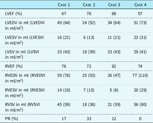
The four cases were assessed by cardiac MRI to evaluate the anatomy of the right ventricular outflow tract, the flow dynamics and the right ventricular outflow tract/main pulmonary artery angle analogous to that described for the aorto-septal angle in the sub-aortic membrane. Reference Shar, Brown, Keswani, Grande-Allen and Sucosky9 Comparing the shape and length of the right ventricular outflow tract infundibulum and the sub-pulmonary area to that of normal subjects showed right ventricular outflow tract narrowing in two cases with acceleration and turbulence of flow on one, and no anatomical narrowing in the other two including the post-operative case. The right ventricular outflow tract angle with the main pulmonary artery did not show obvious change. Three cases showed mild pulmonary regurge; one case had no pulmonary regurge. It is of note that the first case was imaged after the excision of the sub-pulmonary membrane. Cardiac MRI findings are listed in Table 2.
Cardiac MRI findings in the patients

LVEDV = left ventricular end-diastolic volume; LVEF = left ventricular ejection fraction; LVEDVI = indexed left ventricular end-diastolic volume; LVEDVI = indexed left ventricular end-systolic volume; LVESV = left ventricular end-systolic volume; PR = pulmonary regurgitation; RVEDV = right ventricular end-diastolic volume; RVEF = right ventricular ejection fraction; RVEDVI = indexed right ventricular end-diastolic volume; RVEDVI = indexed right ventricular end-systolic volume; RVESV = right ventricular end-systolic volume.
Discussion
The sequence of events that lead to semilunar valve atresia and ventricular hypoplasia needs better understanding, as to which is the primary pathology, and the mechanisms involved. More evidence is available for the left ventricle and outflow tracts; similar influences might be valid for the right ventricle and outflow tract.
Outflow obstruction results in complex secondary responses with disturbed flow dynamics and shear stresses, leading to impaired and disorganised ventricular growth and development. Genetic modifiers might impact subsequent remodelling, including intrinsic myocardial growth signalling pathways. Furthermore, during the latter stages of gestation, cardiomyocytes switch their myogenic capacities and become incapable of mitosis: ventricular hyperplasia cannot occur therefore, and remodelling becomes limited to ventricular hypertrophy. Reference Hickey, Caldarone and McCrindle10
Sub-aortic membranes are well known to recur in a subset of patients after surgical removal; this proposes that an underlying pathology is present and not merely the anatomical condition restored by surgery. Gewillig and colleagues in 1992 studied patients with discrete sub-aortic membranes by echocardiography and showed abnormal flow patterns in these cases and suggested that these chronic flow disturbances trigger endothelial transformation and growth, leading to the development and recurrence of the anatomical stenosis. Reference Gewillig, Daenen and Dumoulin11 It has also been reported that minor changes in the left ventricular outflow anatomy, including the aorto-septal angle, Reference Shar, Brown, Keswani, Grande-Allen and Sucosky9 can produce significant alterations in the dynamic forces and septal shear stress, and thus influencing the development and the progression of sub-aortic obstruction, which is much faster in infants than in older children. Reference Mohan, Shukla, Mohan and Sethi12,Reference Silbiger, Lee, Christia and Perk13 These mechanical theories, however, could not explain the increased familial recurrence rate that is also well-identified in cases with sub-aortic membranes; an underlying genetic factors must therefore be co-existent. Reference Foker8,Reference Gale, Cartmill and Bernstein14-Reference Richardson, Menahem and Wilkinson16
A theory that combines both the mechanical and the genetic hypothesis states that the disturbed flow dynamics might induce the expression of a number of genes responsible for endothelial and smooth muscle cell proliferation, similar to the mechanisms involved in atherosclerosis. Reference Chiu and Chien17 It is important to remember that discrete sub-aortic membrane can occur following a repair of underlying CHD. Irrespective of the gross appearance, the stenosing lesions exhibit five tissue layers beginning from the luminal aspect: endothelium, acid mucopolysaccharide-rich sub-endothelial layer, collagen-rich fibrous layer, fibro-elastotic layer and a smooth muscle layer. Reference Butany, Vaideeswar and David18
Fibrous sub-aortic or sub-pulmonary stenosis is in direct fibrous continuity with the overlying semilunar valve, indicating that this sub-valvar ring-like stenosis really is valvar, not infundibular or conal. If the obstruction becomes muscular, then most probably the origin is infundibular and not valvar. Reference Tomar, Radhakrishnan and Sharma4 Anderson thinks that this is not a “membrane” and appears to look more like a fibrous shelf, is almost certainly an acquired lesion and is a fibrous accretion exacerbating the narrowing at the mouth of the sub-pulmonary infundibulum. Anderson and colleagues have illustrated such fibrous accretions in their studies on tetralogy of Fallot, albeit not specifically commenting on the structure. Reference Anderson and Jacobs5
In our statement, we are suggesting that the sub-pulmonary membrane that was not present before the first intervention might have developed later due to anatomical disturbance such as a long infundibulum forming the right ventricular outflow tract or due to malalignment of the septum in the right ventricular outflow tract reaching the sub-valvular area, either anatomic abnormality resulting in disturbed flow dynamics. Another postulation is that the sub-pulmonary membrane represents a response of the right ventricular outflow tract endothelium to the mechanical trauma during the initial intervention. This, however, does not explain fully the annular nature of the membrane but would have rather resulted in a shelf-like obstruction.
On echocardiography, it is important to differentiate this anomaly from valvar pulmonary stenosis with intact ventricular septum for the management plan. For valvar pulmonary stenosis, balloon pulmonary valvotomy is the treatment of choice with excellent results, while for sub-valvar pulmonary stenosis, surgery is the only option. We have excised the sub-pulmonary membrane in only one patient that had severe obstruction, another has mild right ventricular outflow tract obstruction and the other patients have no significant obstruction either by echocardiography or cardiac MRI. It is time and close follow-up that will show if the sub-pulmonary membrane will act in a similar manner as a sub-aortic membrane and progress into hemodynamic obstruction and recur after surgical excision or not. The rarity of the condition limits the extraction of sold conclusions; however, with the advancement and popularity of interventional treatment for pulmonary atresia with intact interventricular septum, the future might bare more insights.
Conclusion
We report the finding of newly developing sub-pulmonary membrane as a rare cause of right ventricular outflow tract obstruction after relieving pulmonary atresia with intact interventricular septum by radiofrequency perforation and balloon dilatation. Transthoracic echocardiography was helpful to delineate the lesion and was confirmed by cardiac MRI. Differentiating it from the usual valvar, pulmonary stenosis has guided us to formulate for each case a unique management plan. We believe that a careful serial follow-up echocardiography of all pulmonary atresia with intact interventricular septum and neonatal critical pulmonary valve stenosis post-intervention would enable us to learn more about this rare lesion.
Acknowledgements
None.
Compliance with ethical standards
Financial support
This study received no external funding.
Conflicts of interest
All authors declare no conflicts of interest.
Ethical standards
All procedures performed in studies involving human participants were in accordance with the ethical standards of the institutional and national research committee and with the 1964 Helsinki declaration and its later amendments or comparable ethical standards.
Informed consent
Informed consent was obtained from all the caregivers of the individual participants included in the study.