A straddling tricuspid valve is one where a part of its tension apparatus crosses a ventricular septal defect to attach to the septum or a papillary muscle in the left ventricle. Reference Milo, Ho and Macartney1 This rare anomaly has been reported concomitantly with various defects such as isolated ventricular septal defect, atrioventricular discordance as in congenitally corrected transposition of the great arteries, and atrioventricular canal defect. Reference Milo, Ho and Macartney1,Reference van Son, Hambsch and Mohr2 Repair is challenging, with strategies including patching around the straddling tricuspid valve or replacing the straddling valve entirely. Reference Dodge-Khatami, Mavroudis, Frost, Jacobs and Mavroudis3,Reference Pacifico, Soto and Bargeron4 An alternative strategy is transposition of the straddling papillary muscle to the appropriate ventricle, Reference van Son, Hambsch and Mohr2,Reference Reddy, Liddicoat, McElhinney, Brook, van Son and Hanley5 but there are limited reports of this strategy being used in the context of hypoplastic right ventricle.
There is a growing literature in the field of biventricular repair for patients with ventricles of borderline size or function. Reference Andersen, Scherba and Turek6,Reference Emani and del Nido7 In this report, we present the case of a patient with an inlet ventricular septal defect, subpulmonic stenosis, hypoplastic right ventricle, and straddling tricuspid valve – a rare constellation of defects. Reference Arciniegas, Hakimi, Farooki and Green8 The patient underwent successful single-stage biventricular repair with right ventricular rehabilitation and tricuspid valve papillary muscle transposition.
Case report
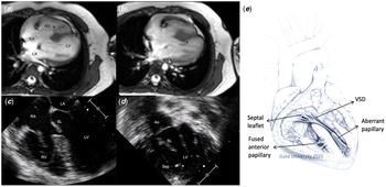
The patient is a 22 kg 5-year-old female child with Wildervanck (cervical-oculo-acoustic) syndrome who was referred for poor exercise tolerance and found to have cyanosis with oxygen saturations in the 80s. Transthoracic echocardiography and cardiac MRI demonstrated an unrepaired complex CHD including a large inlet ventricular septal defect, severe subvalvar pulmonic stenosis, moderately hypoplastic right ventricle with hypoplasia of the sinus portion (right ventricle volume 23–26 mL, right ventricle end diastolic volume index 27.4 mL/m2, z-score between − 8 and − 3 based on method), Reference Buechel, Kaiser, Jackson, Schmitz and Kellenberger9 normal left ventricle dimensions (left ventricle volume 44 mL, left ventricle end diastolic volume index 52.4 mL/m2), straddling tricuspid valve (annulus 25 mm, z-score = +1), and large secundum atrial septal defect (Fig 1). She was taken to surgery for complete biventricular repair including ventricular septal defect closure, relief of pulmonic stenosis, right ventricular rehabilitation, papillary muscle transposition, fenestrated atrial septation, and possible Glenn depending on post-bypass physiology.
Pre-operative MRI showing (a) fusion of anterior papillary muscle to the septum (*), and (b) aberrant papillary muscle (*). Pre-operative transthoracic echocardiogram demonstrating (c) straddling septal leaflet, as well as (d) aberrant papillary on left ventricle septal surface (*). Ao = aorta. Sep = interventricular septum. (e) Pre-operative illustration of anatomy.

At surgery, the anterior papillary muscle of the tricuspid valve was found fused to the septum, restricting right ventricular cavity growth. The septal leaflet of the tricuspid valve was overriding the crest of the ventricular septum, with all chordal attachments coursing to a discrete large papillary muscle on the left ventricular side of the septum. Right ventricular rehabilitation was performed by delaminating the anterior papillary muscle from the ventricular septum and dividing apical trabeculations, thereby enlarging the ventricular cavity. Next, the straddling papillary muscle was amputated from the left ventricle. The large inlet ventricular septal defect was then closed to the true tricuspid annulus using a bovine pericardial patch. The amputated papillary muscle was attached to the right ventricular septal surface close to where the anterior papillary muscle was delaminated, to decrease the risk of conduction system injury. Pledgeted sutures were used to anchor the papillary muscle at a location that achieved the appropriate height of the chordal attachments to avoid prolapse of the septal leaflet. The tricuspid valve was floated and there was no prolapse of the septal leaflet and mild leakage from a cleft in the posterior leaflet. The cleft was closed and the valve appeared competent. The atrial septal defect was closed with a bovine pericardial patch with a central 4 mm fenestration. Lastly, the right ventricular infundibulum was incised across the area of sub-valvar pulmonic stenosis. Right ventricle muscle bundles were resected and the infundibulum was patched with bovine pericardium (Fig 2B).
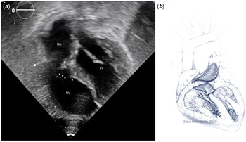
(a) Follow-up transthoracic echocardiogram 2 years post-operatively demonstrating enlarged right ventricle cavity at end diastole, chordae of the septal leaflet (arrows), and transposed papillary muscle (*). (b) Post-operative illustration of anatomy.

Post-bypass transoesophageal echocardiography showed normal biventricular function, trivial-to-mild tricuspid regurgitation, trivial residual ventricular septal defect, no right ventricular outflow tract obstruction, and right-to-left shunting across the atrial septum. The patient was haemodynamically stable with central venous pressure of 15 mmHg, left atrial pressure of 10mmHg, and oxygen saturations of 98-100%. Therefore, a Glenn anastomosis was not thought to be needed. The patient was discharged home on post-operative day 16 with normal oxygen saturations.
The patient has not required further cardiac interventions. Follow-up echocardiography 2 years later shows normal biventricular function, mildly hypoplastic right ventricle, mild tricuspid regurgitation, mild tricuspid stenosis, trivial residual ventricular septal defect, no residual atrial septal defect, and no significant sub-pulmonic stenosis (Fig 2A). The right ventricle pressure was estimated to be normal based on the velocity of the tricuspid valve regurgitant jet (2.1 m/s) and ventricular septal defect gradient (5.2 m/s).
Discussion
This case report of single-stage complex biventricular repair adds to the growing literature on papillary muscle transposition as well as ventricular-level procedures for borderline ventricle rehabilitation. Reference van Son, Hambsch and Mohr2,Reference Reddy, Liddicoat, McElhinney, Brook, van Son and Hanley5–Reference Emani and del Nido7 To our knowledge, complete surgical repair of this constellation of defects including straddling tricuspid valve in the setting of hypoplastic right ventricle has not been reported. An alternative approach would have been staged ventricular recruitment, whereby ventricular rehabilitation and atrial septal defect restriction are performed initially followed by closure of the ventricular septal defect after a period of ventricular growth. The Boston group has found success in this method, with increase in ventricular volume from 31 to 49 mL/m2 in their cohort within a median period of 9 months. Reference Oladunjoye, Piekarski, Banka and etal10 In our case, we opted to perform single-stage repair and utilise the atrial septal defect to offload the right ventricle. Our decision making was influenced by the fact that the patient lived in a rural area with limited access to medical services.
We believe there were several key decisions made in this case. For one, judgement was required as to whether the right ventricle size would be adequate to support nearly an entire cardiac output after septation. The initial size of the ventricle was considered borderline for two-ventricle repair, but we anticipated future growth of the ventricle after delamination of the anterior papillary muscle and aggressive division of apical trabeculations. In addition, we have found that two-ventricle repair in cases of borderline right ventricle are typically quite forgiving, given that an atrial septal defect can be used to offload the right ventricle at the expense of oxygen saturations, or a Glenn anastomosis can be performed to achieve a 1.5-ventricle repair in severe cases. Second, we believe that papillary muscle transposition was necessary to ensure an excellent and durable biventricular repair. Alternate methods such as placing a patch around the straddling valve would have risked leaving residual ventricular septal defect, creating chordal immobility, or producing atrioventricular block. Reference van Son, Hambsch and Mohr2 Similarly, replacing the tricuspid valve would not have been preferable in a growing child. Overall, this case demonstrates that a straddling tricuspid valve can be successfully navigated without resorting to single-ventricle palliation by default.
Acknowledgements
None.
Financial support
This research received no specific grant from any funding agency, commercial, or not-for-profit sectors.
Conflicts of interest
None.