Prenatal diagnosis of transposition of the great arteries and intact ventricular septum is crucial as 4–12% of fetuses will develop major haemodynamic alterations that can cause death shortly after birth.Reference Bonnet, Coltri and Butera 1 , Reference Soongswang, Adatia, Newman, Smallhorn and Williams 2 For these patients, immediate postnatal management – balloon atrioseptostomy – is life-saving.Reference Baylen, Grzeszczak and Gleason 3 Prenatal identification of this at-risk population is a key factor to avoid haemodynamic compromise and possible neurological sequelae, but the ability to predict postnatal hypoxia remains limited. Restriction of the foramen ovale leading to low interatrial mixing as well as restrictive or retrograde flow in the ductus arteriosus have been reported in several prenatal studies.Reference Maeno, Kamenir, Sinclair, van der Velde, Smallhorn and Hornberger 4 – Reference Punn and Silverman 7 Haemodynamic data in human fetuses with transposition of the great arteries and intact ventricular septum, however, are limited, and the prenatal pathophysiology is poorly understood, especially regarding pulmonary output and ductal flow.
Therefore, our study had two main objectives: first, to compare characteristics related to the interatrial communication and the ductus arteriosus between fetuses with transposition of the great arteries and intact ventricular septum showing severe neonatal hypoxia and fetuses with transposition without severe neonatal hypoxia; and, second, to compare ventricular and pulmonary flow patterns between fetuses with transposition of the great arteries and intact ventricular septum and age-matched population controls in order to understand which circulatory modifications lead to the observed neonatal desaturation. We propose that an interplay between the size of the foramen ovale, the pulmonary flow, and the distribution of cardiac output between the left ventricle and the right ventricle leads to an abnormal flow across the arterial duct in the case of transposition of the great arteries and intact ventricular septum.
Methods
Population
Echocardiographic data of 45 fetuses diagnosed with transposition of the great arteries and intact ventricular septum from January, 2000 to July, 2014 at Sainte-Justine University Hospital Center (Montreal, Canada) and Centre Mere Enfant of Quebec (Quebec City, Canada) were retrospectively reviewed. Gestational age was determined by ultrasound examination before 20 weeks of gestation. Saturation was recorded with a Masimo oxygen saturation monitor (Masimo, Irvine, California, United States of America). In order to study the potential prenatal causes of postnatal hypoxia, our cohort was divided into two groups on the basis of pre-ductal oxygen saturation – that is, saturation at the right upper extremity.
We arbitrarily chose a transcutaneous saturation of 50% to separate our patients into two groups. This seems a reasonable threshold to conclude inadequate interatrial mixing. In our institution, 50% is also our threshold to perform an atrioseptostomy in emergency.
Patients with peripheral oxygen saturation ⩽50% were included in group 1, and patients with peripheral saturation >50% were included in group 2. Prenatal and postnatal data were compared between the two groups. In order to correct for potential confounding effects and measurement biases, age-matched control fetuses from an in-house population were included in the analysis of flow patterns. This control group was composed of normal singleton pregnancies referred between 30 and 35 weeks of gestation. The reason for cardiac evaluation was suspicion of arrhythmias, not confirmed on our examination, or late screening for cardiac anomalies in the case of consanguinity or extracardiac malformations.
Exclusion criteria
All patients with transposition of the great arteries and intact ventricular septum had to deliver in the participating recruiting centres. Patients from the three groups were excluded from the study on the basis of the following criteria: chromosomal anomalies, arrhythmia, congenital syndromes, abnormal amniotic fluid, interventricular septal defects, abnormal pulmonary or aortic valves, suspicion of coarctation of the aorta, other associated major intracardiac anomalies, or abnormal flow in the umbilical or maternal uterine arteries.
Echocardiographic study
Fetuses in the three groups underwent echocardiography by experienced echocardiographers following the same study protocol established in our centre since 1998. This protocol includes a zoom on aortic and pulmonary valves in a long-axis view of the vessels and a cine-loop recording of at least three beats. In normal fetuses, aortic flow was recorded on a vertical five-chamber view, the main pulmonary artery’s flow on a sagittal short-axis view, and the ductal flow on a sagittal view at the narrowest diameter. In fetuses with transposition of the great arteries and intact interventricular septum, pulmonary and aortic Doppler were obtained from the transverse vertical view of the heart and ductal flow from a sagittal view of the fetus at the narrowest diameter. Doppler signals were assessed with an angle <15° between the cursor line and the flow, and five consecutive beats had to be recorded. All examinations were then stored on video tapes or DVDs that we could retrospectively use for our measurements and analysis.
Prenatal data
Echocardiographic examinations were performed with a Sequoia sonographic equipment from Acuson (Acuson, Mountain View, California, United States of America), a Hewlett Packard 5500 (Hewlett-Packard, Palo Alto, California, United States of America), or a Philips Sonos 5500 (Philips Healthcare, Andover, Massachusetts, United States of America), with either a 4- or a 6-MHz transabdominal transducer.
The last Doppler study was retrospectively reviewed by two independent observers – L.V. and M.J.R. – blinded to the measurements of the other observer and unaware of the postnatal outcome. Both observers reviewed echocardiograms from the two centres. Complete ultrasounds were reviewed. For all parameters studied, calibration was performed if necessary (video tapes). Doppler measurements were obtained by averaging velocity time integrals of three consecutive cycles on still images. In order to minimise errors in haemodynamic calculations, measurements of the aortic and pulmonary annulus were carried out on a zoomed cine-loop of each valve. Each annulus was measured three times on three consecutive beats, and the average value was used for calculations.
Data were compiled as follows:
Valvular dimensions: the inner diameter of the pulmonary and the aortic annuli were measured using two-dimensional echocardiography with an insonation plane perpendicular to the long axis of the vessels at the maximal opening.
Right and left ventricular output: The aortic and main pulmonary arterial flows, corresponding, respectively, to right and left ventricular output (QRV and QLV), were calculated using the following equation: ventricular output=(π×D2/4)×VTI×HR, where π×D2/4 represents the cross-sectional area of the vessel at the level of the annulus (cm2), VTI the velocity time integral for each vessel (cm), and HR the heart rate (beats per minute).
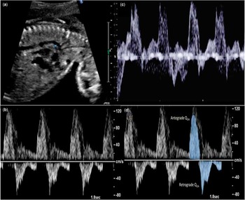
Flow in the ductus arteriosus (Fig 1): the narrowest diameter of the ductus arteriosus was measured in a sagittal view (Fig 1a). We looked for the presence of diastolic retrograde flow by color and pulsed Doppler. Retrograde flow was classified as persistently retrograde if holodiastolic retrograde flow was present for each cardiac cycle (Fig 1b). It was classified as intermittently retrograde if retrograde flow was not constant, variable with fetal respiratory movements, or if it was present only in early or mid-diastole (Fig 1c). A restrictive ductus arteriosus was defined as increased diastolic flow velocity with decreased pulsatility index.Reference Tulzer, Gudmundsson, Sharkey, Wood, Cohen and Huhta 8 , Reference Huhta, Moise, Fisher, Sharif, Wasserstrum and Martin 9
Flow analysis in the ductus arteriosus. ( a ) The ductus arteriosus is measured on a sagittal view at the narrowest diameter. ( b ) Superposition of isthmic and ductal flow with holodiastolic retrograde flow in the ductus arteriosus. ( c ) Intermittent retrograde flow: retrograde flow is seen in early diastole and varies between the cardiac cycles. ( d ) Antegrade and retrograde velocity time integrals were traced from the ductal Doppler.

Owing to the disposition of the two great vessels, antegrade flow in the ductus arteriosus (antegrade QDA) is related to left ventricular output, whereas retrograde flow (retrograde QDA) is from the aorta, and is therefore related to right ventricular output (Fig 1d). For the calculation of ductal flow, we separated antegrade and retrograde blood flow. Antegrade and retrograde flows were calculated using the same formula as described for the right and left ventricular output. Global ductal flow (QDA) was calculated as QDA=(anterograde QDA−retrograde QDA).
Effective pulmonary flow: blood flow to the lungs or effective pulmonary flow (QEP) could finally be obtained from the two previous calculations:
QEP=(QLV−antegrade QDA)+retrograde QDA, taking into account the perfusion from the left ventricle and retrograde flow from the aorta to the lungs through the ductus arteriosus.
Interatrial communication: the size of the fossa ovalis was measured in a horizontal four-chamber view at the level of the septum secundum. On any two-dimensional view centred on the septum – horizontal, vertical, or oblique – we assessed carefully the interatrial septum in order to measure the size of the communication at the level of the septum primum. This measurement was also performed using color Doppler, which allows visualisation of the flow across the septum. The length of the septum primum was measured at its maximal deployment into the left atrium. An aneurysmal interatrial septum bulging into the left atrium or an angle of <30° between the septum primum and the interatrial septal plane were reported when present.Reference Wilson, Rao and Aeschlimann 10
Postnatal data
Data on gestational age at delivery, mode of delivery, weight (g) and height (cm), Apgar scores at 5 minutes, and the need for cardiopulmonary resuscitation were collected from the hospital chart for all neonates with transposition of the great arteries and intact ventricular septum. We collected the first stable pre-ductal saturation (%) in the neonatal ICU and the difference of saturation between the upper and lower limbs. In neonates with transposition of the great arteries, the pulmonary artery is perfused with highly oxygenated blood, and the lower limbs are exposed to a higher saturation in case of right-to-left shunt through the ductus arteriosus. We looked at the rate of atrioseptostomy in each group, as well as at the first lactate level obtained in arterial blood after atrioseptostomy. We noted the preoperative death rate in each group.
Statistical analysis
Descriptive statistics are presented as median±standard deviation [range] for continuous variables and as absolute numbers and percentages for categorical variables. Normality of data was assessed graphically using the Kolmogorov–Smirnov test. In order to compare echocardiographic findings and postnatal data between groups, a Fisher’s exact test was performed for categorical variables. Continuous variables were compared using the Mann–Whitney or t-tests according to their distributions. Spearman’s rank correlation was used to assess the relationship between flows and cardiac measurements. The performance of prediction of the hypoxic status by the prenatal cardiac ultrasound was then determined using receiver operating curves. Area under the curve, detection rate, and false-positive rate were computed. All data analyses were performed using the statistical software framework R, and p values below 0.05 were considered significant. Interobserver variability was determined using both the Bland–Altman method – plot of the difference of means, limits of agreement, and bias – and the concordance correlation coefficient with corresponding 95% confidence intervals and Pearson’s statistic on a subset of six randomly chosen echocardiographs, which were assessed independently by the two observers.
Results
Our study cohort included 45 fetuses diagnosed with transposition of the great arteries and intact ventricular septum in participating centres between January, 2000 and July, 2014. The first group included 13 fetuses that postnatally developed a peripheral transcutaneous saturation ⩽50%. The second group comprised 32 fetuses with saturation above 50%. The control group comprised 50 normal singleton pregnancies referred at 33±2.6 weeks of gestation.
The agreement between the two observers was excellent [Pearson’s R 2 0.98, 95%, confidence interval 0.9–1.0; p<0.0001], and was highly reproducible for echocardiographic measurements for both fossa ovalis and defect sizes with a concordance correlation coefficient of 0.96 (0.94–0.97).
Prenatal data
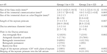
Prenatal cardiac morphological data are presented in Table 1. The last echocardiogram was performed at a median gestational age of 35 [±2.5] weeks for group 1, 35 [±3.5] weeks for group 2, and at 33 [±2.6] weeks for controls. There was no statistical difference in terms of gestational age at the time of echocardiography between the three groups. Measurement of the interatrial communication using two-dimensional and color echocardiograms was missing for one fetus in group 1 and four in group 2.
Interatrial communication and ductus arteriosus characteristics of fetuses with transposition of the great arteries and intact ventricular septum.

Data are presented as median±SD [range] for continuous variables and as absolute numbers (percentages) for categorical variables
* Measurements available for 12 fetuses in group 1 and 28 in group 2
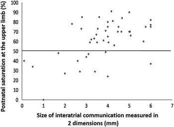
Fetuses in group 1 had smaller interatrial communication on both two-dimensional (2.9 versus 4, p=0.004) and color (2.6 versus 3.8 mm, p=0.005) measurements. All patients with a foramen ovale ⩽2 mm (five patients) had saturation below 50% after birth. The size of the interatrial communication measured by two-dimensional echocardiography against postnatal saturation is given in Figure 2. Length of the septum primum did not vary significantly between groups, even if there was a tendency towards a longer septum primum in group 1 (17 versus 15 mm, p=0.06). There was no difference regarding the size of the fossa ovalis between groups.
Size of interatrial communication measured during the third-trimester echocardiogram of fetuses with transposition of the great arteries plotted against postnatal saturation recorded at the upper limb. The horizontal line separates saturation above and below 50%.

Fetuses in group 1 were more likely to have retrograde diastolic flow in the ductus arteriosus than fetuses in group 2 (92 versus 23%, p=0.02). The majority of fetuses in group 1 (nine patients, 69%) had intermittent retrograde diastolic flow. Holodiastolic retrograde flow was observed among two fetuses from group 1 and three from group 2. Only one fetus from group 1 showed restrictive flow in the ductus arteriosus.
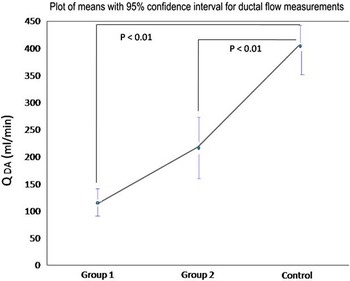
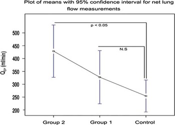
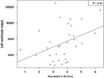
Ventricular and pulmonary flow characteristics were available for 38 fetuses (group 1: n=12; group 2: n=26) (Table 2) with transposition of the great arteries and intact ventricular septum and 50 controls. Owing to some missing data, we could not adequately calculate one of the three flow rates for the rest of the population. Both groups with transposition of the great arteries showed a significant decrease in ductal flow pattern when compared with the control group (Fig 3) (paired Wilcoxson’s rank test with Bonferroni’s correction; p<0.01). Importantly, there was a higher effective pulmonary flow in group 2 patients when compared with the control group (Fig 4). Fetuses in group 1 showed a tendency towards a lower effective pulmonary flow compared with fetuses in group 2. This trend was inversed with significantly lower effective pulmonary flow in controls when compared with group 1. In addition, we found a mild positive correlation between left ventricular flow and size of the interatrial communication (Fig 5) (Spearman’s rank correlation 0.44).
Comparison of ductal flow (QDA) of fetuses with transposition of the great arteries and intact ventricular septum and controls.

Comparison of effective pulmonary flow (QEP) of fetuses with transposition of the great arteries and intact ventricular septum and controls.

Correlation between left ventricular output and interatrial defect measured in two dimension (2D) in fetuses with transposition of the great arteries and intact ventricular septum.

Comparison of ductal flow and effective pulmonary flow at the third-trimester echocardiogram of fetuses with transposition of the great arteries and controls.

QDA=net ductal flow (anterograde−retrograde flow); QEP=effective pulmonary flow
Data are presented as median±SD [range]
Postnatal data
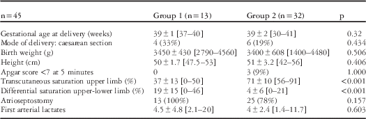
Postnatal data are presented in Table 3. There were no significant differences observed in postnatal data regarding mode of delivery, gestational age, birth weight, height, and Apgar scores. The differential saturation between the upper and the lower limbs was higher in group 1 than in group 2 (19±15 versus 4±6%, p<0.01), indicating right-to-left shunt through the ductus arteriosus in group 1 as well as important aortic desaturation. In group 1, five patients had saturation <30%; two patients from this group needed resuscitation before and during atrioseptostomy. For the first one, saturation was impossible to record after birth, and the neonate experienced immediate bradycardia needing chest compression. This patient was not considered at risk for premature closure of the interatrial communication on the basis of the prenatal echocardiogram, but was delivered during daytime, and atrioseptostomy was performed shortly after birth via the umbilical vein. For the second, saturation was initially <30%, and bradycardia occurred a few minutes after birth. The three other patients with saturation <30% had a normal cardiac rate before atrioseptostomy. Except for the first patient, all other fetuses were considered at risk of postnatal hypoxia before birth, and a senior cardiologist specialised in cardiac catheterisation was present at the time of delivery and could perform the atrioseptostomy immediately after birth. The five children are doing well. There was no preoperative or postoperative death in our study cohort. The patient with a prenatal restrictive ductus arteriosus had a saturation of 44% after birth. His clinical course was uneventful after atrioseptostomy.
Postnatal data of the population with transposition of the great arteries and intact ventricular septum according to postnatal saturation.

Data are presented as median±SD [range] for continuous variables and as absolute numbers (percentages) for categorical variables
Discussion
Premature closure of the foramen ovale leading to refractory hypoxia with transposition of the great arteries and intact ventricular septum was described in a neonate in 1979.Reference Bhatt and Jue 11 Since then, several important fetal studies have reported on parameters to be assessed for evaluation in fetuses with transposition of the great arteries and intact interventricular septum in order to predict postnatal desaturation.Reference Maeno, Kamenir, Sinclair, van der Velde, Smallhorn and Hornberger 4 – Reference Punn and Silverman 7 A small foramen ovale and restrictive or abnormal ductal flow are two main prenatal factors associated with preoperative desaturation and death.Reference Maeno, Kamenir, Sinclair, van der Velde, Smallhorn and Hornberger 4 – Reference Punn and Silverman 7 Postnatally, persistant pulmonary hypertension has been reported for some patients.Reference Masutani, Seki, Taketazu and Senzaki 12 , Reference Avila-Alvarez, Bravo-Laguna, Bronte and Del Cerro 13 Recently, Punn et alReference Punn and Silverman 7 reported that hypermobile septum primum and retrograde diastolic flow in the ductus arteriosus can help predict the need for urgent atrioseptostomy.
Neonates with transposition of the great arteries and intact ventricular septum need adequate interatrial mixing to reach an acceptable saturation. A left-to-right shunt through a large interatrial communication is ideal, but the shunt is frequently observed via a foramen ovale, more limited in size. The shunt through the ductus arteriosus indirectly increases aortic saturation via higher pulmonary flow. Our study found the same risk factors as those reported in the literature. We found a smaller interatrial communication in group 1 (2.9 versus 4 mm, p=0.04), and all fetuses with a defect size <2 mm had postnatal saturation <50%. We did not observe any significant difference between the two groups regarding the length of the septum primum.
Our results are also in agreement with studies describing retrograde ductal flow in fetuses at risk for neonatal desaturation.Reference Maeno, Kamenir, Sinclair, van der Velde, Smallhorn and Hornberger 4 – Reference Jouannic, Gavard and Fermont 6 Diastolic retrograde flow was present in the majority (92%) of fetuses with postnatal hypoxia, intermittently in most cases but during all diastole in 15% of patients. Only one fetus with postnatal desaturation had a normal flow in the ductus arteriosus.
Despite its vital importance, premature closure of the foramen ovale in patients with transposition of the great arteries and intact ventricular septum has not yet been fully understood. Consequently, the ability to identify patients at risk for this complication before birth is limited. The incidence of transposition of the great arteries in the general population remains low, estimated at 1/3500–5000 live births.Reference Martins and Castela 14 Prenatal diagnosis is still less frequent than desired,Reference Quartermain, Pasquali and Hill 15 , Reference Raboisson, Samson and Ducreux 16 and extremely limited information is available about the pathophysiology of transposition of the great arteries during pregnancy. To the best of our knowledge, this is the first attempt to try to understand prenatal risk factors for postnatal hypoxia by studying fetal circulation.
We found that fetuses with transposition of the great arteries and intact ventricular septum have a lower flow through the ductus arteriosus compared with normal fetuses, regardless of whether or not they will present postnatal hypoxia. In this pathology, ductal antegrade flow is related to the left ventricular output and retrograde flow to the right ventricular output. Left ventricular output is dependent on the flow crossing the foramen ovale and on pulmonary venous return. We did not find any difference in terms of left ventricular output between patients with transposition of the great arteries and intact ventricular septum and normal controls, and could not explain the decreased ductal flow in terms of distribution of cardiac output between the left ventricle and the right ventricle. We know that a complex inter-relationship does exist between the size of the foramen ovale, the left ventricular output, and finally the flow to the lungs. In transposition of the great arteries, the lungs receive a higher concentration of oxygenated blood from the placenta via the ductus venosus, crossing the foramen ovale, and flowing to the left ventricle and the pulmonary arteries. A commonly accepted hypothesis is that this relative hyperoxia leads to vasodilatation of the pulmonary vascular bed, lower resistances, and possibly increased pulmonary flow.Reference Rudolph 17 To support this hypothesis, we know that fetal lungs are sensitive to oxygen during the third trimester.Reference Rasanen, Wood, Debbs, Cohen, Weiner and Huhta 18 In our study, we indeed found a higher net pulmonary flow in fetuses without restrictive foramen ovale compared with the normal cohort.
The flow in the ductus arteriosus depends on the left and right ventricular output as well as the respective values of pulmonary and aortic and/or systemic vascular resistances. During fetal life, the resistances of the descending aorta are largely dependent on low placental resistance, whereas pulmonary resistances are higher. In normal fetuses, the usual flow of the ductus arteriosus is therefore directed from the main pulmonary artery to the aorta with a systolic and diastolic antegrade flow. If in transposition of the great arteries and intact ventricular septum, pulmonary resistances are in fact lower, a significant part of the left ventricular blood flow may be re-directed to the lungs, which results in decreased ductal flow.
For fetuses with transposition of the great arteries and smaller interatrial shunt, we found decreased ductal flow compared with normal controls but without increased net lung flow. We also found a weak correlation between the left ventricular output and the size of the foramen ovale. As left ventricular output depends on pulmonary venous return and flow crossing the foramen ovale, a small foramen ovale may constitute a limiting factor for increasing effective pulmonary flow, which would explain why no difference was found between this group and the normal cohort.
We are aware that calculation of cardiac output is limited by multiple technical limitations; however, the difference observed in the size of the foramen ovale (2.9 versus 4 mm) may be too small to cause important left ventricular output variations. We are also aware that the new parameters proposed in our study, mainly ductal and pulmonary flow, can be difficult to perform on routine echocardiogram, but these data should help better understand this complex pathophysiology and maybe to find other additional parameters.
Our study has important limitations. Owing to missing data, it was more difficult to compare risk factors for neonatal hypoxia in a multivariate analysis. Measurement biases and multicentre effects are other confounding factors in retrospective studies. Protocols in participating centres, however, are well established and have been performed over the past 15 years, including systematic study of Doppler flows and zoom recording of semi-lunar valves.
Finally, we focussed on late gestation. Further longitudinal multicentric studies should help in understanding the pathophysiology of this cardiopathy during early pregnancy and just before delivery as prenatal haemodynamic changes could still occur during the last few weeks of gestation.
Conclusion
In late gestation, fetuses at risk for important postnatal desaturation almost always have some retrograde flow in their ductus arteriosus. Fetuses with interatrial shunt <2-mm are particularly at risk. From a haemodynamic perspective, fetuses with transposition of the great arteries and intact ventricular septum experience lower flow through the arterial duct, regardless of the size of their interatrial communication. Fetuses with postnatal saturation >50% have a higher effective pulmonary flow compared with controls. Peripheral pulmonary vasodilatation due to higher oxygen saturation in pulmonary arteries in the case of transposition of the great arteries could be one possible cause.
These preliminary results should encourage further prospective studies to better understand this complex prenatal pathology.
Acknowledgements
We thank Dr Gregor Andelfinger for his help in the analysis and finalisation of this study.
Financial Support
This study received no specific grant from any funding agency or from commercial or not-for-profit sectors.
Conflicts of Interest
None.
Ethical Standards
This research was approved by our institution.