
Introduction
Modern advances in materials science have allowed the creation of the new generation of synthetic nanomaterials whose special properties have caught the attention of the scientific community. The current trend is to find strategies to control the size and morphology of the nanoparticles during their synthesis to obtain a specific orientation and preferential growth. This is widely illustrated in literature, as in the synthesis of Ag-based nanoprisms and octahedral particles;[ Reference Wang, Wan, Xian, Xia, Huang and Xia 1 ] hexagonal Co nanoplates;[ Reference Vargas, Toro, Palma, Escrig, Chanéac, Coradin and Denardin 2 ] Au double-walled nanoboxes;[ Reference Gonzalez, Arbiol and Puntes 3 ] metal oxide nanorods;[ Reference Xie, Li, Liu, Haruta and Shen 4 ] perfect metallic nanocubes;[ Reference Li, Liu, Yang and Qin 5 ] Ni–Pt octahedral nanoparticles,[ Reference Cui, Gan, Li, Yu, Heggen and Strasser 6 , Reference Huang, Zhao, Cao, Chen, Zhu, Lin, Li, Yan, Zettl, Wang, Duan, Mueller and Huang 7 ] nanopolyhedra, and their evolution into nanoframes,[ Reference Nosheen, Zhang, Zhuang and Wang 8 – Reference Chen, Kang, Huo, Zhu, Huang, Xin, Snyder, Li, Herron, Mavrikakis, Chi, More, Li, Markovic, Somorjai, Yang and Stamenkovic 12 ] etc. Beyond their aesthetic beauty, these nanoforms possess special electronic and geometric properties intrinsically related to their superficial atomic arrangement. The physicochemical mechanism that gives rise to nanoparticles with a specific crystallographic orientation and characteristic shape is not yet fully understood. However, theoretical studies based on computational thermodynamic models constitute an adequate approach to predict nanoparticle properties and phase diagrams for different materials and alloys.[ Reference Guisbiers, Mendoza-Pérez, Bazán-Díaz, Mendoza-Cruz, Velázquez-Salazar and Yacamán 13 , Reference Guisbiers, Mendoza-Cruz, Bazán-Díaz, Velázquez-Salazar, Mendoza-Pérez, Robledo-Torres, Rodríguez-López, Montejano-Carrizales, Whetten and Yacamán 14 ] There is also evidence that the use of capping agents (also called as surfactants or stabilizing agents) such as Oleylamine (Oam) and Oleic acid (Oac) are the key to obtain ‘nanoforms’, as these compounds can selectively adhere to certain atoms of the nanoparticle seeds during their formative stage; thus ensuring, a slow growth rate of these facets through blockage effects.[ Reference Mourdikoudis and Liz-Marzán 15 – Reference Humphrey, Sadasivan, Plana, Celorrio, Tooze and Fermín 17 ]
The Oam/Oac system is often used because it can simultaneously dissolve, stabilize, and reduce the metallic precursors. Particle stability is achieved because of the system's long alkyl chain and affinity to metals in their functional groups. The reduction is possible because the system can donate electrons at elevated temperatures.[ Reference Georgiadou, Kokotidou, Le Droumaguet, Carbonnier, Choli-Papadopoulou and Dendrinou-Samara 18 , Reference Xu, Shen, Hou, Gao and Sun 19 ] The double bond (C9=C10) located in the middle of the Oam/Oac chains promotes a specific morphology and crystallinity when used in the synthesis of nanoparticles.[ Reference Mourdikoudis and Liz-Marzán 15 ] The mixture's ratio has been previously studied to obtain Ni–Pt,[ Reference Niu, Zhou, Yang, Park, Lu, Wang, Kim, Wang and Xia 20 ] gold,[ Reference de la Presa, Multigner, de la Venta, García and Ruiz-González 21 ] and NaLa(MoO4)2 nanoparticles, where the Oam/Oac combination promotes a selective capping on the particle's surface during the nucleation stage, enabling a preferential growth orientation based on a cooperative-controlled crystallization mechanism.[ Reference Bu, Chen, Chen and Shi 16 ]
High monodispersity control remains a key factor to improve because it is necessary to maintain adequate stage separation between the nucleation and the growth of the nanoparticles during synthesis.[ Reference Chikan and Mclaurin 22 ] In this sense, the hot-injection method is effective to obtain monodisperse nanomaterials where a solution of metallic precursors is rapidly injected and reduced in the presence of a hot Oam/Oac mixture, leading to the formation of early nuclei that grow over time.[ Reference Chikan and Mclaurin 22 – Reference Razgoniaeva, Acharya, Sharma, Adhikari, Shaughnessy, Moroz, Khon and Zamkov 24 ] Some problems in the hot-injection method are reagent mixing time and hot–cold temperature controls of the reaction.
This study reports the effect of the injection temperature parameters needed to produce Ni–Pt polyhedral nanoparticles with high-index facets through the use of an Oam/Oac mix. The optimal temperature value attained, resulted in smaller nanoparticles that exhibited Pt segregation toward the corners and edges, potentially widening the applications of this method towards the production of finely nanostructured catalysts for fuel cells and other ends.
Experimental overview
Synthesis of Ni–Pt polyhedral nanoparticles
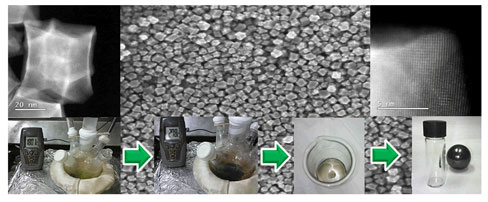
In a typical synthesis, 19.2 mM of Oleylamine (Oam, Sigma technical grade, 70%) was poured into a flask containing 2.8 mM of Oleic acid (Oac, Sigma technical grade, 90%); the mixture (≈90:10 mM%) was deoxygenated with a stream of N2, and magnetically stirred. Five syntheses were performed using a solution of 0.118 mM of Ni(NO3)2·6H2O (J.T. Baker, 98%) and 0.083 mM of H2PtCl6·6H2O (Sigma ≥ 37.50% Pt basis) dissolved in 400 µL of deionized water (Fig. S2b) and rapidly injected into the Oam/Oac mix at different temperatures: 180, 200, 250, 270, and 285 °C. Every reaction employed the same amounts of metallic precursors and Oam/Oac ratio compounds. After injection, the temperature was kept constant for a couple of minutes at the specified value, while water vapor was removed by vacuum. Then, the temperature was increased to 270–280 °C (except for cases with injection temperatures of 270 and 285 °C), resulting in a change of the mixture's color from pale green to dark-black (Fig. S3), indicating the beginning of the reduction process. The particles were collected by centrifugation, washed several times with a mixture of organic solvents, and finally dried by evaporation (Fig. S4a). Details on the synthesis and purification procedures can be found in Electronic Supplementary Information section SI-2; physical characterization details are found in section SI-3.
Results and discussions
A control synthesis was carried out by injection of the Ni–Pt precursor solution at an initial temperature of 180 °C (labeled HIT@180 °C) that was subsequently increased to observe the reaction's progress. One of the first observations was the formation of white fumes and violent flash-boiling in the flask caused by the mixture of organic and aqueous phases after the precursor solution was injected into the hot organic mix. This condition was present throughout the synthesis if the injected water from the precursor solution and the Oleylamine's moisture was not removed by reduced pressure through a vacuum. Different color changes in the mixture were observed as the temperature increased and reached the reduction point, around 270–280 °C (Fig. S3). Aliquots taken at different temperatures and times were analyzed by UV-vis spectroscopy (Fig. S5).
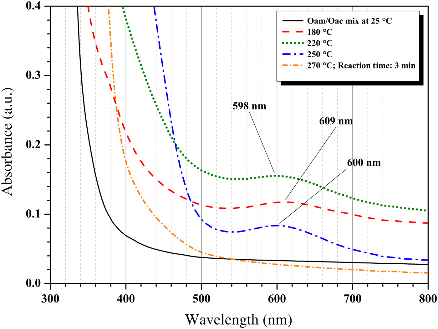
Figure 1 shows the UV–vis spectrum (300–800 nm) obtained with a band at 609 nm, which appears when the metallic precursors added by hot-injection into the Oam/Oac mixture dissolve at 180 °C. This band is evidence of a complex formation by metal–ligand interactions between the Oam/Oac molecules and the metallic precursors, as reported by previous studies.[ Reference Ahrenstorf, Heller, Kornowski, Broekaert and Weller 25 – Reference Long, Ohtaki, Uchida, Jalem, Hirata, Chien and Nogami 27 ] The band is slightly displaced to 598 nm and shows increased absorbance for the aliquot taken at about 220 °C, which revealed that the concentration of the metal–ligand species increases with temperature. For aliquot taken at about 250 °C, the band (600 nm) decreases in absorbance as evidence of the decrease in the concentration of the metal–ligand complex by the metal particle formation. This temperature corresponds with the gradual darkening in color from yellow to brown observed during the synthesis (Fig. S3). For aliquot taken at 270 °C after 3 min of reaction (reaction timer started, then the solution reached a dark-black color at about 270 °C), the band associated with the metal–ligand complex disappears. Thus, the evolution of the reaction can be followed by the increase/decrease of the absorbance values of the band associated with the metal–ligand complex that appears around 598–609 nm. Qualitatively, this is a measure of the degree of conversion of the metal complexes.[ Reference Ahrenstorf, Heller, Kornowski, Broekaert and Weller 25 ] UV–vis analysis of the pure Oam, Oac, and Oam/Oac organic compounds, as shown in Fig. S6, reveals two spectrum regions of interest, one without the presence of absorption bands (360–800 nm), and a lower wavelength region (200–360 nm) with maxima peaks. Maximum peaks for Oam appeared at 210, 212, 247 nm, and a shoulder at 281 nm; maximum peaks for Oac appeared at 212, 247, and 293 nm, with the latter being possibly associated with the n → π* transition in C=O. It is worth noting that the mixed Oam/Oac sample presents all the bands observed for pure compounds. Since the pure Oam, Oac, Oam/Oac organic compounds did not present bands between 500 and 800 nm, the bands observed in this region must be associated with the metal–ligand interaction shown in Fig. 1.
UV-vis spectra for aliquots taken at different temperatures from a control synthesis.

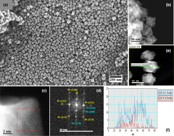
Figure 2a shows a scanning electron microscope (SEM) micrograph for Ni–Pt polyhedral nanoparticles obtained from synthesis HIT@180 °C with an average size of 27.4 ± 7.3 nm (see histogram of Fig. S7b). Figure 2b and S7-a show high-angle annular dark-field (HAADF) micrographs where Z-contrast imaging permits the visualization of bright regions on the edge and corners of the particles, which correspond to higher concentrations of Pt, this was confirmed by an energy-dispersive x-ray spectroscopy (EDS) line profile (Figs. 2e, f). Figure 2c shows a high-resolution HAADF image where a random arrangement of bright (Pt) and dark (Ni) atoms was observed, indicating the presence of a Ni–Pt alloy on the faces of the nanoparticle. Particles have a preferential cubic shape with faceting in different crystallographic orientations, as confirmed by the fast Fourier transform (FFT) image (Fig. 2d) corresponding to Ni–Pt-based polyhedral nanoparticles. Elemental characterization by SEM–EDS on a selected area of the sample revealed the presence of Ni: 72.0 wt.% and Pt: 28.0 wt.% (Figs. S7c,d). Powder samples of this material possess a glittering appearance (Fig. S4a) and paramagnetic behavior (Fig. S4b). This paramagnetic quality is thought to be caused by the presence of nickel, even though De la Presa et al., reported that for Au spherical nanoparticles capped with Oam/Oac, the covalent bond between the organic molecules and the superficial atoms of gold is what induces a ferromagnetic-like behavior.[ Reference de la Presa, Multigner, de la Venta, García and Ruiz-González 21 ] For Ni–Pt nanoparticles, deeper and more extensive studies are required to explain this magnetic phenomenon.
(a) SEM micrograph, (b) HAADF micrograph, (c) HAADF high-resolution micrograph, (d) Indexed FFT image, (e) HAADF micrograph with EDS line profile, and (f) Intensity profile corresponding for Ni–Pt polyhedral nanoparticles obtained from synthesis with precursors hot injected at 180 °C.

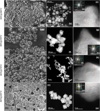
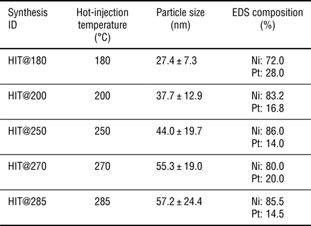
Research experience notes that in the implementation of the hot-injection method, metallic precursors are injected at 180 °C, but do not reduce until the temperature is increased to about 270–280 °C. According to UV–vis results, it was theorized that if the reduction process begins at 250 °C and the maximum reduction occurs at 270 °C, then there was an interval of 20 °C where the reduction was slow, an effect attributed to the gradual increase in temperature by the heating mantle used in the synthesis. A slow reduction process promotes slower nucleation of the early seeds, making the growth rate of those seeds dominant over the nucleation stage, and leading to higher polydispersity.[ Reference Chikan and Mclaurin 22 ] From this observation, four additional syntheses were performed where the hot-injection temperature was set at 200, 250, 270, and 280 °C to study the effect of this parameter on the size of the particles. It was theorized that a hot-injection temperature closer to the reduction temperature (about 270 °C) might promote a faster nucleation rate, minimizing the growth stage, and allowing for smaller nanoparticle sizes. Equal amounts of organic and metallic precursor mixtures were used in all syntheses. Table 1 shows hot-injection temperature, particle size, and elemental composition parameters for the syntheses performed. SEM micrographs revealed the formation of Ni–Pt polyhedral nanoparticles for all hot-injection temperatures under study, see Figure 3a for HIT@200 °C, Fig. 3e for HIT@250 °C, Fig. 3i for HIT@270 °C, and Fig. 3m for HIT@285 °C. SEM imaging shows differences in particle size distribution, corresponding to the different injection temperatures studied (Table 1). Therefore, this experiment proves that the increase of the hot-injection temperature promotes an increase in particle size, as opposed to the theorized outcome.
(a) SEM micrograph, (b) HAADF micrograph, (c) HAADF high-resolution micrograph and (d) indexed FFT images corresponding for Ni–Pt polyhedral nanoparticles obtained from synthesis with precursors hot injected at 200 °C; (e) SEM micrograph, (f) HAADF micrograph, (g) HAADF high-resolution micrograph and (h) indexed FFT images corresponding for Ni–Pt polyhedral nanoparticles obtained from synthesis with precursors hot injected at 250 °C; (i) SEM micrograph, (j) HAADF micrograph, (k) HAADF high-resolution micrograph and (l) indexed FFT images corresponding for Ni–Pt polyhedral nanoparticles obtained from synthesis with precursors hot injected at 270 °C; (m) SEM micrograph, (n) HAADF micrograph, (o) HAADF high-resolution micrograph and (p) indexed FFT images corresponding for Ni–Pt polyhedral nanoparticles obtained from synthesis with precursors hot injected at 285 °C.

Summary of all syntheses performed with corresponding specific values for hot-injection temperature (HIT) of the precursors, particle size distribution, and elemental composition by EDS.

The average size reported in Table 1 comes from the Gaussian fit of the distribution histograms for each synthesis (Figs. S8a, S9a, S10a, and S11a, respectively). However, all obtained materials contain particles whose size range from 100 to 550 nm, which corroborate a polydispersity grade between 37 and 44%. A possible explanation would be that the temperature decreases by approximately 10–20 °C after injection and takes a few minutes to be reestablished; thereby, promoting longer reaction times where the nucleation and growth stages occur simultaneously. The high temperature possibly causes the accelerated dissolution of atoms on the surface of the nanoparticles and their deposition on faces of low surface energy,[ Reference Xiong and Xia 28 , Reference Xia, Wang, Ruditskiy and Xia 29 ] i.e., a growth by an Ostwald ripening process.[ Reference Murray and Kagan 30 ] This leads to a decrease in the number of particles, but an increase in their size and polydispersity. Rapid heating through microwaves or magnetic induction can prevent this phenomenon.[ Reference Chikan and Mclaurin 22 ]
HAADF micrography (Figs. 3b, f, j, n), high-resolution HAADF micrography (Figs. 3c, g, k, o), and FFT imaging (Figs. 3d, h, l, p) facilitate the observation of particle polydispersity and revealed the formation of a Ni–Pt alloy, with superficial Pt concentration dependent on the injection temperature. As the temperature of the precursors increases, the Pt content on the surface decreases (in contrast with the synthesis HIT@180 °C). This was confirmed through EDS-line profiling for each material (Figs. S8d, S9d, S10d, and S11d, respectively). Moreover, Table 1 shows that the elemental composition obtained by EDS analysis (Figs. S8f, S9f, S10f and S11e) varies without a clear trend between 80–86 wt.% for Ni and 14–20 wt.% for Pt, which does not correspond to the Ni: 72 wt.% and Pt: 28 wt.% obtained for the material synthesized with a hot-injection temperature at 180 °C. Energetic differences in the formation of the Ni–ligand and/or Pt–ligand could explain that a specific time is needed for the formation of the metal-ligand complex. It is hypothesized that when the metallic precursors were injected at higher temperatures (closer to the reduction temperature), precursors may not get sufficient time to disperse in the bulk solution and form the metal–ligand complex in a homogeneous concentration. The local concentration of precursor causes faster nucleation of primitive seeds, which rapidly grow at different rates enabling the synthesis of materials with high polydispersity. In this experiment, the gradual increase of the hot-injection temperature from 180 to 220 °C provides enough time to obtain more homogeneous particles with the theoretically estimated metallic composition.
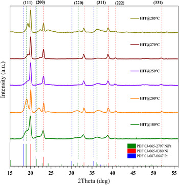
Finally, x-ray powder diffraction (XRD) measurements were used for qualitative identification purposes. Figure 4 shows the XRD patterns obtained for each material. For all synthesized materials, the co-existence of a Ni phase (PDF: 03-065-0380) and a Ni–Pt alloy phase (PDF: 03-065-2797) with a tetragonal crystal structure (P4/mmm) were identified. A Pt phase (PDF: 01-087-0647) with a face-centered cubic crystal system (Fm-3m) is shown as a reference. No peaks associated with organic impurities were found. An ascending analysis from the patterns in Fig. 4 reveals intense peaks (111), (200), and (220) associated with a majority Ni phase and to a Ni–Pt phase in lesser proportion for all materials. This is consistent with the composition found by EDS, where a higher proportion of the Ni phase was observed. The injection-temperature dependence is what enables the co-existence of particles with different sizes (high polydispersity) as was seen through HAADF. Polydispersity occurs due to parallel nucleation and growth stages, leading to the production of many types of particles, some with higher concentrations of Pt and with varied sizes; thus, there is no clear tendency in the ratio of associated phase peaks. The variation of the hot-injection temperature directly impacts on the crystallinity and particle size of the materials and their corresponding XRD patterns. For materials where the hot-injection temperature was increased, XRD patterns showed more intense and narrower peaks, corresponding to an increase in particle size and crystallinity, as confirmed through SEM and HAADF imaging.
XRD patterns corresponding for Ni–Pt polyhedral nanoparticles obtained from syntheses with precursors hot injected at 180, 200, 250, 270, and 285 °C.

Conclusions
Ni–Pt Polyhedral nanoparticles were synthesized through the hot-injection method using Oleylamine and Oleic acid as solvents, reducing, and stabilizing agents. It was theorized that an injection temperature closer to the reduction temperature (about 270 °C) promotes a faster nucleation rate, minimizing the growth stage, and allowing for smaller nanoparticle sizes. The effect of the injection temperature on the particle size distribution was studied by performing additional synthesis where the injection temperature of the metallic precursor was varied. Results revealed that more uniform particles (27.4 ± 7.3 nm) with an elemental composition of Ni: 72 wt.% and Pt: 28 wt.% were obtained using an injection temperature of 180 °C. HAADF electron micrographs confirmed higher concentrations of Pt on the edges and corners of the nanoparticles for the HIT@180 °C synthesis. EDS-line profile analysis showed that superficial Pt content decreases as the injection temperature rises; the corresponding variation in the Ni–Pt composition was found through SEM–EDS, and in this case, Ni concentration increased up to 80–86 wt.%. XRD revealed that all materials are associated with the co-existence of a majority Ni phase and to a Ni–Pt phase. High polydispersity can be explained as a consequence of the injection of both the effect of the injection temperature and reaction times, which promotes simultaneous nucleation and growth stages. It is hypothesized that when the metallic precursors were injected at higher temperatures (closer to the reduction temperature), precursors did not have sufficient time to disperse in the bulk solution to form the metal–ligand complex in a homogeneous concentration. Local concentrations of precursor cause faster nucleation of primitive seeds, which rapidly grow at different rates and enable the synthesis of materials with high polydispersity. Thus, it is evidenced that the hot-injection temperature influences the size properties of the nanoparticles.
Supplementary material
The supplementary material for this article can be found at https://doi.org/10.1557/mrc.2017.122
Acknowledgments
The authors gratefully acknowledge the financial support of the Mexican council for science, Consejo Nacional de Ciencia y Tecnología, CONACYT, (grant 245920) and the Laboratorio Avanzado de Nanoscopía Electrónica (LANE) at CINVESTAV for the technical support in the physical characterization of the nanomaterials. J.L. Reyes-Rodríguez gratefully acknowledges the support of Marco Antonio Leyva for XRD measurements and thanks CONACYT for the doctoral fellowship with registry number, 282205.