Introduction
Originally described by the prominent French physician Prosper Ménière in 1861, Ménière's disease is an inner-ear disorder that primarily affects balance and hearing. It is typically episodic in nature, classically characterised by ‘waves of attacks’. During attacks, cardinal symptoms may include: severe rotary vertigo, fluctuating hearing loss, aural fullness and rushing tinnitus. Furthermore, episodes are often preceded by an ‘aura’, or a specific set of warning symptoms, such as an abrupt increase in tinnitus, aural fullness or hearing loss. Ménière's disease can develop at any age, although it is more likely to occur in females aged 40–60 years. The natural history of Ménière's disease is variable in intensity and frequency, possibly evolving over a period of months to years.Reference Thorp and James 1 Bilaterality of the condition increases in direct conjunction with disease duration; up to 35 per cent of patients will have both ears affected within 10 years and up to 47 per cent of patients will have both ears affected within 20 years.Reference Huppert, Strupp and Brandt 2 Estimates of incidence range between 10 and 150 per 100 000 persons, and prevalence for Ménière's disease has been reported as 190 per 100 000 persons in the USA.Reference Alexander and Harris 3
The aetiology and pathophysiology of Ménière's disease remain controversial even after a century of investigation and research. Currently, the leading theory points toward an abnormal volume or composition of fluid within the inner ear, referred to as endolymphatic hydrops. Outside of post-mortem histological evaluation of temporal bones, there remains no definitive clinical test or procedure to ultimately establish the diagnosis of Ménière's disease. As a result, the diagnosis and treatment of inner-ear maladies have been heavily reliant upon the patient's clinical history and a thorough physical examination. Based on criteria published by the American Academy of Otolaryngology Head and Neck Surgery (AAO-HNS) in 1995, patients considered for Ménière's disease can be graded as having definite, probable or possible disease. Additionally, a complete audiological evaluation and complementary vestibular diagnostic measures can be helpful.
Magnetic resonance imaging (MRI) or computed tomography (CT) scans are often used as ancillary tests to exclude retrocochlear pathologies such as vestibulocochlear nerve tumours or multiple sclerosis, both of which could mimic Ménière's disease. Specifically, MRI of the inner ear has been performed with the use of high-strength magnets and various contrast agents in recent years.Reference Zou, Zhang, Poe, Zhang, Ramadan and Pyykkö 4 Continued advancement in magnetic field strength, spectral resolution, signal-to-noise ratio, contrast-to-noise ratio and image sequencing has allowed for the ultrastructural definition of anatomical structures on radiographical imaging.Reference Teranishi, Yoshida, Katayama, Hayashi, Otake and Nakata 5 These advances may improve the ability to investigate inner-ear diseases, where many pathological changes have previously eluded radiographical diagnosis.Reference Carfrae, Holtzman, Eames, Parnes and Lupinetti 6 This study aimed to elucidate MRI findings in relation to Ménière's disease, which include congenital malformations, white matter disease, posterior circulation abnormalities and temporal bone structural aberrations.
Materials and methods
Ethical considerations
The study was approved by the institutional review board at Pennsylvania State University, College of Medicine (STUDY00000161) on 26 February 2014. All procedures contributing to this work complied with the ethical standards of the relevant national and institutional guidelines on human experimentation, Penn State Hershey Medical Center Human Subjects Protection Office, and with the Helsinki Declaration of 1975, as revised in 2008.
Subject selection
This retrospective study reviewed a total of 220 ears from 113 patients seen between 1 January 2003 and 1 January 2014. Patients were examined by both the Division of Otolaryngology – Head and Neck Surgery and the Division of Neuroradiology at the Pennsylvania State University, College of Medicine, Hershey, USA.
A database query of the electronic medical records was performed on 27 February 2014, using the International Classification of Diseases (ninth revision) code 386.0x, to identify all patients diagnosed with possible, probable or definite Ménière's disease based on criteria published by the AAO-HNS in 1995. 7 Parameters analysed included: age, gender, ethnicity, associated co-morbidities and audiometry findings. Exclusionary criteria included: patients aged less than 18 years, lack of a corresponding MRI study, pregnant women, cognitively impaired adults and prisoners. Seventy-eight patients met the criteria for the case cohort.
A separate database query using Current Procedural Terminology code 70553 was also performed to identify control group subjects who underwent MRI of the brain (with and without contrast) for non-Ménière's disease related complaints. Data parameters paralleled those collected for the Ménière's disease group. Exclusionary criteria were identical to the Ménière's disease group, but additionally included: a history of hearing loss, stroke or intracranial neoplasms. From the abstracted imaging records, 35 patients were selected via random cluster sampling (by study completion year) and met criteria for the control group.
Magnetic resonance imaging protocol
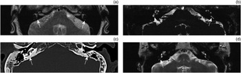
Magnetic resonance imaging of the brain with and without intravenous contrast was performed by utilising an internal auditory canal protocol on either a 1.5 T or 3 T system. The 3 T system replaced the 1.5 T system during the study period. A T2-weighted gradient echo sequence without contrast was performed to evaluate the inner-ear structures with following parameters: field of view = 200 mm, section thickness = 1.0 mm, repetition time = 7.68 ms, echo time = 3.45 ms, number of excitations = 1, and matrix 512 × 464 (Figure 1a).
(a) Normal vestibular aqueduct: axial, high-resolution, T2-weighted magnetic resonance imaging (MRI) scan at the level of inner-ear structures, showing normal T2-weighted hyperintense vestibular aqueducts (arrows) posterior to the semicircular canals. (b–d) Abnormal vestibular aqueducts (arrows) in a patient with bilateral symptoms: (b) axial, high-resolution, T2-weighted MRI scan showing non-visualisation of the vestibular aqueduct and endolymphatic duct bilaterally; (c) axial, high-resolution, temporal bone computed tomography scan confirming the finding of bilateral vestibular aqueduct stenosis; and (d) axial, high-resolution, T2-weighted MRI scan revealing a normal vestibular aqueduct and endolymphatic duct on the right (arrow), and non-visualisation of the left vestibular aqueduct.

Magnetic resonance imaging evaluation
All MRIs were analysed qualitatively by a single neuroradiologist who was blinded to the clinical information. Parameters analysed included: migrainous changes, vestibular aqueduct anomalies, Chiari malformations, basilar artery changes, vertebral artery changes, other posterior circulation changes, multiple sclerosis, dural venous sinus thrombosis, dural venous sinus dilation, dural venous sinus stenosis, jugular vein abnormalities, intracranial hypotension, posterior fossa hypoplasia, pseudotumour cerebri and age-related microvascular disease. Using T2-weighted axial images of the temporal bone, the size of the endolymphatic sac and vestibular aqueduct was evaluated using the current standard of less than 6 mm at the level of the external aperture.Reference Takeda, Sawada, Kakigi and Saito 8 The vestibule and semicircular canals were also assessed for secondary signs of endolymphatic hydrops.
Statistical analysis
A logistical regression analysis assessed odds ratios for all group comparisons. Statistical significance was defined as p < 0.05.
Results
Patient characteristics
A total of 78 patients with Ménière's disease underwent an ancillary MRI study. The average age (± standard deviation) of the cohort was 61.5 ± 12.9 years (range, 27–93 years), with a female-to-male ratio of 1.9:1. Audiometry revealed hearing loss in 60.0 per cent of ears, with slightly more laterality on the right than left.
Of the patients, 91.0 per cent were Caucasian, 5.1 per cent were Asian, 1.3 per cent were Hispanic, 1.3 per cent were African American, and 1.3 per cent were two or more races. These findings confirm a previously known ethnic bias in susceptibility to Ménière's disease. This disorder primarily remains a significant disease burden for Caucasians (83 per cent); it is sporadically prevalent in those of Asian ancestry and is extremely rare in individuals of African descent.Reference Ohmen, White, Li, Wang, Fisher and Zhang 9
The Ménière's group exhibited a wide array of medical co-morbidities; two disease processes are of notable interest. First, initial studies reported 3 per cent of Ménière's disease patients with a positive history for hyperthyroidism. Further research revealed a higher prevalence of association (17 per cent) between Ménière's disease and hypothyroidism. This link is presumed to be autoimmune in aetiology; sera from Ménière's disease patients have been shown to contain positive anti-thyroid microsomal antibody titres.Reference Evans, Baldwin, Bainbridge and Morrison 10 This corroborates our findings, where 15.4 per cent of Ménière's disease patients were found to have concurrent thyroid disease. Second, studies have revealed that 15 per cent of Ménière's disease patients have concomitant obstructive sleep apnoea. This is believed to occur because of vertebrobasilar insufficiency during sleep, which is postulated to cause hydropic distension of the endolymphatic system.Reference Nakayama and Kabaya 11 In our study, 9.0 per cent of Ménière's disease patients were also found to have a diagnosis of obstructive sleep apnoea.
Control group imaging findings
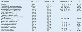
Twenty-five controls (71.4 per cent) underwent a 1.5 T MRI study, whereas 10 (28.6 per cent) underwent a 3 T MRI study. The vestibular aqueduct was clearly visualised bilaterally in 97.1 per cent of controls, with no observed cases of structural abnormalities. Additionally, 11.4 per cent of controls exhibited signs of multiple sclerosis, 11.4 per cent showed changes consistent with age-related microvascular disease and 5.7 per cent exhibited changes consistent with migraine (Table I).
Magnetic resonance imaging findings

*n = 78; † n = 35. ‡Statistical significance was defined as p < 0.05. MRI = magnetic resonance imaging; CI = confidence interval
Ménière's group imaging findings
Sixty-three Ménière's disease patients (80.8 per cent) underwent a 1.5 T MRI study, whereas 15 (19.2 per cent) underwent a 3 T MRI study. Given the high reported frequency of bilateral Ménière's disease, each ear was individually analysed on MRI for associated imaging findings.
There was clear visualisation of the left and right vestibular aqueducts in 38.7 per cent and 46.7 per cent of the Ménière's disease patients, respectively (left p = 0.0001, odds ratio = 0.02; right p = 0.0004, odds ratio = 0.03) (Figure 1b–d). Loss of vestibular aqueduct visualisation was found to correlate with Ménière's disease sidedness in 11.5 per cent of all cases.
The left and right vestibular aqueducts were of an abnormal size in 20.0 per cent and 5.9 per cent of Ménière's disease patients, respectively, albeit statistically significant on the left only (left, p = 0.0079, odds ratio = 10.91; right, p = 0.4925, odds ratio = 2.47). Specifically, seven patients were found to have a narrowed vestibular aqueduct and one patient was found to have an enlarged vestibular aqueduct. Of these eight patients, the enlarged vestibular aqueduct of 25.0 per cent was found to correlate with Ménière's disease sidedness.
Additionally, 18.0 per cent of patients exhibited signs consistent with microvascular disease, 10.3 per cent showed changes consistent with migraine, 3.6 per cent exhibited signs of jugular vein abnormalities, 2.6 per cent had signs of dural venous sinus stenosis, 2.6 per cent showed signs of spontaneous intracranial hypotension and 1.3 per cent exhibited signs of pseudotumour cerebri (Table I).
Discussion
The inner ear, which is housed in dense bone and subdivided into different fluid-filled compartments, often makes routine diagnostic imaging challenging. Technical improvements and high-resolution imaging are providing detailed depictions of membranous structures inside the temporal bone.
Impaired function of both the endolymphatic duct and sac is thought to play a key role in the pathogenesis of endolymphatic hydrops.Reference Kimura 12 Current research has shown that a narrowed endolymphatic duct, an obstructed endolymphatic sac and direct visualisation of endolymphatic hydrops are consistently observed on MRI of patients with Ménière's disease.Reference Muzzi, Rinaldo and Ferlito 13 Additional findings which may be central in the aetiopathogenesis of Ménière's disease are outlined below.
Migraine
The clinical association between vestibular symptoms and migraine has gained increasing recognition during the last two decades.Reference von Brevern and Neuhauser 14 The incidence of migraine in patients with Ménière's disease is estimated at 4.5 per cent, slightly higher than the overall US incidence of 3.8 per cent.Reference Gopen, Viirre and Anderson 15 Our study revealed eight Ménière's disease patients (10.3 per cent) who had MRI findings consistent with migraine (multiple, small, deep white matter changes, or small white matter changes periventricular in location) (Table I).Reference Swartz and Kern 16
Vestibular aqueduct
Histopathological studies of temporal bones confirm that small, tube-like vestibular aqueducts are more prevalent in Ménière's disease, and that there is an association between the size of the vestibular aqueducts and the intraosseous portion of the endolymphatic sac.Reference Yamamoto, Mizukami, Isono, Ohmura and Hirono 17 In this study, 38.7 per cent of the left and 46.7 per cent of right vestibular aqueducts were identified and visualised in Ménière's disease patients. We have included a CT scan from one patient for illustrative purposes (Figure 1c). Both the left and right vestibular aqueducts were of abnormal size, albeit significant on the left side only (Table I). However, of the 38 patients in the Ménière's disease group in whom vestibular aqueducts were visualised, 8 had a vestibular aqueduct of abnormal size; this accurately correlated to Ménière's disease sidedness in 25.0 per cent of patients. These findings are consistent with those reported previously by Welling et al., where 29 per cent of Ménière's disease patients were found to have a visible vestibular aqueduct.Reference Welling, Clarkson, Miles, Schmalbrock, Williams and Chakeres 18
Chiari malformations and posterior fossa hypoplasia
Many patients with cerebrospinal fluid (CSF) abnormalities as seen in Chiari malformations have a Ménière's disease like syndrome of aural fullness, dizziness, disequilibrium, tinnitus and hearing loss. These changes are the result of a significant reduction of CSF volume in the posterior cranial fossa; newly formed CSF is displaced from the compressed subarachnoid spaces into available spaces within the supratentorial and spinal compartments.Reference Milhorat and Milhorat 19 Recently, Guerra Jiménez et al. reported a series of nine patients with Chiari malformations who presented with Ménière's disease like symptoms.Reference Guerra Jiménez, Mazón Gutiérrez, Marco de Lucas, Valle San Román, Martín Laez and Morales Angulo 20 This study revealed no instances of Chiari malformations in either study group (Table I).
Arterial abnormalities
Arterial anomalies including basilar artery dolichoectasia and thrombosis of either the vertebral or basilar artery have been reported to cause acute disturbances in hearing and/or vestibular function.Reference Otterstedde, Tischendorf and Reisser 21 This study observed no instances of posterior arterial abnormalities in either group (Table I).
Multiple sclerosis
Vertigo, at times mimicking Ménière's disease, is a presenting symptom in less than 10 per cent of patients with multiple sclerosis; interestingly, one-third of patients will go on to develop vertigo during their disease course.Reference Bovo, Ciorba and Martini 22 Patients may also present with hearing loss in cases with brainstem involvement. The diagnosis of multiple sclerosis is made based on MRI findings such as periventricular demyelinating plaques (Dawson's fingers) and lesions involving the corpus callosum.Reference Ge 23 This study observed no changes consistent with multiple sclerosis in the Ménière's disease group (Table I).
Venous abnormalities
Impaired venous outflow, also known as chronic cerebrospinal venous insufficiency, is a syndrome characterised by stenosis of the internal jugular or azygos vein, with disturbed flow, insufficient drainage and opening of collateral venous channels. Filipo et al. analysed the presence of chronic cerebrospinal venous insufficiency in Ménière's disease, noting significant intracranial venous reflux and internal jugular vein stenosis with haemodynamic changes on echo-colour Doppler.Reference Filipo, Ciciarello, Attanasio, Mancini, Covelli and Agati 24 Furthermore, a magnetic resonance venography study by Di Berardino et al. revealed cerebrocervical drainage abnormalities in 83.3 per cent of patients with Ménière's disease.Reference Di Berardino, Alpini, Bavera, Cecconi, Farabola and Mattei 25 In our study, two Ménière's disease patients (2.6 per cent) had signs consistent with dural venous sinus stenosis and three patients (3.6 per cent) had signs consistent with jugular vein abnormalities (Table I). Venous abnormalities likely play an important role in the development of inner-ear anomalies and this arena is still under investigation.
Spontaneous intracranial hypotension
The hydromechanical hypothesis, described by Carlborg et al., states that when CSF pressure falls, the perilymphatic pressure falls in parallel, inducing a subsequent state of endolymphatic hydrops in patients with spontaneous intracranial hypotension. These changes have been found to be clinically indistinguishable; the signs and symptoms can completely overlap and perfectly mimic Ménière's disease.Reference Carlborg and Farmer 26 On MRI fluid-attenuated inversion recovery (FLAIR) sequences, changes consistent with spontaneous intracranial hypotension include diffuse pachymeningeal enhancement and gadolinium diffusion into the subdural space.Reference George, Rathore, Pandian and Singh 27 In our study, two Ménière's disease patients (2.6 per cent) exhibited imaging findings consistent with spontaneous intracranial hypotension (Table I).
Pseudotumour cerebri
Patients with a history of pseudotumour cerebri can present with symptoms of tinnitus, aural fullness, hearing loss and vertigo.Reference Murphy 28 Given the considerable overlap of symptoms with Ménière's disease, ancillary studies such as CT or MRI are often performed; appreciable findings may include an empty sella (50 per cent), small ventricles or flattening of the posterior sclera.Reference Wakerley, Tan and Ting 29 In our study, one Ménière's disease patient (1.3 per cent) exhibited imaging characteristics consistent with pseudotumour cerebri (Table I).
Microvascular disease
In a study on the co-morbidities of vertiginous diseases, conducted by Warninghoff et al., the prevalence rates of hypertension and diabetes amongst Ménière's disease patients were reported as 63.6 per cent and 18.2 per cent, respectively.Reference Warninghoff, Bayer, Ferrari and Straube 30 This study observed 14 Ménière's disease patients (18.0 per cent) with age-related microvascular disease (Table I).
-
• High-resolution magnetic resonance imaging (MRI) is key in diagnosing Ménière's disease and helps rule out retrocochlear disease
-
• A number of MRI findings are associated with Ménière's disease
-
• Loss of vestibular aqueduct visualisation correlated with Ménière's disease sidedness in 11.5 per cent of cases
-
• The left vestibular aqueduct was of abnormal size in 20 per cent of Ménière's disease patients
-
• As imaging techniques advance, vestibular aqueduct and endolymphatic sac features may provide new diagnostic information
-
• The study suggests that MRI could be a valuable diagnostic tool during initial care and investigation of potential Ménière's disease
Limitations
Over the study period, both the MRI and imaging protocol underwent technological improvements to enhance the quality and resolution of radiographical images (i.e.1.5 T vs 3 T). This could have affected how well changes in the vestibular aqueduct correlated with Ménière's disease sidedness. Furthermore, although this investigation had a moderate sample size, both the power and significance of this study could be enhanced with a larger patient population. This is particularly pertinent in the analysis of brain MRIs, where some rarer conditions identified in this study are challenging to capture in the general population. Discerning the category of Ménière's disease could have also provided interesting data in order to determine the likelihood of true Ménière's disease when including detailed MRI findings; however, this was not possible because of limited patient data.
Conclusion
Magnetic resonance imaging evaluation has proved worthwhile, as it aids the exclusion of other cerebrocervical disease states and pathologies that mimic Ménière's disease. Further research with advanced imaging techniques is needed to discriminate changes observed in the vestibular aqueduct and endolymphatic sac; unique features of these structures could provide enhanced diagnostic information. Our study reveals that MRI could be a valuable diagnostic tool during the initial care and investigation of patients presenting with signs and symptoms consistent with Ménière's disease.