INTRODUCTION
Infection with the protozoan parasite Leishmania can lead to 3 different forms of disease: cutaneous, mucocutaneous, and visceral leishmaniasis. Visceral leishmaniasis (VL), also called Kala-azar, is the most severe form of leishmaniasis caused by species of Leishmania (L.) donovani complex. The disease can appear either symptomatically or asymptomatically and symptomatic VL is usually fatal if left untreated (D'Oliveira et al. Reference D'Oliveira, Costa, Barbosa, Orge and Carvalho1997). VL is caused by L. donovani in the Indian subcontinent and eastern Africa, and 2 closely related species, L. infantum and L. chagasi, are the causative agents of VL in the Mediterranean area and Latin America, respectively. VL affects more than 2 million people in 47 countries with an estimated 500 000 new cases annually worldwide (Guerin et al. Reference Guerin, Olliaro, Sundar, Boelart, Croft, Desjeux, Wasunna and Bryceson2002). Zoonotic VL is endemic in many Mediterranean basin countries including Iran, where the causative agent is L. infantum (Mohebali et al. Reference Mohebali, Taran and Zarei2004). There is no efficient vaccine available against VL and development of rapid and accurate diagnostic methods is a research priority for disease control.
Diagnosis of VL is made classically by demonstration of Leishmania amastigotes in aspirates from lymph nodes, bone marrow or spleen (Burns et al. Reference Burns, Shreffler, Benson, Ghalib, Badaro and Reed1993). However, the technique is invasive, needs skilled personnel and equipped hospitals and is limited by low sensitivity (Sundar and Rai, Reference Sundar and Rai2002). Anti-leishmanial antibody titres are typically high during the acute disease, and this feature has been exploited for the serodiagnosis of VL using different methods (Bogdan et al. Reference Bogdan, Stosiek, Fuchs, Rollinghoff and Solbach1990; Millesimo et al. Reference Millesimo, Zucca, Caramello and Savoia1996; Kumar et al. Reference Kumar, Pai, Pathak and Sundar2001). The direct agglutination test (DAT) is popular in most countries (Zijlstra et al. Reference Zijlstra, Ali, el-Hassan, el-Toum, Satti, Ghalib and Kager1991) but variation between batches of antigen and cross-reaction with some other infectious agents are limiting factors. Furthermore, the test may remain positive for several years after cure and does not distinguish between past and present infections (Sundar and Rai, Reference Sundar and Rai2002). Different recombinant antigens have been attempted for use in serodiagnosis of VL (Carvalho et al. Reference Carvalho, Charest, Tavares, Matlashewski, Valente, Rabello, Gazzinelli and Fernandes2002; Maalej et al. Reference Maalej, Chenik, Louzir, Ben Salah, Bahloul, Amri and Dellagi2003; Rosário et al. Reference Rosário, Genaro, Franca-Silva, da Costa, Mayrink, Reis and Carneiro2005) among them; rK39 antigen has shown promising results in most studies (Burns et al. Reference Burns, Shreffler, Benson, Ghalib, Badaro and Reed1993; Rosati et al. Reference Rosati, Ortoffi, Profiti, Mannelli, Mignone, Bollo and Gradoni2003). The k39 gene has been cloned from L. chagasi (Burns et al. Reference Burns, Shreffler, Benson, Ghalib, Badaro and Reed1993), the causative agent of American VL and rK39 protein has been used to develop ELISA and rapid immunochromatographic tests for diagnosis of VL. Although the application of L. chagasi rK39 protein for serodiagnosis of New World VL produced reliable results (Bern et al. Reference Bern, Jha, Joshi, Thakur and Bista2000; Sundar et al. Reference Sundar, Pai, Sahu, Kumar and Murray2002), its use for diagnosis of VL caused by L. infantum and L. donovani in Old World VL infections has shown variable results and the sensitivity and specificity of the test were not as high as reported for diagnosis of New world VL (Jelinek et al. Reference Jelinek, Eichenlaub and Löscher1999; Sivakumar et al. Reference Sivakumar, Sharma, Chang and Singh2006). Recently, another promising diagnostic antigen, known as K26 antigen, identified and characterized from Leishmania chagasi (Bhatia et al. Reference Bhatia, Daifalla, Jen, Badaro, Reed and Skeiky1999), showed that rK26 ELISA has greater sensitivity than rK39, especially for detection of VL in the early stage of infection (Sundar et al. Reference Sundar, Pai, Sahu, Kumar and Murray2002; Rosati et al. Reference Rosati, Ortoffi, Profiti, Mannelli, Mignone, Bollo and Gradoni2003). However, there are no available data about K26 antigen in other VL agents, including L. infantum.
The objectives of this study were to characterize the k26 gene of L. infantum and to prepare and evaluate the performance of rK26 ELISA for serodiagnosis of VL caused by L. infantum in endemic areas of Iran.
MATERIALS AND METHODS
Study groups
The study was conducted from January 2005 through March 2007 at the Tabriz University of Medical Sciences, Iran. The study was reviewed and approved by the Ethical Committee of the University and signed, informed consent was obtained from all participants. Three different groups of sera were screened in this study. Group A: sera from 93 parasitologically confirmed VL cases; group B: sera from 100 healthy controls, and group C: sera from patients with tuberculosis (n=10), brucellosis (n=10) and toxoplasmosis (n=10). All patients were from East Azerbaijan and Ardabil provinces in the northwest of Iran that are endemic areas for VL. Among the VL patients, 82 cases (88·2%) belonged to children under 5 years of age, 9 cases (9·6%) to age group 5–10 years and 2 cases (2·1%) to adults. Forty out of 93 cases (43%) were females and 53 cases (57%) were males. The healthy control group consisted of age-matched subjects without any history of Kala-azar. Two ml of peripheral blood was taken from each subject, and sera were isolated and stored at −20°C until used.
Parasite
L. infantum LON49, isolated from a patient with VL, was maintained by serial passages in Syrian hamsters. The promastigotes were cultured at 26°C in RPMI-1640 medium with glutamine (Gibco BRL, Maryland, USA) supplemented with 10% heat-inactivated fetal calf serum (Sigma-Aldrich, Dorset, UK). Organisms were harvested in the logarithmic phase, washed with phosphate-buffered saline (PBS), pH 7·2 and used for DNA isolation.
Preparation of DNA
Genomic DNA was isolated as described previously (Farajnia et al. Reference Farajnia, Alimohammadian, Reiner, Karimi, Ajdari and Mahboudi2004). Briefly, logarithmic-phase promastigotes were disrupted in lysis buffer (50 mm NaCl, 50 mm EDTA, 1% SDS, 50 mm Tris-HCl, PH 8·0) and incubated overnight with proteinase K (100 μg/ml, Sigma-Aldrich, Dorset, UK) at 37°C. DNA was purified further by phenol-chloroform extraction and ethanol precipitation.
PCR amplification of L. infantum k26 gene
The k26 open reading frame of L. infantum was amplified by PCR using oligonucleotide primers, forward, F1, 5′-CGGGATCCATGGGAGCCTCTGCACGAAG-3′ which contained a BamHI site and reverse, R1, 5′-TTAAGCTTCGTTCCCGGCAACCTGCTC -3′ which contained a HindIII site. Primers were designed based on k26 gene sequences reported from L. chagasi (GeneBank Accession number AF131228). An Eppendorf DNA thermal cycler and a proof reading DNA polymerase, pfu (Fermentas, Lithuania) were used for PCR amplification of desired fragments. The reaction mixture included 0·4 μm of each primer, 200 μm dNTPs and 2 mm MgCl2. PCR conditions were: primary denaturation at 94°C for 4 min, followed by 30 cycles of denaturation at 94°C for 1 min, annealing at 65°C for 1 min, extension at 72°C for 1 min, and a final extension for 5 min.
Cloning of PCR products and DNA sequencing
The PCR product was purified and blunt-end ligated into the EcoRV site of the Bluescript vector which resulted in the plasmid clone pBsc-Li-k26. The plasmid was transformed into E. coli, TOP10f competent cells and 3 positive clones containing inserts were submitted for sequencing with M13 forward and reverse primers. Nucleotide and predicted protein sequences were compared to data available by the BLAST search method.
Expression and purification of recombinant Li-K26
The coding region for the L. infantum k26 gene was isolated by digestion of pBsc-Li-k26 clone with BamHI-HindIII restriction enzymes and subcloned into the BamHI-HindIII site of the PQE 80L vector, yielding the plasmid subclone PQ-Li-k26. Sequencing with vector-based primers demonstrated that the His-tag fusion was in frame. The expression construct was transformed into E. coli DH5a strain, cultured at 37°C in LB medium containing 100 μg ml−1 ampicillin to an absorbance at 600 nm (A 600) of 0·5 unit. Protein expression was induced by adding isopropyl-β-d-thiogalactopyranoside (IPTG) to a final concentration of 1 mm and analysed by SDS-PAGE.
For purification of recombinant protein, 500 ml LB media containing 100 μg ml−1 ampicillin was inoculated with 5 ml of an overnight culture of recombinant bacteria and incubated at 37°C with vigorous shaking (200 rpm). Cultures were induced at mid-log phase by adding 1 mm IPTG and growth was continued for another 4 h. Bacteria were harvested (10 000 g, for 10 min), resuspended in 8 ml of lysis buffer A (50 mm NaH2PO4, pH8, 300 mm NaCl, 10 mm imidazole, 1 mg/ml lysozyme) and disrupted by sonication (5×45 sec pulses interrupted with cooling on ice). Soluble and insoluble fractions were separated by centrifugation at 12 000 g for 15 min at 4°C and the recombinant protein was found to be mainly in the soluble fraction.
Briefly, 8 ml of Ni-NTA resin equilibrated with lysis buffer was added to the soluble fraction of E. coli lysate and binding was conducted with constant mixing for 1 h at 4°C. The mixture was then passed over a gravity column, washed with buffer B (50 mm NaH2PO4, pH8, 300 mm NaCl) containing 20 and 50 mm imidazole and bound material was eluted with buffer B containing 500 mm imidazole. The purified recombinant protein was dialysed against 3 changes of PBS, analysed by SDS-PAGE (Towbin et al. Reference Towbin, Staehelin and Gordon1979) and stored at −20°C. The purified recombinant protein was shown by SDS-PAGE analysis to be free of significant amounts of E. coli protein.
Preparation of promastigote crude antigen for ELISA
The stationary-phase promastigotes of L. infantum harvested after the third passage were washed 3 times in ice-cold PBS (pH 7·2) and suspended in 10 mm Tris-HCl buffer (pH 7·5) at a concentration of 1×10⁷/ml. The suspension was sonicated 6 times for 30 sec each time, with 30 sec intervals on ice and stored in aliquots at −70°C until use. Total protein was measured by the Lowry method (Lowry et al. Reference Lowry, Roseborough, Farr and Randall1951).
ELISA assay
The ELISA assay was carried out according to the standard method (Malla et al. Reference Malla, Sengupta, Dubey, Sud, Ansari and Salotra2003) with some modification. Briefly, the optimum concentration of rK26 and whole cell antigens and serum dilutions were determined by a checkerboard titration method. ELISA plates (Nunc, Maxisorb) were coated overnight at 4°C with 100 ng of rK26 or 500 ng of promastigote lysate in 100 μl of 0·1 m bicarbonate buffer (pH 9·0). The wells were blocked with 3% bovine serum albumin at 4°C overnight, washed 3 times with PBS containing 0·05% Tween 20 (PBS-T), and incubated for 1 h at 37°C with 100 μl of patients' sera at a 1/200 dilution. Wells were washed 3 times with the same buffer and incubated with 100 μl of rabbit anti-human IgG conjugated with horseradish peroxidase for 1 h. This step was followed by 3 rinses with PBS-T and developed using tetramethylbenzidine (Sigma) as substrate. The reaction was stopped with 50 μl of 3 n H2SO4 and the optical density (OD) of each well was measured at 450 nm in an ELISA reader. The cut-off values for each antigen were calculated by adding 3 s.d. values to the mean absorbance of control sera. Each sample was assayed in triplicate, along with appropriate controls. A sample was considered positive if its mean OD value was greater than the cut-off value.
Direct Agglutination Test (DAT)
The DAT was performed essentially as described previously (el Harith et al. Reference el Harith, Kolk, Leeuwenburg, Muigai, Huigen, Jelsma and Kager1998). Two-fold serial dilutions of sera were made in physiological saline (0·9% NaCl) containing 0·2% gelatin and 0·78% 2-mercaptoethanol, starting with a dilution of 1:50 and going up to 1:102 400. Then 50 μl of DAT antigen were added to each well containing 50 μl of diluted serum in a V-shaped microtitre plate, and the results were read after 20 h of incubation at 25°C. An antibody titre of 1:3200 and above was considered positive.
Statistical analysis
The sensitivity and specificity of tests were determined as follows: Sensitivity=TP/(TP+FN)×100 and Specificity=TN/(TN+FP)×100, Positive Predictive Value=TP/(TP+FP)×100, Negative Predictive Value=TN/(TN+FN)×100 and Accuracy=TP+FP/(TP+FP+TN+FN)×100. TN represents true negative, TP true positive, FN false negative and FP false positive. The sensitivity of tests was assessed using sera from VL patients (n=93). Sera of healthy controls (n=100) and sera from patients with other infectious diseases (n=30) were used to determine the specificity of the tests. The degree of agreement between rK26 ELISA, DAT and whole-cell ELISA was determined by calculating Kappa (κ) values with 95% confidence intervals (Altman, Reference Altman1991). Statistical significance of sensitivity and specificity between tests was evaluated using the Chi-Square test. To estimate the reliability of tests, a correlation analysis was carried out using the Cronbach Coefficient Alpha.
RESULTS
Amplification and sequencing of L. infantum k26 gene
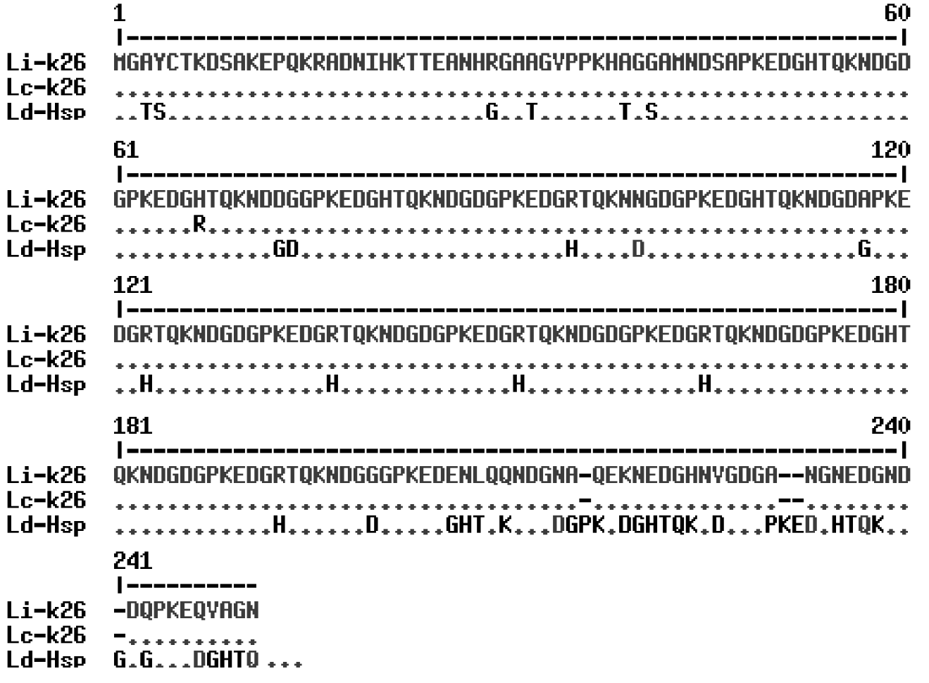
PCR amplification of the coding region for L. infantum k26 gene resulted in a 750 bp product. The PCR product was purified and cloned on Bluescript vector and sequenced. The nucleotide sequence was submitted to the GeneBank/NCBI Data Base under Accession number DQ192034. The nucleotide sequence of the L. infantum k26 gene was found to contain a single ORF of 745 bp long encoding a protein of 247 amino acid residues. Comparison of the predicted amino acid sequence of L. infantum K26 protein with the sequences in the database using BLAST program revealed that L. infantum k26 gene has 99% homology to L. chagasi k26 sequence, whereas its homology to the related gene from L. donovani is only 85% (Fig. 1).
Comparison of the amino acid sequence of the Leishmania infantum K26 antigen (Li-k26) with those from L. chagasi (Lc-k26) and L. donovani (Ld-Hsp). Alignment of the amino acid sequences was performed using the MultAlign program. Dots correspond to identical amino acids.

Expression and purification of L. infantum rK26 protein
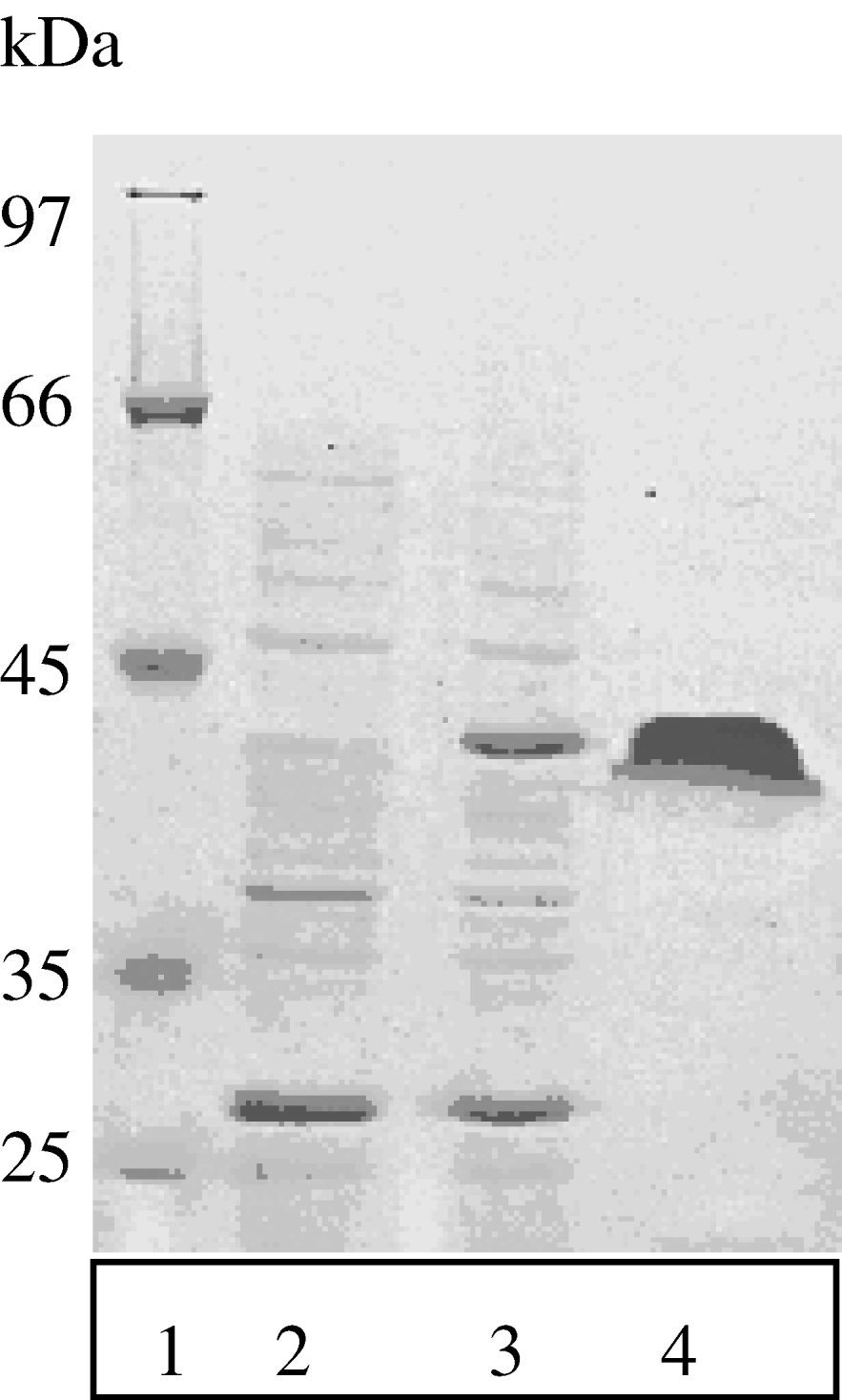
The insert containing the complete ORF of L. infantum k26 gene was subcloned into the PQE 80L (Qiagen) expression vector in frame with an amino-terminal His-tag fusion for affinity purification. Induction with IPTG of E. coli DH5a transformed with expression cassettes resulted in a highly soluble recombinant protein as demonstrated by SDS-PAGE analysis of a lysate of induced bacteria (Fig. 2). The recombinant protein was purified from soluble fractions of E. coli lysate by affinity chromatography over a Ni-NTA agarose matrix with a yield ranging from 200 to 300 mg of purified protein per litre of induced culture.
Expression and purification of Leishmania infantum rK26 protein. Lane 1, size marker; Lane 2, lysate of E. coli containing pQE-Li k26, before induction with IPTG; Lane 3, lysate of E. coli containing pQE-Li k26, after induction with IPTG; Lane 4, purified rK26.

Evaluation of reactivity of VL patients' sera with L. infantum rK26 antigen
To determine the serodiagnostic performance of L. infantum rK26 antigen, the sensitivity and specificity of rLi-K26 ELISA were analysed and compared to the sensitivity and specificity of whole-cell ELISA and DAT. A total of 93 confirmed VL sera along with 100 healthy control samples and 30 samples from patients with other cross-reactive infectious diseases were tested.
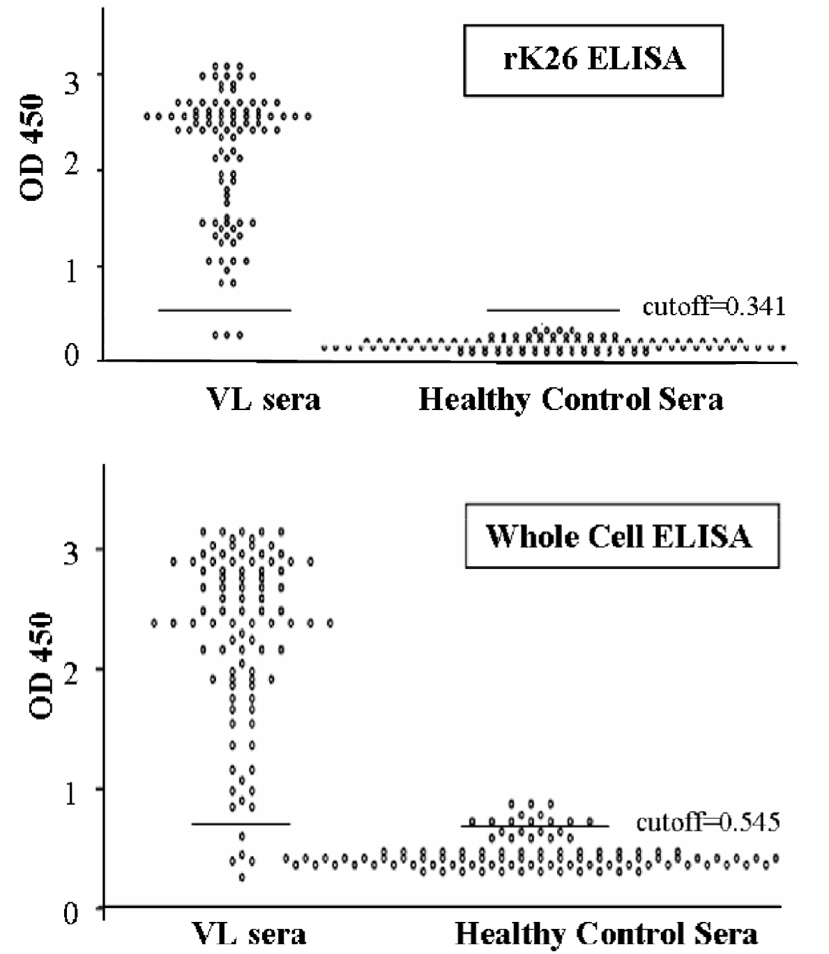
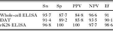
As shown in Tables 1 and 2, the sensitivity of rK26 ELISA (90/93, 96·8%) was higher than whole-cell ELISA (89/93, 95·7%) and especially DAT (85/93, 91·4%), but differences were not statistically significant (P>0·05). Analysis of performance of the 3 tests for serodiagnosis of VL indicated the presence of a good agreement between rK26 ELISA and whole cell ELISA (Κ=0·739, P=0·000), Whole-cell ELISA and DAT (Κ=0·753, P=0·000) and a moderate agreement between rK26 ELISA and DAT (Κ=0·526, P=0·000). However, significant differences were observed in comparing specificity of rK26 ELISA with specificity of whole-cell ELISA (P=0·012) and DAT (P=0·016). rK26 ELISA values were negative in all 130 non-VL cases (specificity=100%), whereas whole-cell ELISA was negative in 114 cases (specificity=87·7%) and DAT was negative in 116 cases (specificity=89·2%). The optical density (OD) distribution of rK26 and whole-cell ELISAs in visceral leishmaniasis patients and healthy control subjects are shown in Fig. 3.
The optical density distribution of rK26 and whole-cell ELISAs in visceral leishmaniasis patients and healthy control subjects. The cut-off value was defined as the mean optical density plus three standard deviations for the values obtained from sera of healthy control subjects.

Reactivity of sera from symptomatic VL patients, healthy controls and patients with other infectious diseases with different antigen preparations
(The results indicate that compared to DAT and whole-cell ELISA, the rK26 ELISA prevents false positive and false negative reactions.)

VL, Visceral Leishmaniasis; DAT, Direct Agglutination Test.
The percentage sensitivity, specificity, positive predictive value, and negative predictive value of DAT, whole-cell ELISA and rK26 ELISA tests for serodiagnosis of symptomatic VL

Sn, Sensitivity; Sp, Specificity; PPV, Positive predictive value; NPV, Negative predictive value; Ef, Efficiency.
Analysis of reliability of tests was carried out by replicate assay of serum samples with each antigen. rK26 ELISA displayed excellent reliability (alpha=0·953) followed by whole cell ELISA (alpha=0·888) and DAT (alpha=0·872) indicating high reproducibility of all assays.
DISCUSSION
The diagnosis of VL is complex because its clinical features are shared by a host of other commonly occurring diseases, such as cutaneous leishmaniasis, malaria, tuberculosis and toxoplasmosis, some of which can be present along with VL in tropical and subtropical regions. Thus, the laboratory diagnosis plays a critical role in the confirmation of clinical diagnosis of VL and in the discrimination of VL from other infectious diseases. The currently used method of laboratory diagnosis is based on the finding of parasites in bone marrow biopsies or in spleen aspirations. These are invasive methods that are not readily applicable in most rural settings. Furthermore, these methods suffer from low sensitivity and fail to detect numerous cases (Chappuis et al. Reference Chappuis, Rijal, Soto, Menten and Boelaert2006). Serological methods have proven to be useful non-invasive methods that could have epidemiological applications for determining carrier rates among asymptomatic subjects and to map out the geographical distribution of the VL (Kar, Reference Kar1995). However, the performance of these methods is highly dependent on the type, source, and purity of the antigens employed. In our study, DAT was positive for only 91·4% of confirmed VL cases, whereas promastigote whole-cell ELISA produced positive results with 95·7% of VL sera. This finding is in agreement with the results of Pal et al. (Reference Pal, Mukerji, Basu, Naskar, Mallick and Ghosh1991) and indicates that ELISA is more sensitive than DAT for diagnosing visceral leishmaniasis. However, numerous false-positive reactions were observed with both methods. DAT was positive with 11 of 100 healthy sera, 2 of 10 tuberculosis sera and 1 of 10 toxoplasmosis sera, with overall specificity of 89·2%. Whole-cell ELISA was positive with 12 of 100 healthy sera, 2 of 10 tuberculosis sera and 1 of 10 toxoplasmosis sera, with overall specificity of 87·7%. This observation is in agreement with several reports (Choudhry et al. Reference Choudhry, Guru, Saxena, Tandon and Saxena1990; Singh et al. Reference Singh, Gilman-Sachs, Chang and Reed1995; Kumar et al. Reference Kumar, Pai, Pathak and Sundar2001) and confirms that application of whole-cell Leishmania antigens in diagnostic tests can result in false-positive results due to the non-specific cross-reactivity. When ELISA was performed using rK26 antigen, 96·8% of sera from confirmed VL cases were positive. Sera from healthy controls and non-VL patients did not show any reaction with rK26 ELISA and the specificity of the test was 100%. These results are consistent with previous studies (da Costa et al. Reference da Costa, Franca, Mayrink, Nascimento, Genaro and Campos-Neto2003; Rosati et al. Reference Rosati, Ortoffi, Profiti, Mannelli, Mignone, Bollo and Gradoni2003) and show that K26 protein is a highly antigenic protein that is specific to VL agents. Very recently, Sundar et al. (Reference Sundar, Singh, Bimal, Gidwani, Mishra, Maurya, Singh, Manandhar, Boelaert and Rai2007) reported a high specificity but low sensitivity for a rapid immunochromatographic test prepared from L. chagasi rK26 for diagnosis of VL in the Indian subcontinent. However, this report is in disagreement with our findings and several other reports from New World geographical regions (Bhatia et al. Reference Bhatia, Daifalla, Jen, Badaro, Reed and Skeiky1999; da Costa et al. Reference da Costa, Franca, Mayrink, Nascimento, Genaro and Campos-Neto2003; Rosário et al. Reference Rosário, Genaro, Franca-Silva, da Costa, Mayrink, Reis and Carneiro2005). The reason for such controversy is unclear but low sensitivity of the rapid immunochromatographic method used and differences in the K26 antigen of VL agents in these geographical regions with that of L. chagasi might be considered as the possible causes. Our result on sequencing of L. infantum k26 gene indicates that K26 antigen is highly similar in L. infantum and L. chagasi (99% identity), whereas the homology of L. infantum K26 antigen with that of L. donovani (which is the causative agent of VL in India) is only 85%. These results suggest that the antigen preparation used for the serodiagnosis of VL should preferably be of local origin.
In conclusion, the results of this study showed a high prevalence of antibody against K26 antigen in Old World VL patients that could be used for diagnosis in regions where L. infantum is the causative agent of VL.
This investigation received financial support from joint WHO Eastern Mediterranean Region (EMRO), Division of Communicable Disease (DCD) and the WHO Special Program for Research and Training in Tropical Disease (TDR): The EMRO/TDR Small Grants Scheme for Operational Research in Tropical and other Communicable Disease.