A fistula connecting the right pulmonary artery – exceptionally the left pulmonary artery – to the left atrium is an uncommon pathology; a report about this was published for the first time by Friedlich et alReference Friedlich, Bing and Blount1 in 1950 regarding a patient with this pathology who was operated on by Alfred Blalock. By 2005, only 59 cases had been described;Reference Chowdhury, Kothari, Airan, Subramaniam and Venugopal2 however, Bockeria et alReference Bockeria, Podzolkov, Makhachev and Kim3 have mentioned in a recent report that 72 cases have been published to date. In spite of the few published cases, this pathology is so interesting that De Souza et alReference De Souza e Silva, Giuciani, Ritter, Davis and Pluth4 made an anatomical classification involving three different types and a fourth type has been added by Ohara et alReference Ohara, Ito and Kohguchi5 (Table 1). This pathology is more frequent in males than in females, with a ratio of 3:1. Most of these cases have been described after the first decade of life and the oldest patient recorded so far was 60 years old; fewer patients have been diagnosed during childhood and/or during the neonatal period, and only 10 cases have been published for the latter group.Reference Chowdhury, Kothari, Airan, Subramaniam and Venugopal2 This pathology has been considered a variant of pulmonary arteriovenous fistula,Reference Russell, Lucas, Lund and Edwards6 and delay in diagnosing it leads to severe complications caused by hypoxia, mainly by neurologic complications such as cerebral abscesses, cerebrovascular accidents, or systemic embolisation caused by paradoxical embolism.Reference Faizal, Sankar, Murthy and Cherian7, Reference Küçükosmanoglu, Poyrazoglu, Topçuoglu, Erdem, Erman and Ozbarlas8 As remarked in several publications, diagnosis can be easily missed, and this – associated with the few published cases – explains why this pathology is not taken into consideration.
Classifying the right pulmonary artery to left atrium communication.

The above-mentioned factors thus led to justifying the description of the next two cases emphasising a diagnostic approach; the clinical findings and the importance of the echocardiography will be emphasised. Both can facilitate the early diagnosis of this rare pathology, and treatment is simple – once the diagnosis has been made – either by surgery as in the two cases described below or by transcatheter closure described in some publications.Reference Francis, Sivakumar and Kumar9–Reference Francis, Sivakumar and Krishna Kumar14
Case 1
A 4-year-old male patient presented with cyanosis and was sent to our cardiology service.
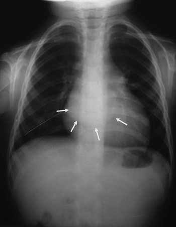
Clinical findings: a cyanotic patient with normal pulses, no hepatomegaly, cardiac hyperactivity, ongoing permanently splitting second heart sound, protosistolic click, and ejective systolic murmur grade 2/6 in the upper part of the left sternal border. Thorax X-ray: prominent pulmonary artery segment and double atrial contour were seen (Fig 1). Normal peripheral pulmonary markings were considered.
Thorax X-ray of case 1: the dense image in the right border of the cardiac silhouette corresponding to the left atrium (arrows) can be observed. Pulmonary trunk dilation can also be observed.

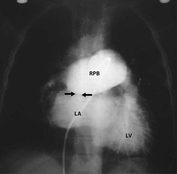
Echocardiography revealed an ostium secundum atrial septal defect of 1 centimetre in diameter and mild pulmonary valve stenosis with 30 millimetres of mercury gradient. Catheterism confirmed the echocardiographic findings of a large atrial septal defect and mild pulmonary valve stenosis; however, there was an important desaturation in the left atrium and left ventricle, which was not explained by these defects. Angiography in the right pulmonary branch revealed a huge right pulmonary branch and a fistula directly connecting the right pulmonary branch with the left atrium (Fig 2). The patient was referred for surgery and made a good recovery afterwards.
Angiography of case 1 showing contrast injection in the right pulmonary branch. The aneurismatic pulmonary branch and the contrast crossing directly to the left atrium through the fistula (arrows) can be observed (RPB = right pulmonary branch; LA = left atrium; LV = left ventricle).

Case 2
The patient was a 16-hour-old male neonate whose mother was 16 years old.
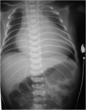
Clinical findings: a cyanotic and tachypnoeic patient, with hyperdynamic pulses, hepatomegaly, precordial hyperactivity, tachycardia with intense second heart sound, and a continuous murmur in all the precordial area. Thorax X-ray: important cardiomegaly was seen with enlargement of the left atrium and left ventricle (Fig 3). The initial diagnosis was cardiac failure secondary to a cyanotic congenital cardiac disease.
Thorax X-ray of case 2: great cardiomegaly can be observed mainly for enlargement of the left atrium and ventricle. Venocapillary congestion can also be seen.

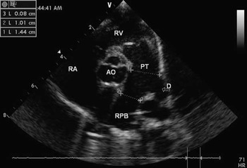
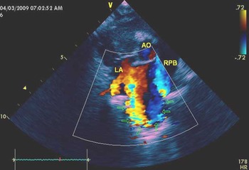
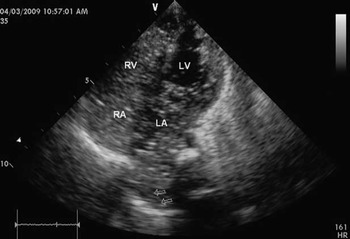
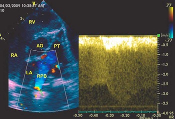
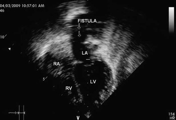
The echocardiographic study was performed by one of the authors (Marquez A.) who found a 4-millimetre-diameter ductus arteriosus in the usual location (Fig 4), with important dilation of the right pulmonary branch (Fig 4). A fistula originating in the distal portion of the dilated right pulmonary branch was visualised by bidimensional echo and was followed by colour Doppler from its origin until its drainage into the upper portion of the left atrium (Figs 5 and 8). A contrast echocardiography was made and micro-bubbles appeared very quickly in the left atrium (Fig 6). The pulsed and continuous Doppler showed continuous high flow rate through the fistula (Fig 7). It was impossible to visualise the left pulmonary branch and there was dilation of the left atrium and left ventricle. The patient was sent for surgery. A 1.5-centimetre-diameter fistula was found during surgery; it connected the right pulmonary artery to the left atrium through a collector structure into which the pulmonary veins were also draining, corresponding to type 3 for this pathology according to De Souza et al's classification (Table 1). The left pulmonary artery was hypoplastic. Saturation was normal after the fistula was ligated and the post-operation was uneventful.
Bi-dimensional echocardiogram of case 2: Observe the huge RPB and the origin of D (RA = right atrium; RV = right ventricle; PT = pulmonary trunk; AO = aorta; RPB = right pulmonary branch; D = ductus arteriosus).

Colour Doppler for case 2: the course of the fistula (arrows) from the distal part of the RPB to LA can be seen (AO = aorta; RPB = right pulmonary branch; LA = left atrium).

Venous contrast echocardiography for case 2: this frame shows that while the contrast still persists in the right cavities (RA and RV), it quickly appears in the LA. The arrows show the connection of the fistula with the left atrium. LV = Left ventricle; LA = left atrium; RV = right ventricle; RA = right atrium.

Continuous Doppler mode for case 2. In the left frame the doppler sample is in the fistula's origin in the distal portion of the right pulmonary branch (arrow). In the right frame the continuous flow is observed. (RV = right ventricle; PT = pulmonary trunk; AO = aorta; RPB = right pulmonary branch; LA = Left atrium; RA = right atrium).

Comments about the diagnostic approach
Diagnosis is delayed in most patients suffering from this rare pathology as can be seen in different publications, with 47% of cases being diagnosed in the second decade of life or after. This delay can be explained by the difficulty encountered in making a clear diagnosis. This paper thus emphasizes on the findings that can help in diagnosing this interesting pathology.
Clinical aspects: It is very important to be aware of the possibility of this pathology occurring in spite of its rareness. On the other hand, it must be borne in mind that although the fistula's origin is almost always in the right pulmonary branch, it is possible to find it originating from the left pulmonary branch; however, this possibility is very rare.Reference Karnik, Nilsson, Vijaayaraghavan, Hashmi and Shuhaiber15, Reference Sambasivam, Dhanawade and Krishnaswami16
Analysing the cases published to date and the pertinent literature on this topic – confirmed by our two cases – there are two types or variants of clinical manifestations. The first type includes severe cardiac failure and cyanosis as in case 2.Reference Krishnakumar, Krishna and Tharakan17–Reference McBrien, Sands and Gladstone21 This group is found during the neonatal period and the severity and precocity of clinical findings depends on how big the shunt is and therefore on the size of the fistula. Hyperdynamic pulses, hepatomegaly, and tachycardia associated with the cyanosis are frequently found in this group and diagnosis is easier when a continuous murmur is present;Reference Karnik, Nilsson, Vijaayaraghavan, Hashmi and Shuhaiber15 however, this finding is not always present. The second type includes cyanosis and chronic hypoxia as in case 1. The patients in this group are older, mainly in the second decade of life or after. The main clinical findings are cyanosis and dyspnoea. The possibility of this pathology should thus be considered in neonates suffering cardiac failure and cyanosis without apparent causeReference Krishnakumar, Krishna and Tharakan17–Reference McBrien, Sands and Gladstone21 or in any child or adult having important cyanosis without a cause that can explain it or in children having a cardiac disease, whose haemodynamics cannot explain the severity of the cyanosis as shown in case 1 because the atrial septal defect and the mild pulmonary valve stenosis could not explain the severe cyanosis that the child was suffering from.
The electrocardiogram did not reveal specific findings regarding this pathology.
Thorax X-ray
In some cases, thorax X-ray can help in diagnosis as has been described in some publications showing a dense retrocardiac image corresponding to the left atrium, as seen in the description of case 1; this image is located in the right-hand part of the cardiac silhouette (Fig 1).Reference Chowdhury, Kothari, Airan, Subramaniam and Venugopal2, Reference Mohanty, Yadav, Kothari and Airan22, Reference Chowdhury, Airan, Kothari, Pandey, Subramaniam and Venugopal23 The dilated right pulmonary branch can be seen as well. The pulmonary trunk can be dilated and abnormal pulmonary flow may be seen, mainly in the lower part of the right lung when there is agenesis of the right lower pulmonary lobule.Reference Chowdhury, Kothari, Airan, Subramaniam and Venugopal2, Reference Ohara, Ito and Kohguchi5 Pulmonary flow is reduced in this part of the lung in such cases, as happens in type 2 De Souza et al's classification of this malformation (Table 1).Reference De Souza e Silva, Giuciani, Ritter, Davis and Pluth4
Echocardiography
Echocardiography is undoubtedly an essential tool for diagnosing this malformation. Transthoracic echocardiography enables a precise diagnosis to be made in infants and children, as in case 2; however, it is different in adults. Transoesophageal echocardiography is necessary in adults because in them this pathology can be missed with transthoracic echocardiography.Reference Veldtman, Bloackburn and Parsons24–Reference Krishnamoorthy and Rao28 Transoesophageal echocardiography could not be used in case 1 because this technology was not available when the diagnosis was made (1985).
Different modes of echocardiographic diagnosis can be used: bidimensional echocardiography (Figs 4 and 8), colour Doppler (Fig 5), contrast echo (Fig 6), pulsed and continuous Doppler (Fig 7), and transoesophageal echo when necessary.
Bidimensional echocardiography allows the origin and course of the fistula to be seen, as in case 2 (Fig 8), the dilation of the pulmonary branch where the fistula originates (Fig 4), which can be aneurismatic as in case 1, and dilation of the left atrium and left ventricle. Colour Doppler enables a fistula to be followed from the pulmonary branch to the left atrium (Fig 5) and the conjunction of these two modes allows the size of the fistula, flow turbulence, and left cavities enlargement to be evaluated. Flow characteristics through the fistula could be evaluated with pulsed and continuous Doppler (Fig 7).
Bi-dimensional echocardiogram of case 2: this frame shows the fistula's distal portion connecting to the LA (arrows) (RV = right ventricle; RA = right atrium; LV = left ventricle; LA = left ventricle).

Venous contrast echocardiography can provide grounds for suspicion about the presence of an arteriovenous fistula, although the origin or place of a fistula cannot be specified with this technique.Reference Faizal, Sankar, Murthy and Cherian7, Reference Sambasivam, Dhanawade and Krishnaswami16, Reference Saatvedt, Gunnar Stake and Lindberg19, Reference Liu, Xiang and Tiecheng26 This is why it is very important to consider the speed with which micro-bubbles appear in the left atrium. As can be seen in case 2 (Fig 6), the bubbles appeared almost simultaneously in the right atrium, right ventricle, and left atrium. This is proved by injecting a shaken solution – micro-bubbles – into a vein of the right arm.
Transoesophageal echocardiography has great usefulness in diagnosing this pathology, mainly in adult or stout patients who do not have a good echocardiographic window.Reference Krishnamoorthy and Rao27, Reference Krishnamoorthy and Rao28
Echocardiographic study also helps to detect associated pathologies such as the atrial septal defect, which is the pathology most frequently associated with this type of fistula.Reference Saatvedt, Gunnar Stake and Lindberg19
Catheterism and angiocardiographic studies are made even though a patient may be directly submitted to surgery following precise echocardiographic studies (as shown in case 2).Reference Chowdhury, Kothari, Airan, Subramaniam and Venugopal2, Reference Krishnakumar, Krishna and Tharakan17, Reference Meskishvili, Dâhner, Ovroutski and Hetzer29, Reference Zeebregts, Nijveld, Lam, van Oort and Lacquet30 The characteristic finding in catheterism is important desaturation in the left atrium and left ventricle. Angiography in the pulmonary branch leads to defining the characteristics of a fistula, as in case 1 (Fig 2).
There has been little experience with nuclear magnetic resonance and computer tomography, because very few cases have occurred with this pathology; however, these techniques can be of great use in studying these types of fistulas.Reference Ivert, Fathi, Forssell, Svane and Lindblom25, Reference Stuckey31–Reference Margaryan, Arcieri, Cantinotti and Murzi33