Caval vein defects are the least common forms of atrial communications, forming only 2–10% of all interatrial communications.Reference Swan, Kirklin and Becu1, Reference Davia, Cheetlin and Bedynek2 Of these, the superior form of caval vein defects constitutes the majority and also the most well recognised. The inferior-type caval vein defect is extremely rare and finds very brief or no mention in paediatric cardiology textbooks or on literature search, often restricted to single case reports or to a very small number of cases in reviews of caval vein defects as a whole.Reference Ettedgui, Siewers, Zuberbuhler and Anderson3–Reference Arnheid, Andre, Rene and Peter8 Inferior-type caval vein defect is most often diagnosed with fossa ovalis atrial septal defect in our experience. This is because attention is not given to details while defining the morphology of these defects.
There is a strong argument that a caval vein defect is not a true atrial septal defect because the defect itself lies outside the confines of the true atrial septum. Thus, these defects are more aptly named caval vein defects.Reference Al Zaghal, Li, Anderson, Lincoln, Shore and Rigby5
In this study we present a large series of patients diagnosed echocardiographically – transthoracic and transoesophageal – as inferior-type caval vein defect and confirmed at surgery in all patients. The echocardiographic features and morphological variations are described.
The aim of this study is to define inferior-type caval vein defect by echocardiography and comparing with surgical findings.
Method
We retrospectively reviewed the clinical data, chest roentgenogram, electrocardiogram, echocardiography, and surgical notes of all 19 patients who were preoperatively diagnosed as inferior-type caval vein defect. This was out of a total number of 514 patients with atrial septal defect who underwent surgical closure during the 9-year period, from June 2000 to October 2009.
Their age varied from 18 months to 27 years with a male-to-female ratio of 13:6. All patients had clinically large left-to-right shunt. On chest roentgenogram – postero-anterior and left lateral views – there was cardiomegaly with increased pulmonary blood flow in all. The electrocardiogram showed normal sinus rhythm in all, and the QRS axis varied from +90 to +120 with rSR pattern in the chest leads.
Echocardiography was performed using the Philips 5500 and 7500 machine (Philips Medical System, Andover, MA, USA) using broadband transducers. Multiple views starting from the subxiphoid views were used to define the atrial septum. In one patient – 27 years of age – poor transthoracic windows necessitated transoesophageal echocardiography.
The following criteria were used to define inferior-type caval vein defect:
• Overriding of inferior caval vein over the defect.
• Intact fossa ovalis area of the atrial septum. If there was an additional fossa ovalis atrial septal defect, then an intact muscular border surrounding the fossa ovalis needs to be demonstrated.
• Anomalies of pulmonary vein(s) (optional criteria).
Echocardiographic examination
Transthoracic echocardiography
Subxiphoid views, that is, both the coronal and sagittal views, were used.
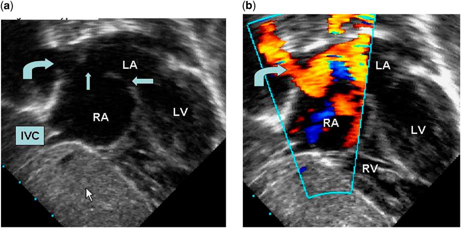
Subxiphoid coronal view (Fig 1a and b). In the coronal section, the defect was seen in the most posterior sweep of the transducer adjacent to the inferior caval vein. As the transducer was swept anteriorly, the septum started reforming, showing the intact fossa ovalis area. The anomalous pulmonary venous drainage of the right pulmonary vein is seen in this view.
Subxiphoid coronal view with posterior, inferior, and rightward tilt from a 2-year-old child with inferior-type caval vein defect. (a) Two-dimensional echocardiography showing the defect (thin arrow) and normal position of the right lower pulmonary vein (curved arrow). Fossa ovalis area of interatrial septum is intact (thick arrow). (b) On colour flow mapping, left to right shunt across the defect and drainage of right lower pulmonary vein to right atrium is seen (IVC = inferior caval vein; LA = left atrium; LV = left ventricle; RA = right atrium; RV = right ventricle).
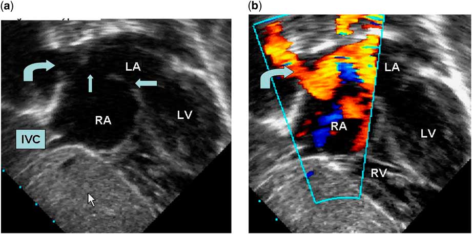
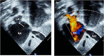
Subxiphoid sagittal view (Fig 2a and b). This is equivalent to the bicaval view of the transoesophageal echocardiography. The superior part of the atrial septum near the superior caval vein and the adjacent fossa ovalis area was intact. The most inferior–posterior part of the septum was deficient. The overriding inferior caval vein was best seen in this view.
Subcostal sagittal view of an adult patient with inferior-type caval vein defect. (a) Two-dimensional echocardiography showing inferior-type caval vein defect (arrow) in the most posteroinferior plane of the septum, inferior caval vein override over the defect is seen. (b) The same view with colour flow mapping showing left-to-right shunt across inferior-type caval vein defect (IVC = inferior caval vein; LA = left atrium; RA = right atrium; SVC = superior caval vein).

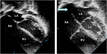
Apical four chamber (Fig 3a and b). This view shows best the intact fossa ovalis area and the defect in the same scan. The defect lies at the level of the crux and further inferior scan. Further superior scan shows the septum to reform. The right lower pulmonary vein can be seen to drain anomalously to the right atrium in this view.
Two-dimensional echocardiography in apical four-chamber view from the same child in Fig 1. (a) Standard apical four-chamber view showing dilated right atria, right ventricle with impression of intact inter atrial septum. (b) Apical four-chamber view in the posterior plane from standard four-chamber view showing defect in the inferior and posterior most part of the atrial septum (arrow) at the site where the inferior caval vein enters the right atrium. Fossa ovalis area of atrial septum is intact (LA = left atrium; LV = left ventricle; RA = right atrium; RV = right ventricle).

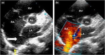
Parasternal short-axis view (Fig 4a and b). This view shows the defect in its most posterior part. The anomalous right upper and lower pulmonary vein drainage is well seen in this view.
Parasternal short-axis view at the level of the great vessels from same child as in Fig 1. (a) Two-dimensional echocardiography showing the defect (thick arrow) and right lower pulmonary vein at its normal location (yellow arrow). (b) On colour flow mapping, the right lower pulmonary vein draining to right atrium is seen (Ao = aorta; LA = left atrium; RA = right atrium; RV = right ventricle).

Transoesophageal echocardiography
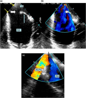
Basal short-axis view. To define inferior-type caval vein defect, the transducer is kept at the lower oesophagus level and the defect is seen in the postero-inferior part of atrial septum where the inferior caval vein joins the right atrium (Fig 5a and b).
Transoesophageal echocardiography from a 27-year-old patient with inferior-type caval vein defect. (a) Short-axis view with probe at the lower oesophagus level showing inferior-type sinus venosus defect (thick arrow) in the most posterior and inferior part of septum; inferior caval vein straddling over the defect is also seen (marked by yellow arrows). The fossa ovalis area is intact (thin arrow). (b) The same view with colour flow mapping showing left-to-right shunt. (c) The same view with anti-clock rotation of the transducer on colour flow mapping showing anomalous drainage of the right lower pulmonary vein into the right atrium (arrow) (LA = left atrium; RA = right atrium; RLPV = right lower pulmonary vein).

Basal long-axis (bicaval) view. Inferior-type caval vein defect is seen related to inferior caval vein. In addition, inferior caval vein overriding over atrial septum, anomalous drainage of right-sided pulmonary vein(s) to right atrium, and intact fossa ovalis area should be defined from this view.
Results
By using the above-mentioned criteria, 19 patients were diagnosed to have inferior-type caval vein defect on echocardiography. In addition, one patient had an additional small fossa ovalis atrial septal defect and one had a patent foramen ovale. The left pulmonary veins were normally connected in all patients. The right pulmonary veins appeared in their usual position in all patients. However, anomalous drainage of right pulmonary vein(s) to right atrium was observed in 18 of the 19 patients. Both right upper and right lower pulmonary veins were anomalously draining in 10 patients and only the right lower pulmonary vein was anomalous in eight patients. No patient had isolated anomalous right upper vein drainage.
Surgical technique
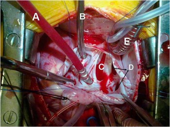
Surgical closures for inferior-type caval vein defect proceeds in a manner similar to that for secundum defects except for certain procedural modifications. The inferior caval vein is disserted free from the pericardial reflection and is cannulated as far away as feasible from the inferior caval vein – right atrium junction. Low cannulation is not required in secundum atrial septal defect, as the lower end of the patch is sutured to the floor of the atrium. The right inferior pulmonary veins are located more caudally than usual and need to be identified as they enter the atrial chamber. Care must be taken to avoid injuring the veins while looping the inferior caval vein. After opening the right atrium, the diagnosis of inferior-type caval vein defect is confirmed by the presence of an intact fossa ovalis, with or without patency or a separate fossa ovalis type of defect. The defect straddles the inferior caval vein opening and has a sharp crescent-shaped superior margin (Fig 6). There is no inferior rim. The right lower pulmonary vein may appear to drain into the right atrium or the left atrium depending on the location of the posterior extent of the superior rim. When the rim courses anterior to the orifice of the pulmonary veins, the drainage appears to be in the normal manner into the left atrium, and when the rim courses posterior to the pulmonary vein orifices the drainage appears to be anomalous. It is not possible to ascertain anomalous drainage from the external appearance, as in both instances the right lower pulmonary veins appear to join the heart at the same location. The defect is closed with a patch of autologous pericardium or synthetic material. The suture line runs along the right margin of the pulmonary veins and inferiorly along the floor of the atrium at the inferior caval vein–atrial junction, taking care to the prevent obstruction of either the inferior pulmonary veins or the inferior caval vein.
Surgical findings of a child with inferior-type caval vein defect. Opened-up right atrium showing inferior-type caval vein defect (C) with inferior caval vein overriding the defect, straddling over the inferior caval vein (E), anomalous drainage of the right-sided pulmonary veins (D), bullet sucker in patent foramen ovale (A) showing the intact fossa ovalis area, and metallic sucker in coronary sinus (B).

Surgical findings
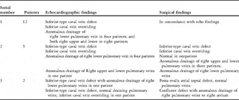
Echocardiographic and surgical findings are given in Table 1.
Comparison of echocardiographic and surgical findings in patients diagnosed as inferior-type caval vein defect.

Atrial communication
Surgical diagnosis of inferior-type caval vein defect was confirmed in 17 of the 19 patients. In two patients, the surgical diagnosis was that of a large fossa ovalis atrial septal defect. In one patient with a large fossa ovalis atrial septal defect, the surgical notes mentioned a large confluent defect, whereas another patient was having fossa ovalis atrial septal defect with deficient inferior caval vein rim.
Pulmonary veins
The surgical notes clearly made a distinction between anomalous connection and anomalous drainage. Normal connection implied that the pulmonary veins externally appeared to connect to the left atrium. Normal drainage of the pulmonary vein is present when the crescent-shaped lower rim of the defect coursed anterior to the orifice of the pulmonary vein. However, if the lower rim courses posterior to the orifice of the pulmonary vein, then the veins are anomalously draining to the right atrium.
In all cases, the pulmonary veins externally appeared to be connecting to the left atrium, that is, normally connected, but the surgeon had to divert the patch to direct the pulmonary veins to the left atrium in all except one case. Thus, anomalous drainage of pulmonary vein was present in 16 of the 17 cases.
Thus, our echocardiographic impression of the pulmonary veins appearing in its normal position but showing abnormal drainage to right atrium was in agreement with the surgical notes.
Discrepancy was found in the number of pulmonary veins draining anomalously. In 16 patients with surgically confirmed anomalous drainage, there was concordance between echocardiography and surgery in 12 and discordance in four. The discordance was related to overdiagnosis of anomalous drainage in three out of four patients. Thus, echocardiography reported both right pulmonary veins draining anomalously, whereas surgery detected only the right lower pulmonary vein draining anomalously in three cases. In one, only the right lower pulmonary vein was diagnosed to be anomalous by echocardiography, whereas both right upper and lower pulmonary veins were draining anomalously.
Discussion
Previously published studies suggested inferior-type caval vein defect as the least common variety of atrial communication.Reference Van Praagh, Carrera, Sanders, Mayer and Van Praagh9–Reference Ferreira Martins and Anderson14 Most often, these patients were wrongly diagnosed with secundum atrial septal defect. However, attention to detail anatomy reveals that their morphology is very different. We describe here one of the largest series of patients diagnosed echocardiographically with inferior-type caval vein defect and further detailed at surgery in all cases.
Definition dilemmas
Characterisation of the atrial septum would aid in understanding the morphology of the communications that exist between the atrial chambers, not all of which are true septal defects. Anderson et alReference Anderson, Ettedgui and Devine10 and Al Zaghal et alReference Al Zaghal, Li, Anderson, Lincoln, Shore and Rigby5 defined a true septal structure as one that can be removed without exiting the cavity of the heart. The flap valve of the fossa ovalis, along with its muscular rim, fulfils this criterion. The remainder of the posterior atrial septum is no more than an unfolding between the walls of the right and left atria and the drainage of systemic and pulmonary venous, incorporating a core of extracardiac fibro adipose tissue. True defects of the atrial septum therefore exist because of deficiency, perforation, or absence of the flap valve or muscular rim of the fossa ovalis. The other interatrial communications are outside the confines of the atrial septum. Recognition of this feature is key to the diagnosis of the caval vein defect and to its differentiation from true atrial septal defects.Reference Ettedgui, Siewers, Zuberbuhler and Anderson3, Reference Li, Al Zaghai and Anderson13, Reference Ferreira Martins and Anderson14
It was suggested by International Nomenclature Committee that, rather than representing an interatrial communication, the defect was an anomalous venoatrial communication, the essential feature being anomalous pulmonary venous drainage to the inferior caval vein with retention of pulmonary venous connection to the left atrium. This combination produces an extraseptal cavopulmonary conduit, permitting shunting of blood from the left to the right atrium. Such a definition excludes the inferior-type caval vein defect from interatrial communications existing through the mouth of the coronary sinus or true septal defects within the oval fossa, extending posteroinferiorly into the mouth of the inferior caval vein. Even with the use of this anatomic criterion, appropriate diagnosis of the defect remains difficult during life, as noted in studies comparing echocardiographic findings with anatomic specimens.Reference Van Praagh, Carrera, Sanders, Mayer and Van Praagh11
Al Zaghal et alReference Al Zaghal, Li, Anderson, Lincoln, Shore and Rigby5 reported that the path gnomonic feature of caval vein defect is the overriding of the intact rim of the fossa ovalis by the superior caval vein or inferior caval vein and the presence of a shunt at the atrial level.Reference Al Zaghal, Li, Anderson, Lincoln, Shore and Rigby5 Although a defect within the true atrial septum can coexist with a caval vein defect, the most reliable and unequivocal diagnosis is made by confirming that the muscular border of the fossa ovalis is intact.
Inferior caval vein overidding and anomalous pulmonary venous drainage
Owing to the fact that the defect overrides the inferior caval vein, it is best seen in the most posterior scan planes of the subxiphoid, apical, and parasternal short-axis views. Another important two-dimensional echocardiographic feature of these defects is the complete absence of septal tissue between the defect and the atrial wall. We wrongly diagnosed inferior-type caval vein defect in two patients who at surgery had a large fossa ovalis atrial septal defect. A review of the echocardiogram in these two patients revealed that the mistake lies in not correctly identifying the absence of the rim of the fossa ovalis.
Previous reports state that pulmonary venous abnormalities are uncommon in inferior-type caval vein defect as compared with the superior forms of caval vein defect.Reference Van Praagh, Carrera, Sanders, Mayer and Van Praagh11 Our findings were opposite. Perhaps the previous studies were based on a fewer number of cases. Here, we would like to highlight the difference between abnormal connection and abnormal drainage of pulmonary veins in this setting. The impression on two-dimensional echocardiography in all these patients was that the pulmonary veins appear to be in their normal position. But colour flow clearly showed that the right lower, and less commonly both the right upper and lower pulmonary venous flow, was draining into the right atrium. This impression was confirmed by our surgeon who had to direct the patch to close the inferior-type caval vein defect in such a way that the pulmonary venous flow was finally directed into the left atrium in all except one patient.
The embryological mechanism of the anomalous pulmonary venous connection differs in the two types of caval vein defects. Some authors believe that the superior-type caval vein defect is due to unroofing of the right upper pulmonary vein; therefore, partial anomalous pulmonary venous connection in this setting is always present.Reference Ettedgui, Siewers, Zuberbuhler and Anderson3, Reference Van Praagh, Carrera, Sanders, Mayer and Van Praagh11, Reference Li, Al Zaghai and Anderson13 In inferior-type caval vein defect, the wall separating the inferior caval vein from the left atrium normally does not form a roof over the pulmonary vein, and anomalies of the pulmonary vein is one of abnormal drainage.Reference Al Zaghal, Li, Anderson, Lincoln, Shore and Rigby5 Thus, a deficiency in this extraseptal wall results in overriding of the inferior caval vein but not in the anomalous pulmonary vein connection. The most frequent partial anomalous pulmonary vein connection to the inferior caval vein is the Scimitar syndrome, but an interatrial communication is not usually found in adult patients with this syndrome.Reference Dupuis, Charaf, Breviere, Abou, Rémy-Jardin and Helmius15 However, we agree with other authors in considering the overriding of the intact rim of the fossa ovalis by the superior caval vein or inferior caval vein in the presence of an intact fossa ovalis area to be the path gnomonic feature of caval vein defect.Reference Ettedgui, Siewers, Zuberbuhler and Anderson3, Reference Al Zaghal, Li, Anderson, Lincoln, Shore and Rigby5, Reference Anderson, Ettedgui and Devine10, Reference Li, Al Zaghai and Anderson13, Reference Ferreira Martins and Anderson14
There is a trend towards more invasive and expensive testing for patients with this diagnosis because accuracy of all features remains a challenge. Transoesophageal echocardiographic analysis offers excellent acoustic windows and is ideally suited to defining the posterior features and connections but is limited in its accessibility to patients, especially children in the pre-operative awake setting.Reference Arnheid, Andre, Rene and Peter8, Reference Van Praagh, Carrera, Sanders, Mayer and Van Praagh11 Intraoperative transoesophageal echocardiographic analysis at completion of surgical repair allows for immediate and early detection of residual defects, obstructed caval or pulmonary venous pathways, and confirmation of cardiac anatomy. However, in our series, only one adult patient required pre-operative transoesophageal echocardiography for diagnostic evaluation. A good subxiphoid window is essential for delineating some of the features of the defect – caval vein override and pulmonary venous abnormality. This window is almost always available in children in our experience. We have also never felt the need for an intraoperative transoesophageal echocardiography in children for further clarification of anatomy.
Accurate echocardiographic diagnosis of anomalous pulmonary venous drainage is more difficult than diagnosis of inferior-type caval vein defect per se. Although we did diagnose anomalous drainage in all cases, there was discordance between echocardiographic and surgical findings on the number of pulmonary veins draining anomalously. The incorrect diagnosis on the echocardiogram is mainly related to the overdiagnosis of anomalous drainage of the right upper pulmonary vein. This underscores the difficulty in accurately diagnosing anomalous pulmonary venous drainage in this setting.
Surgical implications
A number of authors have outlined different approaches to surgical correction of this rare anomaly. Re-implantation of the anomalously connected pulmonary vein or veins directly into the left atrium, combined with suture closure of the venoatrial communication, has been successfully reported.Reference Dupuis, Charaf, Breviere, Abou, Rémy-Jardin and Helmius15 Others have reported the detachment of the posterior edge of the atrial septum, suturing it into position to the right of the anomalously connected pulmonary vein and leftward of the caval veins, thereby closing the left-to-right defect and thus redirecting the anomalous pulmonary venous return to the left atrium without requiring a patch.Reference Sturm and Ankeney16, Reference Hiramatsu, Takanashi, Imai, Hoshino, Seo and Terada17 In our institution, a strategy towards simple repair with a patch or baffle avoids the potential long-term venous obstruction associated with the aforementioned approaches. We used a baffle rather than a simple patch when we considered the insertion of the patch might induce either caval or pulmonary venous obstruction.
Following surgical repair of an inferior-type caval vein defect, residual right-to-left shunt has been reported when the lower portion of the defect is not closed because a biatrial connection of the inferior caval vein is not seen. Occasionally, the Eustachian valve of the inferior caval vein has been mistaken for the lower margin of the defect and blood flow was deferred from the inferior caval vein to the left atrium.Reference Al Zaghal, Li, Anderson, Lincoln, Shore and Rigby5, Reference Blom, Gittenberger-deGroot, Jongeneel, DeRuiter, Poelmann and Ottenkamp12, Reference Weber, Markowitz, Hellenbrand, Kleinman and Kopf18–Reference Becker, Buss, Sebening, Meisner and Döhlemann19 Correct recognition of the anatomy of the caval vein defect and intraoperative transoesophageal echocardiography-guided intervention may avoid these surgical mistakes.
Conclusion
We report a large series of patients with the inferior-type caval vein defect, which is one of the rarest forms of anomalous venoatrial communication diagnosed by echocardiography. Inferior caval vein straddling and an intact fossa ovalis are prerequisites for diagnosis. Anomalous pulmonary venous drainage of the right pulmonary veins is very common in our series, although accurate diagnosis of the number of pulmonary veins was not possible in all cases. More often, echocardiography over-diagnosed the number of anomalously draining pulmonary venous drainage.
It is important to be aware of this rare type of venoatrial communication, as well as to not wrongly diagnose it as a fossa ovalis atrial septal defect. One postulated surgical mistake in unrecognised cases lies in the incorrect placement of the atrial septal defect patch, resulting in inferior caval vein blood being directed to the left atrium or obstruction of the right lower pulmonary vein(s).