Introduction
An iatrogenic pseudoaneurysm of the internal carotid artery secondary to transsphenoid or intrasphenoid endoscopic surgery is uncommon and may present with delayed, life-threatening epistaxis.Reference Hollis, McGlashan, Walsh and Bowdler1–Reference Lister and Sypert3 False aneurysms of the internal carotid artery can also develop after a head injury.Reference Tampieri, Atkinson, Daniel, Teitelbaum and Shemie4–Reference Celil, Engin, Orhan, Barbaros, Hakan and Adil6 Delayed epistaxis resulting from iatrogenic trauma to the sphenopalatine branch of the internal maxillary artery (as it crosses the anterior wall of the sphenoid sinus) during transsphenoidal surgery has also been reported.Reference Cockroft, Carew, Trost and Fraser7
Case report
A 65-year-old woman who had previously undergone a left maxillectomy for paranasal sinus malignant melanoma was being followed up with a magnetic resonance imaging (MRI) scan. This scan showed a mass in the pituitary fossa (Figure 1a), which was subsequently explored and biopsied endoscopically in another unit.
(a) Axial computed tomography scan at the level of skull base and (b) angiogram (lateral view), showing a large right internal carotid artery pseudoaneurysm pointing into the right sphenoid sinus.

Four days after the endoscopic surgery, the patient presented with profuse epistaxis.
Nasal packing and inflated balloons in the nasopharynx failed to control the bleeding, and the patient was referred urgently to our unit. After transfer and resuscitation, the bleeding was stopped by packing the sphenoid sinus with oxidised cellulose. This was done ‘blind’ as the quantity of bleeding prevented the use of an endoscope or head light. The site to be packed was located by palpating the ‘shoulder’ of the posterior choana and packing approximately 1 cm above this.
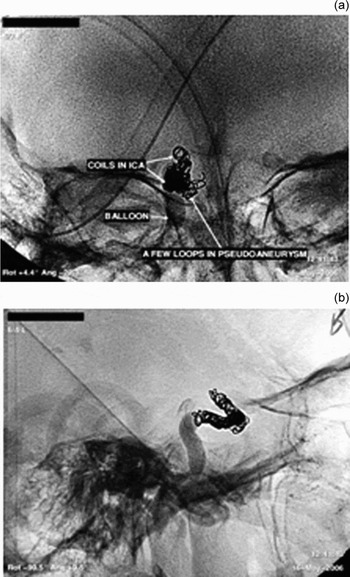
Subsequent angiography showed a large right internal carotid artery pseudoaneurysm pointing into the right sphenoid sinus (Figure 1b). This was managed with multiple coils to occlude the internal carotid artery from just below the anterior choroidal artery (Figure 2). At the end of the procedure, the cross-flow from the left internal carotid artery was poor. The patient developed a right hemispherical stroke in evolution post-operatively; however, fortunately she recovered rapidly from her neurological deficit.
(a) Angiography images in the antero-posterior view and (b) lateral view, showing multiple coils in situ, used to occlude the internal carotid artery (ICA).

Discussion
Most carotid aneurysms are secondary to head trauma.Reference Tampieri, Atkinson, Daniel, Teitelbaum and Shemie4, Reference Bavinzski, Killer, Knosp, Ferraz-Leite, Gruber and Richling5 The sudden onset of life-threatening epistaxis following sphenoid surgery should prompt suspicion of rupture of a carotid aneurysm.Reference Reddy, Lesiuk, West and Fewer2, Reference Lister and Sypert3, Reference Kai, Hamada, Nishi and Ushio8, Reference Vergez, Folia, Michenet, Rose, Lacroix-Loubes and Percodani9 Excessive bleeding during trans-sphenoidal surgery also raises this possibility, and imaging will help to establish the diagnosis.Reference Kai, Hamada, Nishi and Ushio8 Early involvement of an experienced rhinologist, an interventional radiologist and possibly a neurosurgeon is needed, after initial resuscitative measures. Angiography is required, both for diagnosis and for treatment (by endovascular occlusion or embolisation or stenting of the aneurysm).Reference Tampieri, Atkinson, Daniel, Teitelbaum and Shemie4–Reference Celil, Engin, Orhan, Barbaros, Hakan and Adil6 Angiography performed in patients with refractory bleeding should also include the external carotid artery if nothing abnormal is found in the internal carotid artery territory.Reference Cockroft, Carew, Trost and Fraser7 The optimal management is carotid occlusion, provided the patient has been able to tolerate a trial of occlusion to show an adequate cross-flow through the circle of Willis.Reference Bavinzski, Killer, Knosp, Ferraz-Leite, Gruber and Richling5, Reference Kai, Hamada, Nishi and Ushio8, Reference Vergez, Folia, Michenet, Rose, Lacroix-Loubes and Percodani9 Bypass surgery is required where the patient cannot tolerate the balloon occlusion test, although in an emergency situation a trial occlusion is not possible.Reference Bavinzski, Killer, Knosp, Ferraz-Leite, Gruber and Richling5, Reference Kai, Hamada, Nishi and Ushio8
• This paper describes a rare case of iatrogenic pseudoaneurysm of the internal carotid artery secondary to endoscopic sphenoid surgery, resulting in profuse epistaxis
• Any episode of torrential epistaxis after sphenoid surgery should raise the possibility of damage to the internal carotid artery
• Early diagnosis and treatment is needed in order to avoid exsanguination and to maintain circulation until interventional radiology can be undertaken
Conclusion
Any episode of torrential epistaxis after sphenoid surgery should raise the possibility of damage to the internal carotid artery. Awareness of this complication is important in order to recruit the appropriate help as soon as possible. Early diagnosis and treatment is needed in order to avoid exsanguination and to maintain circulation until interventional radiology can be performed.