Introduction
Eosinophilic oesophagitis is a disease in which upper intestinal symptoms are associated with dense eosinophilic infiltration of the squamous oesophageal epithelium or deeper oesophageal tissue. Neither symptoms nor eosinophilia respond to administration of a proton pump inhibitor. The pathophysiological mechanisms of the condition are likely to be related to allergic inflammation rather than an underlying motility defect (as in gastroesophageal reflux disease).Reference Potter, Saeian, Staff, Massey, Komorowski and Shaker1, Reference Fox, Nurko and Furuta2
The prevalence of eosinophilic oesophagitis appears to be increasing. In a demographic study, Noel et al. reported that the annual incidence of paediatric oesophagitis had risen from one in 10 000 in the year 2000, to 4.3 cases per 10 000 in 2003.Reference Noel, Putnam and Rothenberg3
Eosinophilic oesophagitis is recognised as an important, independent clinicopathological entity in the gastroenterological literature, but it is far less reported in the ENT literature.Reference Dauer, Ponikau, Smyrk, Murray and Thompson4–Reference Goldstein, Putnam and Dohar10 We report two cases presenting to an otolaryngology department, as an illustration to raise awareness of the condition within our specialty.
Case one
A two-year-old boy presented with food aversion, and a history of intermittent ‘choking’ episodes with food since commencing solids. This was initially attributed to a behavioural aetiology by a community paediatrician. However, the child's episodes became more severe. The catalyst prompting his family doctor to refer him to a paediatric otolaryngologist was an episode of high oesophageal food bolus impaction, characterised by aphasia and drooling, which lasted for one hour before resolving spontaneously on arrival at the local hospital emergency department. On further questioning in the ENT clinic, there was a history of swallowing aversion: the child would hold food in his mouth and chew for prolonged periods of time. However, there seemed to be no correlation with the type or consistency of food. Non-inflammatory adenotonsillar hypertrophy was noted on examination.
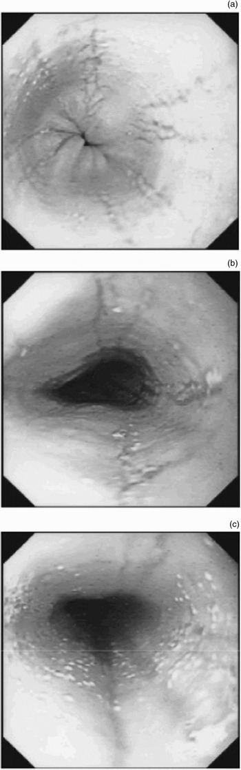
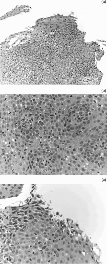
Paediatric speech and language assessment, including videofluoroscopy with a range of food consistencies, showed normal swallow but gagging when holding larger boluses in the mouth. Adenoidal nasal obstruction and tonsil hypersensitivity were considered as contributing factors. However, the child developed further symptoms of recurrent, mid-oesophageal, momentary food impaction. A barium swallow examination was normal. However, rigid and flexible oesophagoscopies, performed by a paediatric otolaryngologist and a paediatric gastroenterologist, respectively, showed pronounced vertical lines, mucosal blebs of whitish exudate and a ringed oesophagus along the entire length (Figure 1). A lower oesophageal pH probe was left in situ for 24 hour monitoring: no significant reflux was demonstrated. Histological examination of mucosal biopsies showed a dense eosinophilic infiltrate (Figure 2).
Endoscopic views of the proximal oesophagus, showing (a) vertical lines, (b) loss of vascular pattern and (c) whitish exudates.

Photomicrographs showing: oesophageal epithelium with a dense infiltration of eosinophils (i.e. more than 50 per high power field); eosinophils layered along the superficial surface of the epithelium; eosinophilic microabscess (i.e. more than four eosinophils clustering adjacent to each other); eosinophilic degranulation; and basal zone hyperplasia. (Published by courtesy of Dr Ruth Nash, Consultant Histopathologist, St Georges's Hospital.) (H&E; original magnification ×300–400 approximately)

The child was referred for a paediatric immunology opinion. A mild eosinophilia was found. There were no positive reactions to radioallergosorbent testing (RAST) or skin prick allergy testing for milk, eggs, peanuts, shellfish, peas, beef, fish, rye, tomato or wheat.
The child's symptoms improved with a dairy-free diet. Although there was a concomitant, dramatic improvement in the macroscopic appearance of the oesophagus on endoscopy, histological analysis of biopsies still showed a dense eosinophilic infiltrate. Subsequently, a topical steroid was added to the child's treatment regimen.
Case two
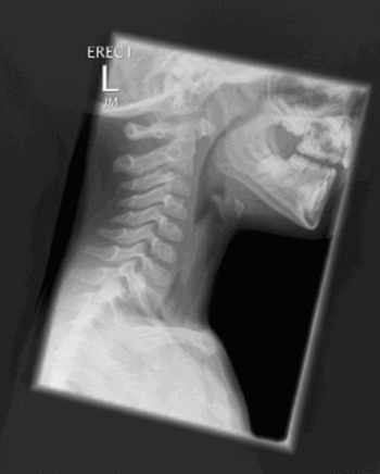
A four-year-old boy presented with a history initially of reflux unresponsive to proton pump inhibitor and, more recently, of recurrent food impaction occurring approximately every two weeks and lasting no longer than 30 minutes. Each episode resolved with enforced vomiting. Similar to the previous case, the child was referred to a tertiary care centre for a paediatric otolaryngology opinion, following an episode of prolonged impaction which led to emergency department attendance. A lateral soft tissue neck X-ray taken while the patient was symptomatic showed an obstruction at the level of the cricopharyngeus (Figure 3).
Lateral soft tissue radiograph showing food bolus impaction at the ricopharyngeus. L = left

Rigid oesophagoscopy performed by a paediatric otolaryngologist showed a very narrow cricopharyngeus inlet, too narrow to pass a small, rigid oesophagoscope. Flexible endoscopy (performed by a paediatric gastroenterologist) showed inflammatory changes as impressive as those of the previous case. Mucosal biopsies demonstrated a dense eosinophilic infiltrate.
A pH probe was inserted for 24 hours, showing significant gastroesophageal reflux. However, the density of eosinophilic infiltration (i.e. 50 eosinophils per high power field) was too great to be attributed to gastroesophageal reflux alone.
This patient also had mild eosinophilia, and no positive RAST or skin prick reactions to the same range of test agents use in patient one.
However, on closer questioning, the child had a history of dairy product allergy. His symptoms improved dramatically with a dairy-free diet alone.
Discussion
We report two children with symptoms of oesophageal dysphagia presenting to a paediatric otolaryngologist. By involving a paediatric gastroenterologist, a diagnosis of eosinophilic oesophagitis was considered, and subsequently confirmed with mucosal biopsies. The patients were appropriately and expediently investigated and managed.
Both our patients were boys. Eosinophilic oesophagitis is distinctly more common in males. Previously, there had been a preponderance of reports in the paediatric population, but the condition has recently emerged as a disease also affecting adults worldwide.Reference Potter, Saeian, Staff, Massey, Komorowski and Shaker1
In a recent report of 103 children, 12.5 per cent presented with feeding difficulties (mean age 3.5 years), 25 per cent with vomiting (mean age 7.9 years), 25 per cent with abdominal pain (mean age 12 years), 26 per cent with dysphagia (mean age 13 years) and 6.8 per cent with food impaction (mean age 16 years).Reference Noel, Putnam and Rothenberg3 Interestingly, both of the patients in our report, aged two and four years, presented with intermittent dysphagia and food bolus impaction. These patients also had histories of feeding difficulties and vomiting, respectively.
It is important to distinguish eosinophilic oesophagitis from gastroesophageal reflux disease. Heartburn is often present, particularly in older children and adults, making differentiation from gastroesophageal reflux disease difficult.Reference Dauer, Freese, El-Youssef and Thompson8 Although there may be partial improvement in symptoms after acid blockade, eosinophilia persists.Reference Liacouras and Ruchelli11
Many patients with eosinophilic oesophagitis have been previously treated, unsuccessfully, for gastroesophageal reflux disease, with some patients even receiving antireflux surgery (e.g. fundoplication).Reference Fox, Nurko and Furuta2, Reference Kelly, Lazenby, Rowe, Yardley, Perman and Sampson12, Reference Liacouras13
Both our patients underwent pH probe monitoring. Multiple studies have shown that patients with eosinophilic oesophagitis usually have normal pH monitoring results in the distal oesophagus.Reference Fox, Nurko and Furuta2, Reference Walsh, Antonioli, Goldman, Fox, Bousvaros and Leichtner14–Reference Stevoff, Rao, Parsons, Kharilas and Hirano18 The presence of severe oesophagitis found after aggressive acid blockade in the setting of normal pH monitoring results is virtually diagnostic of eosinophilic oesophagitis. Normal pH monitoring results nearly eliminate gastroesophageal reflux disease as the aetiology of intractable oesophagitis. However, in recent years it has become evident that there are patients with eosinophilic oesophagitis and some concomitant, underlying gastroesophageal reflux. This finding may explain why some patients may have a partial response to acid blockade.Reference Fox, Nurko and Furuta2
One of the patients in our report had significant reflux demonstrated on pH probe monitoring. Of course, eosinophilic inflammation of the oesophagus can be found in a number of diseases, including gastroesophageal reflux disease, inflammatory bowel disease, parasitic infection and collagen vascular diseases.Reference Noel, Putnam and Rothenberg3, Reference Liacouras and Ruchelli11, Reference Walsh, Antonioli, Goldman, Fox, Bousvaros and Leichtner14 One might question how a distinction can be made between these conditions (particularly gastroesophageal reflux disease) and eosinophilic oesophagitis. To date, no studies have clearly defined the upper limit of the normal range for eosinophils visualised within the epithelium of adults and children with gastroesophageal reflux disease; however, this number is thought to be less than five per high power field, with the eosinophilia being limited to the distal oesophagus.Reference Chang and Anderson19
The histological examinations for both our patients showed proximal oesophageal eosinophilic infiltration. The epithelium contained a dense eosinophilic infiltrate, greater than 50 per high power field, a density too high to be attributable to gastroesophageal reflux alone. In addition, there were other histological features typical of eosinophilic oesophagitis, as demonstrated in Figure 2. The eosinophils were seen to layer along the superficial surface of the epithelium. There were eosinophilic microabscess (i.e. more than four eosinophils clustering adjacent to each other). Abscesses on the surface formed the histological correlate of the whitish exudates or pinpoint papules seen at endoscopy. Eosinophilic degranulation could be seen. These patterns of eosinophilia were present on a background of basal zone hyperplasia.
Neither of our patients presented with other allergic symptoms or airway symptoms. However, epidemiological studies have reported concomitant allergic symptoms, such as eczema, allergic rhinitis and bronchospasm, in 30–62 per cent of patients, and several series have reported airway manifestations.Reference Potter, Saeian, Staff, Massey, Komorowski and Shaker1, Reference Dauer, Ponikau, Smyrk, Murray and Thompson4, Reference Thompson, Arora, Romero and Dauer6, Reference Liacouras and Ruchelli11, Reference Walsh, Antonioli, Goldman, Fox, Bousvaros and Leichtner14, Reference Orenstein, Shalaby, Di Lorenzo, Putnam, Sigurdsson and Kocochis15, Reference Straumann, Spichtin, Bucher, Herr and Simon20
Both our patients had mild eosinophilia. Other studies have found eosinophilia to be common but usually not universal, appearing in 30 to 100 per cent of patients.Reference Esposito, Marinello, Paracchini, Guidali and Oderda21
The exact aetiology of eosinophilic oesophagitis is not known. Most investigators agree that eosinophilic oesophagitis is driven by an aberrant, immune-mediated response.Reference Liacouras and Ruchelli11 The established association between eosinophils and food allergies suggests that food antigens may cause eosinophilic oesophagitis, and this assumption is true in many patients. The most common food allergens that have been identified include milk, soy, egg and wheat.Reference Sundaram, Sunku, Nelson, Sentongo, Melin-Aldana and Kumar22
Neither of our patients had a positive RAST or skin prick test result. In a series of 30 children, 60 per cent showed either a positive RAST or skin prick test result, supporting the theory of an immunoglobulin E mediated process.Reference Liacouras and Ruchelli11 However, recent studies have suggested that eosinophilic oesophagitis may represent a delayed response, and thus allergy evaluations have turned to skin patch testing. When compared with skin prick and RAST testing, skin patch testing was able to identify food allergens in three of 16 patients who were skin prick and RAST negative.Reference Orenstein, Shalaby, Di Lorenzo, Putnam, Sigurdsson and Kocochis15 Unfortunately, skin patch testing was not available in our centre at the time of investigation of the current patients. That withstanding, skin patch testing is currently not considered sufficiently well standardised for routine testing to be recommended.Reference Chang and Anderson19
Truly definitive evidence that a food is causing eosinophilic oesophagitis depends on the demonstration of symptom resolution, and a return to normal histology on biopsy, with an elimination diet, as well as a return of oesophageal eosinophils after reintroduction of the suspected food.Reference Esposito, Marinello, Paracchini, Guidali and Oderda21
• The prevalence of eosinophilic oesophagitis appears to be increasing
• A missed diagnosis may result in oesophageal stricture
• Early involvement of a paediatric gastroenterology team in the diagnosis is recommended in children presenting with oesophageal symptoms
Great effort should be made to identify foods associated with symptoms. If food restriction does not improve symptoms, it has been suggested that an elemental diet should be used for four weeks, followed by addition of individual foods every five to seven days.Reference Spergel, Beausoleil, Mascarenhas and Liacouras23 However, compliance is often an issue with elemental diets. Children who are unresponsive to dietary restriction or who cannot tolerate dietary treatment may benefit from systemic or topical corticosteroids.
Our first patient received topical fluticasone propionate. The steroid delivery technique comprised spraying a metered dose inhaler into the mouth without a spacer. The patient was then asked to swallow, without inhaling first, in order to attempt to deliver the topical anti-inflammatory to the oesophageal mucosa.
Several studies have reported successful treatment of eosinophilic oesophagitis with topical steroids in children and adults.Reference Teitelbaum, Fox, Twarog, Nurko, Antonioli and Gleich17, Reference Stevoff, Rao, Parsons, Kharilas and Hirano18, Reference Croese, Fairley, Masson, Chong, Whitaker and Kanowski24, Reference Noel, Putnam, Collins, Assa'ad, Guajardo and Jameson25 Topical delivery to the oesophageal mucosa has the advantage that only 1 per cent of the drug is absorbed systemically, and this fraction undergoes rapid hepatic processing. The main potential side effect is oral or oesophageal candida infection.Reference Noel, Putnam, Collins, Assa'ad, Guajardo and Jameson25
It is important not to miss a diagnosis of eosinophilic oesophagitis, in order to avoid long-term complications. Several studies have identified stricture as one such long-term complication.Reference Dauer, Freese, El-Youssef and Thompson8, Reference Stevoff, Rao, Parsons, Kharilas and Hirano18, Reference Chang and Anderson19, Reference Arora, Perrault and Smyrk26 In a paediatric series, three of 18 children with eosinophilic oesophagitis had a demonstrable stricture on barium swallow examination.Reference Orenstein, Shalaby, Di Lorenzo, Putnam, Sigurdsson and Kocochis15
Conclusions
Eosinophilic oesophagitis is a clinicopathological disease characterised by upper intestinal symptoms and the finding of more than 15 to 20 eosinophils per high power field in the oesophageal epithelium; these findings are unresponsive to proton pump inhibitor treatment. A diagnosis of eosinophilic oesophagitis must be considered in patients with bolus impaction, reflux not responding to proton pump inhibitor treatment, food aversion or other unusual oesophageal symptoms. We recommend the early involvement of a gastroenterologist for patients presenting with these symptoms, in order to expediate diagnosis and treatment.