Introduction
Inorganic forms of copper (Cu) and zinc (Zn) are generally supplemented in diets for growing and finishing pigs to satisfy the requirements for maintenance and growth. In the case of Cu sulfate, higher levels (125 to 175 mg/kg) can have a growth promotion effect in growing and finishing pigs (Stansbury et al., Reference Stansbury, Tribble and Orr1990; Davis et al., Reference Davis, Maxwell, Brown, de Rodas, Johnson, Kegley, Hellwig and Dvorak2002). However, there are concerns related to the accretion of heavy metals such as Cu and Zn, derived from piggery effluent, which could have adverse effects on the soil microbiota and potentially cause a decline in soil fertility and pasture and crop yields (McGrath et al., Reference McGrath, Chaudri and Giller1995). Banning the use of pharmacological inclusions of Cu and Zn in pig diets is the approach that some countries have enforced, even setting maximum allowable total mineral concentrations in pig feeds (Byrne, Reference Byrne2003).
An alternative to inorganic forms of dietary Cu and Zn is organic complexes, which have been shown to increase mineral absorption and retention in the body of pigs (Apgar et al., Reference Apgar, Kornegay, Lindemann and Notter1995; Schiavon et al., Reference Schiavon, Bailoni, Ramanzin, Vincenzi, Simonetto and Bittante2000) compared with inorganic forms, indicating a higher bioavailability of the organic form of mineral. Furthermore, some studies with organic forms of Cu and Zn have also been shown to reduce mineral excretion (Creech et al., Reference Creech, Spears, Flowers, Hill, Lloyd, Armstrong and Engle2004) and improve performance when included at similar levels to inorganic sources (Zhou et al., Reference Zhou, Kornegay, van Laar, Swinkels, Wong and Lindemann1994; Veum et al., Reference Veum, Bollinger and Ellersiek1995), or can be incorporated at lower dietary levels than those in the inorganic forms without negatively affecting growth performance (Mullan et al., Reference Mullan, Wilson, Harris, Allen and Naylor2002). Results from Smits and Henman (Reference Smits and Henman2000) indicated that Cu could be reduced from 150 mg/kg Cu sulfate to 40 mg/kg in the form of Cu proteinate amino acid chelate in grower diets, while Fremaut (Reference Fremaut2003) showed that Zn, Fe, Mn and Cu could be reduced to 36, 39, 18 and 7 mg/kg (30% of normal levels used in Belgium) if all were included in the proteinate amino acid chelate form. Benefits in terms of reduced mineral excretion would accrue from such interventions.
In view of this information, the hypotheses tested in this experiment was that pig performance and mineral status could be maintained by feeding lower levels of Cu and Zn in the proteinate amino acid chelate form than pharmacological Cu levels in the sulfate form, and that this would reduce mineral concentrations in fecal material. The effect of the inclusion level (IL) and mineral form (MF) of Cu and Zn on carcass and meat quality was also investigated.
Material and methods
Animals and housing
This experiment was conducted at the Medina Research Centre and approved by the Animal Ethics Committee of the Department of Agriculture and Food of Western Australia (AEC No. 2-04-7).
One hundred and sixty female pigs (Large White × Landrace) were used. The pigs were obtained from a commercial supplier and brought to the Medina Research Centre at the time of weaning (approximately 21 days of age). Upon arrival, the pigs were group housed in a shelter having an earthen floor covered with wheat straw and fed a commercial weaner diet until they reached the appropriate BW to start the experiment (approximately 25 kg). The weaner diet contained 14.5 MJ/kg DE, 20% CP, 0.8 available lysine per MJ DE, 0.90% calcium and Cu and Zn at base levels for pigs of that weight. No antibiotics were included in the diet. At approximately 25 kg BW, pigs were randomly allocated to treatments, based on BW (see below), in groups of eight in a naturally ventilated grower shed having 3.6 m× 1.8 m pens with concrete (2/3) and plastic slatted (1/3) floors, a 30-cm-wide plastic individual single-space feeder and two stainless-steel swinging nipple drinkers per pen.
Experimental design and diets
Copper at 27 mg/kg and Zn at 65 mg/kg of diet (‘low’) or Cu at 156 mg/kg and Zn at 170 mg/kg of diet (‘high’) fed in an organic (proteinate amino acid chelate) or inorganic (sulfate) MF were used in a 2 × 2 factorial arrangement of treatments. The Cu and Zn dietary levels were established in accordance with European Union Regulation No. 1334/2003, which currently applies and restricts the maximum total content of Cu and Zn in growing/finishing pig diets to 25 and 150 mg/kg, respectively (Byrne, Reference Byrne2003). These levels were compared with higher levels, which in the case of Cu have demonstrated to have a growth-promoting effect in growing-finishing pigs (125 to 170 mg/kg Cu; Stansbury et al., Reference Stansbury, Tribble and Orr1990; Davis et al., Reference Davis, Maxwell, Brown, de Rodas, Johnson, Kegley, Hellwig and Dvorak2002). The ‘high’ levels of Zn were included to alleviate potential Cu–Zn interactions (Underwood and Suttle, Reference Underwood and Suttle1999).
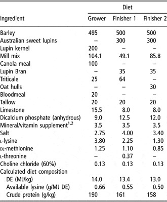
Diets are subsequently expressed as ‘low’ or ‘high’ proteinate amino acid chelate (Bioplex) (LB and HB) and ‘low’ or ‘high’ sulfate (LS and HS). Pigs on each treatment were fed the same base diet within the experiment (Table 1). The mineral supplement incorporated in the diets was similar to commercial practice, but did not contain any Cu or Zn. Copper-sulfate pentahydrate and Zn-sulfate monohydrate (Adisseo P/L, Queensland, Australia), or Cu proteinate amino acid chelate (Bioplex Cu, Alltech Biotechnology P/L, Victoria, Australia) and Zn proteinate amino acid chelate (Bioplex Zn, Alltech Biotechnology P/L, Victoria, Australia), were added according to their required levels in each dietary treatment. The concentration of total Cu and Zn in the mineral products as per Certificate of Analysis for each product was 250, 340, 100 and 150 g/kg for Cu sulfate, Zn sulfate, Cu proteinate amino acid chelate and Zn proteinate amino acid chelate, respectively. The mineral–vitamin premixes, which contained excess amounts of other trace minerals (Co., I, Fe, Mn and Se), were supplied to a commercial feed mill (Wesfeeds P/L, Perth, Australia), where the diets were manufactured.
Composition of experimental diets (g/kg as-fed)

DE=digestible energy.
1Provided the following nutrients (per kg of air-dry diet): Vitamins: A, 1500 IU; D3, 300 IU; E, 37.5 mg; K, 2.5 mg; B1, 1.5 mg; B2, 6.25 mg; B6, 3 mg; B12, 37.5 g; calcium pantothenate 25 mg, folic acid 0.5 mg, niacin 30 mg, biotin 75 g. Minerals: cobalt 0.5 mg (as cobalt sulfate heptahydrate), iodine 1.25 mg (as potassium iodine), iron 150 mg (as ferrous sulfate monohydrate), manganese 100 mg (as manganous oxide), selenium 0.3 mg (as sodium selenite). Premix supplied by Adisseo Australia P/L, Australia.
2Inclusion of copper sulfate pentahydrate, Zn sulfate monohydrate, Cu proteinate amino acid chelate and Zn proteinate amino acid chelate according to treatment levels.
Allocation of pigs, feeding and sample collection
When the average BW of pigs in each pen reached approximately 25 kg, the diets were randomly allocated to the pens, taking into account the location in the shed, and feeding the experimental diets started (day 0). Pigs were fed three diets (Grower, Finisher 1 and Finisher 2) on an ad libitum basis over the BW range of 25 to 55, 55 to 80 and 80 to 107, respectively. Pigs were fed twice per day. Feed remaining was measured and recorded every week. Measurements of individual BW were made weekly throughout the experiment as well as feed consumption. Water was also available at all times. On days 7 and 35 of the experiment, all pigs were vaccinated against Erysipelas as directed by the study veterinarian.
On day 14 of the experiment (at approximately 36 kg BW) blood samples were collected from half of the pigs per pen (four selected randomly pigs), via jugular venipuncture into 10 ml Li-heparin tubes. On day 77 of the experiment (at approximately 97 kg BW), a second blood sample was collected from the same pigs previously sampled. At the same time, a ‘grab’ fecal sample (200 g approximately) was collected from the same set of pigs. Additionally on day 42 of the experiment (at approximately 65 kg BW), four fecal samples were collected from each pen, and thoroughly mixed and sub-sampled. Fecal samples were frozen at −20°C and later analyzed for Cu and Zn concentrations. Blood samples were kept chilled until submission to the Animal Health Laboratories (AHL; Department of Agriculture and Food, WA), where sample preparation and all mineral analyses were performed.
Pigs were slaughtered at a commercial abattoir (Linley Valley, Wooroloo, Western Australia) when they reached a minimum of 104 kg BW. At slaughter, samples of approximately 200 g liver, kidney and bone and a small section of duodenum (20 cm), were taken from the same 80 pigs sampled for blood and fecal measurements during the experiment, placed on ice, and then frozen at −20°C for subsequent mineral analysis.
Carcass weight (AUSMEAT trim 13; head off, flare off, foretrotters off, hindtrotter on) and depth of backfat at the P2 site (Hennessy Grading Probe 4, Hennessy Grading Systems Limited, Auckland, New Zealand) and at three points along the midline, namely shoulder (between the first and second ribs), P2 at the midline and the pelvic area (perpendicular to last true vertebrae), were measured on the hot carcasses of all pigs (calipers). Belly fat thickness between the 9th to the 11th rib approximately 18 cm from the midline was also measured (introscope). After chilling the carcasses at 1°C for at least 24 h, a section of the muscle longissimus thoracis (15 cm× 15 cm, approximately) was removed between the 12th and 13th rib from the set of 80 carcasses and was used for measurements of surface exudates, pH and color.
Analysis
Feed was analyzed on an as-fed basis and fecal samples were freeze-dried (Heto freeze dryer type CD 1.0-110, Heto, Birkerod, Denmark) and ground using a 1 mm screen in a Cyclotec mill (Foss P/L, Sweden) before analysis. Feed and fecal samples were assayed for Cu and Zn concentration by atomic absorption spectrophotometry (AAS) using a Varian Spectra AA 400 Atomic Absorption Spectrophotometer (Varian Techtron P/L, Mulgrave, Australia) at the AHL. Total Hb was measured in whole-blood samples on an automated clinical chemistry analyzer (Cobas Mira S, Roche Diagnostics, Basel, Switzerland), calibrated using an Hb standard (Sigma Cat. No. 525-18) with a concentration of 180 g/l, according to the Sigma Diagnostics Total Hemoglobin Determination Procedure No. 526. The remaining blood was then centrifuged (Clements 2000 benchtop centrifuge, Clements, Sydney, Australia) at 3000 r.p.m. for 15 min at 4°C and the plasma separated.
Since erythrocyte superoxide dismutase activity is highly correlated with RBC Cu and measurement of RBC Cu is less problematic (Underwood and Suttle, Reference Underwood and Suttle1999), blood samples were tested for RBC Cu content by a similar method to that of plasma Cu, but instead of using the plasma it was performed on the erythrocyte pellet. Plasma samples were also tested for alkaline phosphatase (ALP) using the method based on the recommendations of the ‘International Federation for Clinical Chemistry’ (IFCC), with an Olympus AU400 auto-analyzer (Olympus, Hamburg, Germany).
Tissue samples (liver and kidney) were thawed, rinsed with distilled water, thinly sliced and dried overnight at 110°C. Bone samples were cut from the metacarpals with a band saw and all muscular tissue and the bone marrow thoroughly removed. Approximately, 3 g of bone was dried at 110°C overnight, de-fatted with diethyl ether by changing the liquid for three consecutive nights, and then dried again. Determination of Cu and Zn concentration in plasma, liver and kidney samples and Zn in bone samples was performed by AAS using a Varian Spectra AA 400 Atomic Absorption Spectrophotometer at the AHL.
Samples of liver and intestine mucosal cells scraped from the thawed duodenum were analyzed for concentration of the protein metallothionein (MT) by a silver-saturation method described by Scheuhammer and Cherian (Reference Scheuhammer and Cherian1991). The concentration of silver in the final sample supernatant was measured by AAS using a Varian Spectra AA 10/20 Atomic Absorption Spectrophotometer (1985) with an air-acetylene flame. It was assumed that the amount of silver in the supernatant fraction was proportional to the amount of MT present (Scheuhammer and Cherian, Reference Scheuhammer and Cherian1991). The concentration of MT is expressed as micrograms of MT per gram of wet weight tissue.
Collected sections of the muscle l. thoracis were standardized to 20 mm in thickness for measurement of meat quality. After allowing the surface of the meat exudates for 10 min, surface exudate was measured using the filter paper method (Kauffman et al., Reference Kauffman, Eikelenboom, van der Wal, Merkus and Zaar1986), pH by a portable pH meter (Model 6027, Jenco Electronic Ltd, San Diego, USA) and surface L*(lightness), a* (redness) and b* (yellowness) using a chromameter (CR-400, Minolta, Japan) using D65 lighting, a 2° standard observer and an 8 mm aperture in the measuring head, standardized to a white tile.
Statistical analyses
Data were analyzed using the ANOVA procedures of StatView (StatView 5.0 for windows, AddSoft Pty. Ltd, Wooden, Vic., Australia). Two-way ANOVA was conducted to compare the responses of pigs to two MF of Cu and Zn at two IL. The experimental unit for all measurements was the pen of pigs; however, data were collected and/or analyzed on an individual basis and results were then pooled per pen. Statistical significance was accepted at P < 0.05. Fishers’-protected LSD comparisons were used (at 5% significance level) for comparisons between significantly different mean values of different variables. Carcass weight was included as a covariate in the ANOVA for subcutaneous fat depth to eliminate its effect on this parameter.
Results
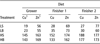
The concentration of Cu and Zn in the diets as measured (Table 2) was reasonably close to that which was planned for in the experimental design.
Total concentrations of Cu and Zn in grower, finisher 1 and finisher 2 experimental diets (analyzed values)
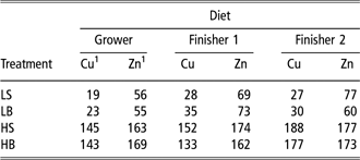
LS = low sulfate; LB = low proteinate amino acid chelate (Bioplex); HS = high sulfate; HB = high proteinate amino acid chelate (Bioplex).
1mg/kg, as-fed.
Pig performance
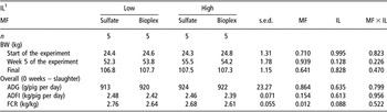
Over the entire experiment (week 0 to slaughter), there was no significant difference between treatments on average daily gain (ADG) or average daily feed intake (ADFI); however, there was a main effect of MF (P = 0.012) on feed conversion ratio (FCR). The FCR was improved by using the proteinate amino acid chelate (2.63 and 2.72 kg/kg for proteinate amino acid chelate and sulfate, respectively) (Table 3).
Comparison between organic v. inorganic Cu and Zn at low and high inclusion levels on performance of growing and finishing pigs

a,bWithin a row means without a common superscript letter differ (P < 0.05).
n = number of pens/treatment, each pen of eight pigs.
1Refer to Table 2 for the concentration of Cu and Zn in the diets.
IL = inclusion level; MF = mineral form; ADG = average daily gain; ADFI = average daily feed intake occurred; FCR = feed conversion ratio.
Carcass and meat quality
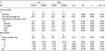
There was a main effect of IL of Cu and Zn on dressing percentage (P = 0.006), with pigs fed more Cu and Zn having a higher dressing percentage (67.6% and 68.6% for ‘low’ and ‘high’ IL, respectively) (Table 4). There was no significant difference between experimental diets on hot carcass weight, subcutaneous fat thickness at the P2 site, shoulder, and P2 at the midline or meat quality indices. However, there was a main effect of IL of Cu and Zn on the thickness of subcutaneous fat in pelvis (P = 0.029), with pigs fed higher levels of Cu and Zn having more subcutaneous fat than the others. There was also an interaction between MF and IL on belly subcutaneous fat depth (P = 0.049). Pigs fed LB had lower subcutaneous fat in the belly than pigs fed LS or HB, but it was similar to those fed HS.
Comparison between organic v. inorganic Cu and Zn at low and high inclusion levels on carcass and meat quality of finishing pigs

a,bWithin a row means without a common superscript letter differ (P < 0.05).
n = number of pens/treatment, each pen of eight pigs.
1Refer to Table 2 for the concentration of Cu and Zn in the diets.
IL = inclusion level; MF = mineral form; HCW = hot carcass weight.
Fecal mineral concentrations
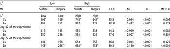
An interaction (P = 0.009) between MF and IL for Cu concentration in feces was observed on day 14 of the experiment (Table 5). Copper content decreased by 5- to 7-fold when the IL in the diet was reduced from ‘high’ to ‘low’. At the ‘high’ IL, it was also lower in pigs fed the proteinate amino acid chelate as compared with the inorganic form. There was a main effect of IL (P < 0.001) on the concentration of Zn in feces. Zinc concentration was about 2.5 times lower with ‘low’ IL compared with ‘high’ (303 v. 801 mg/kg dry matter (DM)).
Comparison between organic v. inorganic Cu and Zn at low and high inclusion levels on mineral concentration in fecal material

a,b,c,dWithin a row means without a common superscript letter differ (P < 0.05).
n = number of pens/treatment, each pen of eight pigs.
1Refer to Table 2 for the concentration of Cu and Zn in the diets.
IL = inclusion level; MF = mineral form.
On day 42 of the experiment, there was a main effect of IL (P < 0.001) on Cu concentration in feces. Copper level in feces was approximately four times lower with ‘low’ IL compared with ‘high’ (127 and 546 mg/kg DM for ‘low’ and ‘high’, respectively). There were main effects of IL and MF (P < 0.001 and P = 0.007, respectively) for Zn excretion. Concentration of Zn in feces was approximately 2-fold less with a lower dietary Zn level (268 and 609 mg/kg DM for ‘low’ and ‘high’, respectively). Lower fecal Zn concentration was also observed in sulfate compared with proteinate amino acid chelate-fed pigs (421 v. 456 mg/kg DM, respectively) (Table 5).
On day 77 of the experiment, there were main effects of IL and MF (P < 0.001 and P = 0.014, respectively) for Cu levels. The concentration of Cu decreased approximately 4.5 times with ‘low’ IL compared with ‘high’ (155 and 704 mg/kg DM for ‘low’ and ‘high’, respectively). Concentration of Cu in feces was also lower in sulfate than in proteinate amino acid chelate-fed pigs (397 v. 462 mg/kg DM, for sulfate and proteinate amino acid chelate, respectively). There was an interaction (P < 0.001) between MF and IL for Zn concentration. At ‘low’ IL, the content of Zn was lower in proteinate amino acid chelate-fed pigs while at ‘high’ IL it was higher compared with sulphate-fed pigs.
Blood levels
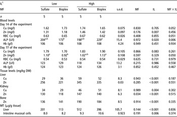
Blood mineral levels in all treatments were maintained within the normal physiological range (1.3 to 3.0 mg/l, 0.7 to 1.5 mg/l, 80 to 269 IU/l and 100 to 140 g/l; Puls, Reference Puls1994; for plasma Cu, Zn and ALP and whole-blood Hb, respectively) (Table 6). On day 14 of the experiment, Cu, RBC Cu and Hb levels were similar between diets. There was a main effect of IL (P = 0.007) on plasma Zn with pigs fed ‘low’ diets having lower Zn levels than pigs fed ‘high’ diets (1.24 v. 1.43 mg/l). There was also an interaction between MF and IL (P = 0.006) for ALP, with pigs fed LB having lower ALP activity than HB-fed pigs, but similar to LS- and HS-fed pigs. On day 77 of the experiment, Cu, Zn, RBC Cu, ALP and Hb levels were similar between treatments (P > 0.05).
Comparison between organic v. inorganic Cu and Zn at low and high inclusion levels on blood and tissue mineral levels and metallothionein concentration in growing/finishing pigs

a,bWithin a row means without a common superscript letter differ (P < 0.05).
n = number of pens/treatment, each pen of eight pigs.
1Refer to Table 2 for the concentration of Cu and Zn in the diets.
IL = inclusion level; MF = mineral form; Hb = hemoglobin; RBC = red blood cells; ALP = alkaline phosphatase; DM = dry matter; MT = metallothionein.
Tissue levels
Copper and Zn concentrations in liver in all treatments were within the normal range (19 to 56 mg/kg dry weight (DW) for Cu and 150 to 337 mg/kg DW for Zn; Puls, Reference Puls1994) (Table 6). The concentration of Cu in kidneys from pigs fed HB and HS can be considered high compared with normal physiological levels (26 to 37 mg/kg DW; Puls, Reference Puls1994), as was the concentration of Zn in kidneys in pigs from all treatments (normal levels are 56 to 112 mg/kg DW; Puls, Reference Puls1994). Pigs from HB and HS treatments had high concentrations of Zn in bone relative to normal levels (95 to 146 mg/kg DW; Puls, Reference Puls1994). The concentration of Cu and Zn in liver, kidney and Zn in bone increased (P < 0.005), with Cu and Zn IL in the diet. There was also a main effect of MF (P = 0.034) on the concentration of Zn in kidney. Pigs fed sulfates had a higher content of Zn in kidneys than proteinate amino acid chelate-fed pigs (139 mg/kg DM v. 129 mg/kg DM for sulfate- and Bioplex-fed pigs, respectively).
The IL of the mineral had a direct effect on the MT concentration in liver and intestinal mucosal cells. The level of MT in the liver (157 v. 454 μg/g tissue for ‘low’ and ‘high’, respectively, P < 0.001) and intestinal cells (8.1 v. 9.9 μg/g tissue for ‘low’ and ‘high’, respectively, P = 0.006) increased significantly with IL in the diet.
Discussion
The hypothesis that pig performance and mineral status could be maintained by feeding lower levels of Cu and Zn in the proteinate amino acid chelate form than pharmacological Cu levels in the sulfate form, and that this would reduce mineral concentrations in fecal material, is supported by the results of this experiment.
Production data over the BW range 25 to 107 kg showed that pigs can be fed total dietary levels of approximately 27 mg/kg Cu and 65 mg/kg Zn, without any reduction in performance, compared with pigs fed 156 mg/kg Cu and 170 mg/kg Zn. There was no difference between mineral forms, except for an improvement in FCR when the proteinate amino acid chelate was included. Our data do not allow us to ascertain the reason(s) for this improvement, but it could be attributable to an improved nutrient digestibility of proteinates by these organic forms of Cu and Zn, as has been described previously (Apgar et al., Reference Apgar, Kornegay, Lindemann and Notter1995; Schiavon et al., Reference Schiavon, Bailoni, Ramanzin, Vincenzi, Simonetto and Bittante2000). Additionally, Cu and Zn are involved as co-factors in myriad of metabolic enzyme systems within the pig (Underwood and Suttle, Reference Underwood and Suttle1999), and their greater availability over the entire study could have caused the improvement in FCR. Further experimentation is required to confirm this finding.
The total Cu and Zn concentrations in the ‘low’ diets of this experiment were below the maximum levels allowed by the European Union Commission Regulation No. 1334/2003 for growing/finishing pigs (25 and 150 mg/kg, for Cu and Zn, respectively) (Byrne, Reference Byrne2003), except for Cu concentration in the ‘low’ finisher diets, which was slightly in excess. However, it is questionable whether reducing the concentration of Cu in the finisher diets by 2 to 10 mg/kg to meet the maximum levels established in the aforementioned regulation would affect pig performance, since this level is well above requirements and nowhere near a growth promotion level.
The level of 65 mg/kg total dietary Zn in this study concurs with NRC (1998) recommendations on requirements for pigs of this weight, unlike the level of Cu (27 mg/kg), which is higher. However, it has been suggested that the mineral requirements for today’s rapidly growing genotypes may be higher than current recommendations (Close, Reference Close2002) and that requirements to optimize immune function may be higher than the requirements for growth (Klasing, Reference Klasing2001; van Heugten et al., Reference van Heugten, Spears, Kegley, Ward and Qureshi2003).
The aforementioned ‘low’ mineral levels (27 mg/kg Cu and 65 mg/kg Zn), independent of the source, reduced fecal mineral concentration by approximately 6-fold for Cu and by 2.5-fold for Zn compared with a diet containing 156 mg/kg Cu and 170 mg/kg Zn. The Cu and Zn concentrations in feces were determined only by their dietary IL, which agrees with results from previous studies in weanling pigs (Apgar and Kornegay, Reference Apgar and Kornegay1996; Case and Carlson, Reference Case and Carlson2002; Carlson et al., Reference Carlson, Boren, Wu, Huntington, Bollinger and Veum2004). The effect of MF on fecal mineral concentration, however, was inconsistent. For example, on day 77 of the experiment, the concentration of Zn in feces was reduced by 40% when proteinate amino acid chelate was included instead of the sulfate at ‘low’ levels, while a similar effect was not observed on day 14. However, and considering the inconsistency in the results obtained, it is important to compare Cu and Zn absorption and retention by pigs fed proteinate amino acid chelate and sulfates by means of carefully controlled balance studies where intake and excretion can be measured more accurately than in the current study.
Feeding ‘low’ levels of Cu and Zn reduced blood and tissue mineral concentration in pigs in this experiment; however, a normal Cu, Zn and Fe mineral homeostasis was maintained. There was no significant difference in the Cu or Zn status of pigs when fed either proteinate amino acid chelate or sulfates. This agrees with previous studies where other organic forms of Cu or Zn were used (Wedekind et al., Reference Wedekind, Lewis, Giesemann and Miller1994; Cheng et al., Reference Cheng, Kornegay and Schell1998). Schiavon et al. (Reference Schiavon, Bailoni, Ramanzin, Vincenzi, Simonetto and Bittante2000), on the other hand, found a higher content of Fe in plasma, hemoglobin and red cells in piglets fed trace elements in the form of proteinates compared with those fed sulfates at 20 and 100 mg/kg, indicating a higher bioavailability of the proteinates. These authors concluded that there was a different metabolism of Fe when sulfates were replaced by proteinates. A possible antagonistic effect among Cu, Zn and Fe might have indirectly increased plasma Fe levels, rather than a higher absorption of Fe.
A different metabolism in liver between Cu sulfate and Cu lysine was also highlighted by Apgar et al. (Reference Apgar, Kornegay, Lindemann and Notter1995), who found higher Cu concentrations in the liver of weanling pigs fed 200 mg/kg Cu lysine compared with those pigs fed sulfate. However, further research in the same laboratory with finishing pigs showed no differences in either absorption or retention between the two sources when fed at 200 mg/kg (Apgar and Kornegay, Reference Apgar and Kornegay1996). The inconsistency in the response of pigs to organic mineral supplementation might be explained by factors including age (young pigs seem to absorb more Cu than do older pigs, Apgar and Kornegay, Reference Apgar and Kornegay1996), initial mineral status of the pigs, and content of mineral antagonists in the diet such as Cu, Zn and Fe (Jondreville and Revy, 2003). It is also clear that there is a major influence of the mineral source. Thus, all ‘organic’ forms are not the same and the processes used to obtain the commercial products might result in products with a similar name (i.e. generic chelate) but containing variable concentrations of bioavailable minerals.
Neither MF nor IL of Cu and Zn consistently influenced subcutaneous fat depth measured at several points along the carcass. These findings concur with results from Henman (Reference Henman2001), who compared Cu proteinate amino acid chelate at 100 mg/kg against 20 and 200 mg/kg Cu sulfate. Interestingly, Kawas et al. (Reference Kawas, Moreno, Sepulveda, Alejo and Garza1996) found less subcutaneous fat thickness in carcasses from pigs fed Cu sulfate at 250 mg/kg compared with no Cu-supplemented pigs, but the mechanism of action was not explored.
To our knowledge, this is the first time that the effect of Cu and Zn proteinate amino acid chelate on meat quality has been assessed, and results indicate no difference with carcasses from sulfate-fed pigs or between the two levels of inclusion studied on surface exudate, pH and color. Jensen et al. (Reference Jensen, Flensted-Jensen, Skibsted and Bertelsen1998) compared the effect of Cu-sulphate supplementation at 25 and 175 mg/kg or no Cu supplementation on meat quality (drip loss, color stability and lipid oxidation), and concluded that Cu-sulphate supplementation did not have any negative effect on the parameters measured. A similar conclusion was drawn by Bosi et al. (Reference Bosi, Cacciavillani, Casini, Lo Fiego, Marchetti and Mattuzzi2000) comparing diets supplemented with 35 or 175 mg/kg Cu sulfate on fresh meat or cured ham color.
Another component of this study was the quantification of MT. Interestingly, Carlson et al. (Reference Carlson, Hoover, Hill, Link and Ward1997) reported lower MT contents in intestinal mucosal cells from pigs fed a basal diet supplemented with 250 mg/kg Zn–methionine or Zn–amino acid complex compared with those fed a basal diet or a diet supplemented with 3000 mg/kg ZnO, suggesting a different response of MT to organic and inorganic Zn forms. In our experiment, however, MF did not influence the MT concentration in enterocytes. However, the dietary levels in our study were lower than those used in the study of Carlson et al. (Reference Carlson, Hoover, Hill, Link and Ward1997); hence intestinal MT levels were around 10 μg/g tissue v. 64 μg/g tissue in the work of Carlson et al. (Reference Carlson, Hoover, Hill, Link and Ward1997).
It appears that the induction of intestinal MT is an initial defense to prevent excessive Zn absorption and a more sustained mechanism might be adopted when high Zn intake is prolonged. In fact, intestinal MT levels declined after high Zn diets were fed for several weeks to rats (Reeves, Reference Reeves1995), and after 9 days to chickens (Cao et al., Reference Cao, Henry, Davis, Cousins, Miles, Littell and Ammerman2002). In the study of Carlson et al. (Reference Carlson, Hoover, Hill, Link and Ward1997) with pigs, MT levels were measured after 20 days of Zn supplementation. Since the pigs in the present study were fed high levels of Cu and Zn for 13 weeks (from 25 to 107 kg BW) and the concentration of MT was measured in samples obtained at slaughter, the length of supplementation could have affected the response of intestinal MT.
Conclusion
Total dietary levels of 27 mg/kg Cu and 65 mg/kg Zn, in either the proteinate amino acid chelate or sulfate form, significantly reduced the excretion of Cu and Zn via feces by approximately 6-fold for Cu and 2.5-fold for Zn compared with a diet containing 156 mg/kg Cu and 170 mg/kg Zn in growing/finishing pigs. Levels of 27 mg/kg Cu and 65 mg/kg Zn also maintained the growth rate and mineral homeostasis in the pigs compared with diets containing 156 mg/kg Cu and 170 mg/kg Zn, under the conditions of this experiment. There was no significant effect of MF on ADG, ADFI, fecal mineral content, mineral status in the pigs, carcass and meat quality indices and MT concentration in enterocytes and liver. Feed efficiency was significantly improved by the inclusion of proteinate amino acid chelate over the entire experimental period.
Acknowledgment
The following people from the Department of Agriculture and Food of Western Australia assisted in the conduct of this project and their input is greatly appreciated: Bob Davis, Jae Kim, Hugh Payne, Megan Trezona, Roland Nicholls, Richard Seward, Gerard Smith, Mark Dolling, Grant Doncon, Maeve Harvey and Malcolm McGrant. The assistance of the following students from the School of Veterinary and Biomedical Sciences at Murdoch University is also gratefully recognized: Jung-Min Heo, Kelly Stanger, Danka Halas and Malcom Boyce.