Introduction
Recurrent bacterial meningitis in the paediatric population represents a serious clinical event that mandates a comprehensive evaluation for underlying anatomical or immunological causes. Although immune deficiencies are frequently considered early in the diagnostic workup, structural abnormalities of the skull base and temporal bone are equally significant and often under-recognised contributors to recurrent infection.Reference Tebruegge and Curtis1 Among these, congenital malformations of the inner ear are well-documented causes of spontaneous cerebrospinal fluid (CSF) leakage and subsequent ascending meningitis.Reference Ohlms, Edwards and EO2
Incomplete partition type II, historically referred to as Mondini deformity, results from arrested cochlear development during the seventh week of gestation. It is characterised by a cochlea with one and a half turns, apical cystic dilatation and an often-dilated vestibule.Reference Sennaroglu and Bajin3 Defects in the modiolar architecture establish an abnormal communication between the subarachnoid space and the perilymphatic space, thereby predisposing individuals to CSF fistula.Reference Jackler, Luxford and House4
Hyrtl’s fissure, also known as the persistent tympanomeningeal fissure, represents incomplete ossification between the petrous and squamous portions of the temporal bone.Reference Hyrtl5 Although often an incidental finding, its persistence reflects aberrant embryological development of the temporal bone. In the context of associated inner-ear malformations, it may serve as a marker of generalised developmental vulnerability of the otic capsule and adjacent skull base.
The embryological basis for these anomalies lies in the complex process of otic capsule ossification and cranial base development, which occurs during the first and second trimesters of gestation. Disruption during this critical period can result in incomplete partition of the cochlea, patent fissures or deficient bony barriers between the intracranial and middle-ear spaces. The coexistence of incomplete partition type II and persistent Hyrtl’s fissure in the same patient suggests a more extensive developmental field defect involving multiple temporal bone structures, rather than isolated arrested organogenesis. This combined anomaly creates multiple potential independent pathways for CSF egress, rather than a single continuous pathway.
The incomplete partition type II malformation facilitates transmission of CSF from the subarachnoid space to the perilymphatic compartment via deficient modiolar architecture. Hyrtl’s fissure, conversely, represents a direct communication between the posterior cranial fossa and the middle-ear cavity. In our patient, the presence of both anomalies provides two independent potential routes for CSF leakage. However, a connection between the perilymphatic space and Hyrtl’s fissure has not been anatomically confirmed.
We present a case of recurrent meningitis in a child with incomplete partition type II deformity and persistent Hyrtl’s fissure, in which surgical exploration confirmed a congenital otogenic CSF fistula and which was notable for an intra-operative CSF gusher.
Case report
A 13-year-old child presented with fever, severe headache, vomiting and nuchal rigidity. Clinical examination revealed positive meningeal signs. Lumbar puncture yielded CSF findings consistent with bacterial meningitis, including neutrophilic pleocytosis, elevated protein and reduced glucose concentration. This episode represented the third documented occurrence of bacterial meningitis over a two-year period. Prior episodes had resolved with intravenous antibiotic therapy.
Otoscopic examination revealed an intact right tympanic membrane with evidence of middle-ear effusion and visible pulsations synchronous with the heartbeat. Audiometry demonstrated profound sensorineural hearing loss in the right ear.
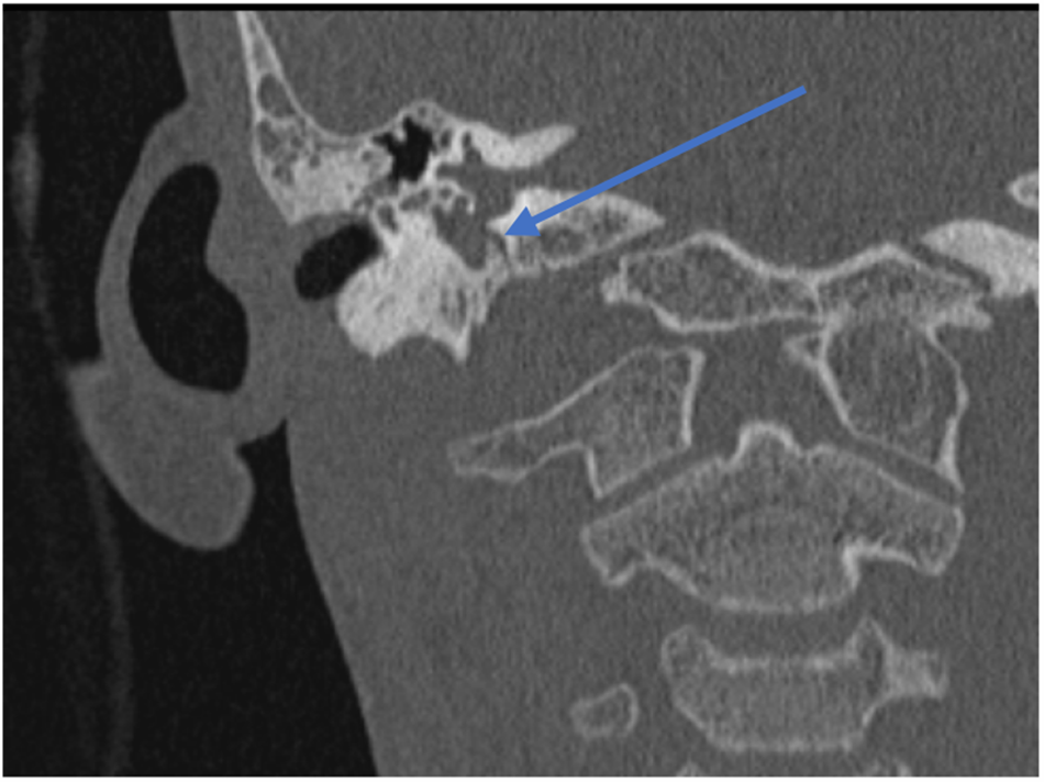
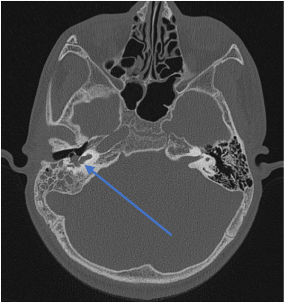
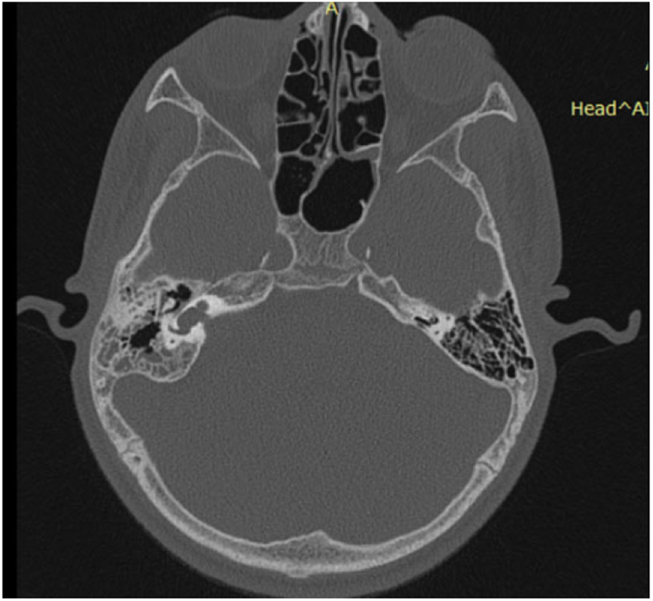
High-resolution computed tomography of the temporal bone confirmed the characteristic morphology of incomplete partition type II and additionally demonstrated a persistent tympanomeningeal suture consistent with Hyrtl’s fissure (Figures 1, 2 and 3).
Coronal view showing Hyrtl’s fissure. The arrow indicates Hyrtl’s fissure seen communicating with the middle ear.

High-resolution computed tomography temporal bone axial view. The arrow indicates soft tissue opacification seen around the round window and hypotympanum, which correspond to the sac filled with cerebrospinal fluid seen on exploratory tympanotomy.

High-resolution computed tomography axial view showing incomplete partition type II deformity.

Based on the constellation of recurrent meningitis, pulsatile middle-ear effusion, profound unilateral sensorineural hearing loss and radiologically confirmed congenital inner-ear anomaly, a congenital otogenic CSF fistula was strongly suspected.
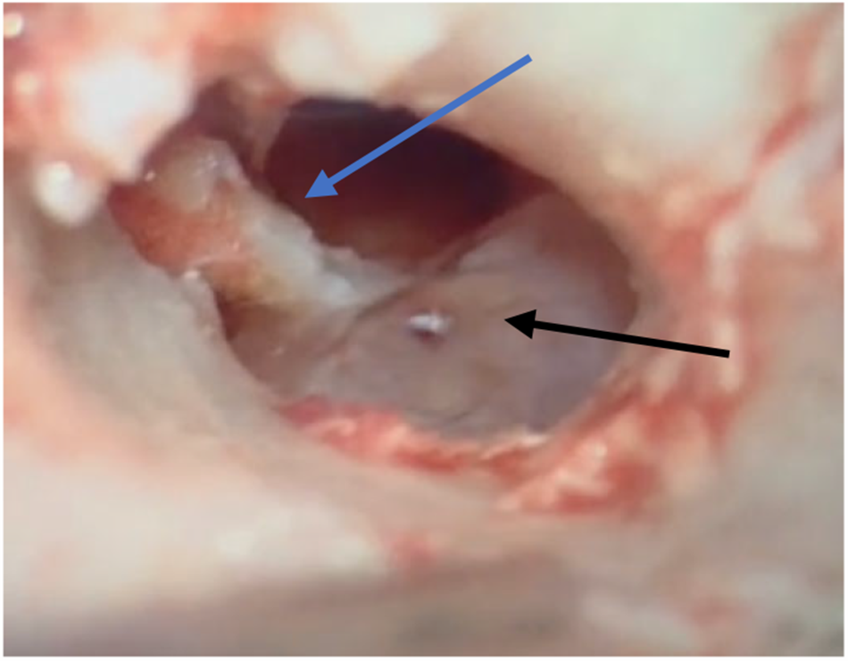
The patient underwent exploratory tympanotomy. Following elevation of the tympanomeatal flap, a translucent, pulsatile cystic sac was identified occupying the middle-ear cavity. The sac appeared tense with visible pulsation, suggestive of a CSF-filled meningeal protrusion (Figure 4).
Cerebrospinal fluid (CSF) sac seen occupying the inferior quadrants touching the handle of malleus. The blue arrow indicates the handle of malleus and the black arrow indicates the CSF-filled sac.

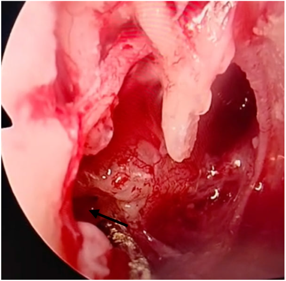
The sac was carefully aspirated and excised. It was also sent for histopathological evaluation. On excision of the cystic sac, a brisk CSF gusher was visualised emanating directly from the bony defect superior to the round window niche. The flow was clearly from the defect site, consistent with a direct communication with the posterior cranial fossa via the Hyrtl’s fissure. There was no visible CSF leakage from the round window, oval window or other inner-ear structures, suggesting that the fistula represented the primary site of CSF egress in this case (Figure 5). The defect was identified superior to the round window niche, an atypical location for classic Hyrtl‘s fissure (classically described as terminating inferior or postero-inferior to the round window), suggesting either anatomical variation or distortion by the CSF-filled sac.
Defect seen superior to round window. The arrow indicates the fissure seen in the superior part of round window.

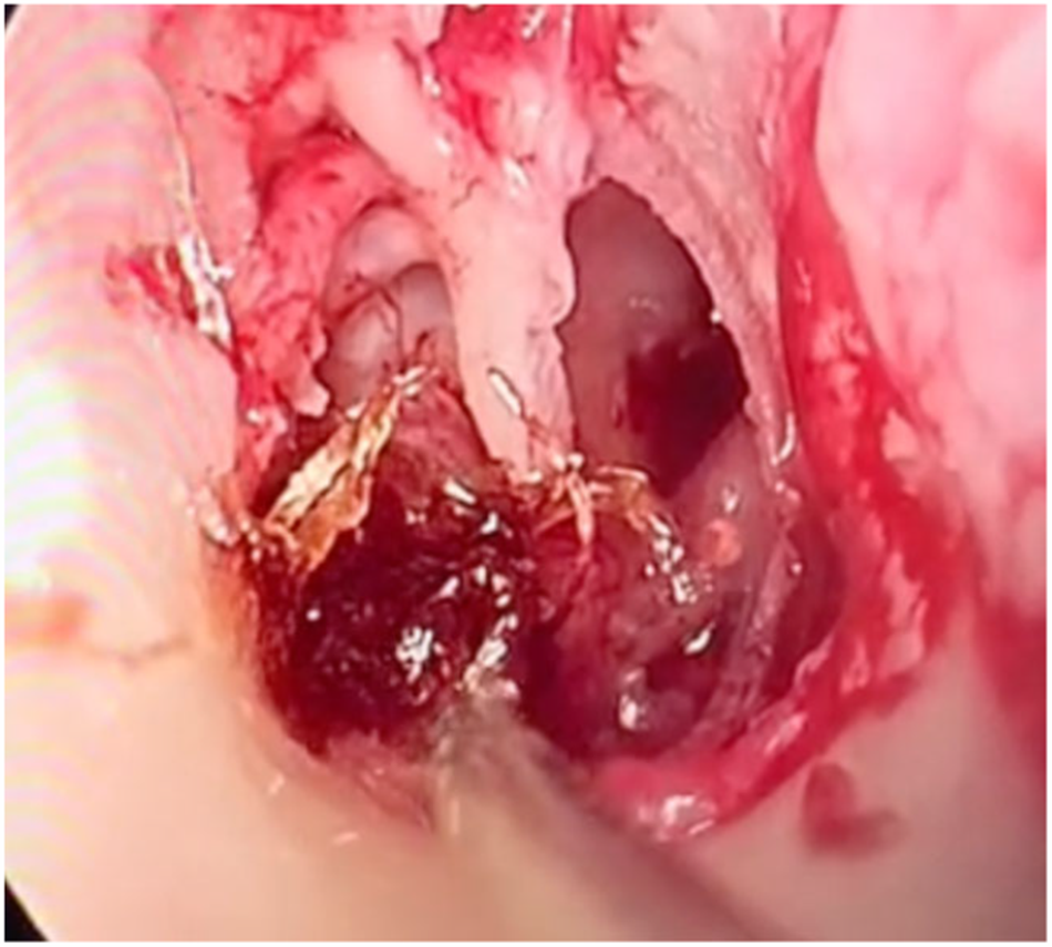
The defect was repaired using a multilayer closure technique. Surgicel was placed directly over the fistulous opening, followed by a temporalis muscle graft and an overlay temporalis fascia graft (Figure 6).
Multilayer closure of the defect being carried out.

The repair was reinforced until complete cessation of CSF flow was achieved. Cortical mastoidectomy with tympanoplasty was then completed in standard fashion. In addition to the temporal bone findings, imaging demonstrated ipsilateral sigmoid sinus thrombosis. The patient was started on therapeutic anticoagulation and remains on oral anticoagulant therapy for a planned six-month duration with follow-up imaging to assess recanalisation.
Methodology
PubMed and Google Scholar searches were performed for all English-language articles from inception to February 2026.
This systematic review was conducted in accordance with the Preferred Reporting Items for Systematic Reviews and Meta-Analyses (‘PRISMA’) guidelines. The new case report was conducted in accordance with the principles of the Declaration of Helsinki. Institutional review board approval was not required for a single retrospective case report at our institution. Written informed consent for publication of the case details and accompanying images was obtained from the patient’s parent or legal guardian.
Search strategy
PubMed and Google Scholar were systematically searched from database inception to 15 February 2026. The search strategy combined MeSH terms and keywords including ‘Hyrtl fissure’, ‘Hyrtl’s fissure’, ‘tympanomeningeal fissure’, ‘tympanomeningeal suture’, ‘persistent Hyrtl fissure’ and ‘congenital CSF otorrhea Hyrtl’. The complete PubMed search string was (‘Hyrtl*’[All Fields] AND ‘fissure’[All Fields]) OR (‘tympanomeningeal’[All Fields] AND (‘fissure’[All Fields] OR ‘suture’[All Fields])) OR (‘congenital’[All Fields] AND ‘CSF’[All Fields] AND ‘otorrhea’[All Fields] AND ‘Hyrtl’[All Fields]).
Inclusion and exclusion criteria
Studies were included if they (1) reported on human cases of Hyrtl’s fissure; (2) were published in the English language; and (3) contained original case data. Exclusion criteria included (1) non-English publications; (2) review articles without original case data; (3) animal studies; and (4) duplicate publications.
Screening process
Two independent reviewers screened titles and abstracts. Full-text review was performed for articles meeting initial screening criteria. Disagreements were resolved by consensus discussion with a third reviewer.
Duplicate handling
When duplicate publications of the same case were identified, the most comprehensive report was selected and the duplicate was excluded. Reference lists of included articles and relevant reviews were manually searched to identify additional cases.
Data extraction
A standardised data extraction form was developed to capture author, year, patient age, gender, laterality, clinical presentation, diagnostic methods, associated radiological findings, surgical approach, repair materials and outcomes.
Search results
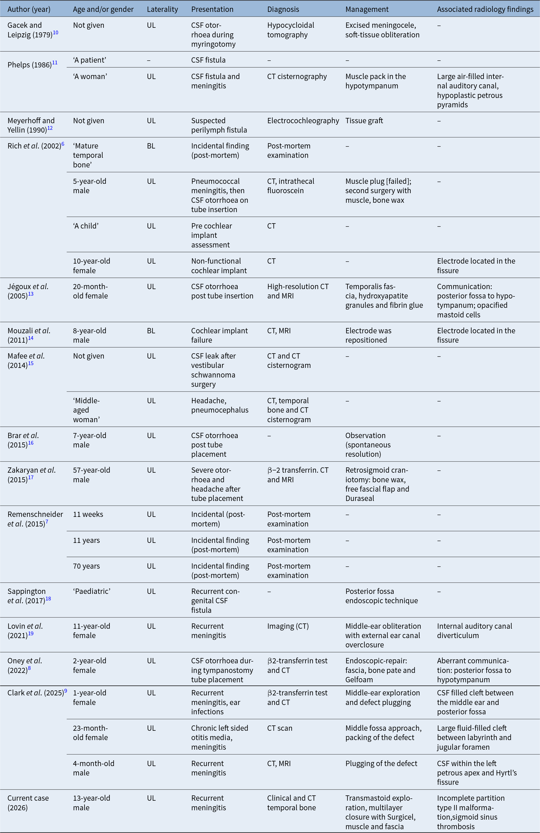
The initial search yielded 47 records from PubMed and 62 from Google Scholar (total n = 109). After removal of duplicates (n = 23), 86 records were screened. Of these, 72 were excluded based on title and/or abstract review. Full-text review of 14 articles was performed. Two additional cases were identified through manual review of reference lists. A total of 19 articles (reporting 23 cases) met the inclusion criteria and are summarised in Table 1. Including our present case, the total number of reported cases was 24.
Summary of all the cases in this review, including the new case

Table 1 Long description
Rows list published reports and one current case, with columns for author and year, age and or gender, laterality, presentation, diagnosis, management, and associated radiology findings. Laterality is usually unilateral, with bilateral cases noted for a mature temporal bone post mortem and an 8 year old male with cochlear implant failure. Presentations include cerebrospinal fluid otorrhoea during or after myringotomy or tympanostomy tube placement, cerebrospinal fluid fistula with or without meningitis, recurrent meningitis, cochlear implant failure or non functional implant, headache with pneumocephalus, and incidental post mortem findings at 11 weeks, 11 years, and 70 years. Diagnosis methods include hypocycloidal tomography, electrocochleography, computed tomography including temporal bone computed tomography, computed tomography cisternography or cisternogram, magnetic resonance imaging, intrathecal fluoroscein, beta 2 transferrin testing, imaging noted as computed tomography, and post mortem examination, with several rows showing no diagnosis method listed. Management includes excision of meningocele with soft tissue obliteration, muscle pack or plug, tissue graft, temporalis fascia with hydroxyapatite granules and fibrin glue, electrode repositioning, observation with spontaneous resolution, posterior fossa endoscopic technique, middle ear obliteration with external auditory canal overclosure, endoscopic repair using fascia bone pate and Gelfoam, middle ear exploration and defect plugging, middle fossa approach with packing of the defect, plugging of the defect, and transmastoid exploration with multilayer closure using Surgicel muscle and fascia, with multiple rows showing no management listed. Associated radiology findings are often not reported, and when present include a large air filled internal auditory canal with hypoplastic petrous pyramids, electrode located in the fissure, communication or aberrant communication from posterior fossa to hypotympanum with opacified mastoid cells, internal auditory canal diverticulum, cerebrospinal fluid filled clefts between middle ear and posterior fossa or between labyrinth and jugular foramen, cerebrospinal fluid within the left petrous apex and Hyrtls fissure, and in the current case an incomplete partition type two malformation with sigmoid sinus thrombosis.
UL = unilateral; CSF = cerebrospinal fluid; CT = computed tomography; MRI = magnetic resonance imaging; BL = bilateral
Results and analysis
The literature search identified 23 previously reported cases of Hyrtl’s fissure in the English language literature.Reference Rich, Graham and Phelps6–Reference Lovin, Appelbaum, Makoshi, Whitehead and Sweeney19 Including our present case, the total number of reported cases was 24. Table 1 provides a comprehensive summary of all 24 cases with detailed clinical information.
Demographic analysis of all reported cases (n = 24)
Analysis of the cumulative 24 cases revealed the following demographic patterns.
Age distribution
Among cases where age was stated or could be inferred (n = 22), presentation occurred in children (≤18 years) in 16 cases (72.7 per cent) and in adults in 6 cases (27.3 per cent). The mean age at diagnosis among living patients was 13.8 years (median, 7.5 years; range, 4 months to 70 years, excluding post-mortem cases).
Gender
Among cases where gender was reported (n = 14), there were 6 males (42.9 per cent) and 8 females (57.1 per cent), suggesting a possible female predominance, although the small sample size limited definitive conclusions.
Laterality
Unilateral involvement was documented in 22 cases (91.7 per cent). Bilateral involvement was documented in only 2 cases (8.3 per cent).Reference Rich, Graham and Phelps6, Reference Jégoux, Malard, Gayet-Delacroix, Bordure, Legent and Beauvillain de Montreuil13
Clinical presentations
The most common clinical presentations were cerebrospinal fluid otorrhoea following tympanostomy tube placement, meningitis, cochlear implant-related presentation, incidental findings and spontaneous CSF leak without prior surgery.
Cerebrospinal fluid otorrhoea following tympanostomy tube placement occurred in 10 cases (41.7 per cent). In six of these cases, the otorrhoea was immediate and profuse at the time of myringotomy,Reference Oney, Sloneker, Bloom, Avillion, Crawford and Chen8–Reference Gacek and Leipzig10, Reference Jégoux, Malard, Gayet-Delacroix, Bordure, Legent and Beauvillain de Montreuil13, Reference Brar, Vijendren, Salam and Picken16 while in the remaining cases it was delayed or the timing was not specified. Meningitis occurred in 8 cases (33.3 per cent). Including our case with three episodes, eight patients presented with bacterial meningitis as either the initial manifestation or part of the clinical course. Cochlear implant-related presentation occurred in 3 cases (12.5 per cent). Two cases involved cochlear implant electrode mis-insertion into the fissure,Reference Rich, Graham and Phelps6, Reference Mouzali, Ouennoughi, Haraoubia, Zemirli and Triglia14 and one case was identified during pre-operative computed tomography (CT) assessment for cochlear implantation.Reference Rich, Graham and Phelps6 Incidental findings occurred in 4 cases (16.7 per cent), including three post-mortem histopathological identificationsReference Remenschneider, Lookabaugh and Aliphas7 and one imaging finding during workup for an unrelated condition.Reference Mafee, Kumar, Heffez, Mafee, Valvassori and Becker15 Spontaneous CSF leak without prior surgery occurred in 2 cases (8.3 per cent).Reference Gacek and Leipzig10, Reference Phelps11
Diagnostic methods
High-resolution CT of the temporal bone was the primary imaging modality in 19 of 24 cases (79.2 per cent). The characteristic finding was a linear bony defect or communication between the posterior cranial fossa and the hypotympanum, typically located inferior or posteroinferior to the round window niche.
β2-transferrin testing confirmed the presence of CSF in middle-ear fluid in six cases.Reference Oney, Sloneker, Bloom, Avillion, Crawford and Chen8, Reference Clarke, Manov, Ndolo, Githu, Saigal and Saigal9, Reference Brar, Vijendren, Salam and Picken16, Reference Zakaryan, Poulsgaard, Hollander and Fugleholm17 Magnetic resonance imaging (MRI) was obtained in five casesReference Clarke, Manov, Ndolo, Githu, Saigal and Saigal9, Reference Jégoux, Malard, Gayet-Delacroix, Bordure, Legent and Beauvillain de Montreuil13, Reference Zakaryan, Poulsgaard, Hollander and Fugleholm17, Reference Lovin, Appelbaum, Makoshi, Whitehead and Sweeney19 and provided complementary information by demonstrating fluid-filled spaces and ruling out other inner-ear anomalies, but was not essential for diagnosis of the fissure itself.
Intra-operative diagnosis occurred in several cases where the fissure was directly visualised during tympanotomy or mastoid exploration,Reference Clarke, Manov, Ndolo, Githu, Saigal and Saigal9, Reference Gacek and Leipzig10, Reference Meyerhoff and Yellin12, Reference Jégoux, Malard, Gayet-Delacroix, Bordure, Legent and Beauvillain de Montreuil13 including our case.
Associated radiological findings
Our case is unique in demonstrating the coexistence of Hyrtl’s fissure with incomplete partition type II (Mondini) malformation. Other associated findings reported in the literature include CSF within the left petrous apex and Hyrtl’s fissure,Reference Clarke, Manov, Ndolo, Githu, Saigal and Saigal9 internal auditory canal diverticulum communicating with jugular bulb,Reference Lovin, Appelbaum, Makoshi, Whitehead and Sweeney19 demineralised bone around jugular bulb,Reference Lovin, Appelbaum, Makoshi, Whitehead and Sweeney19 bilateral petrous pyramid hypoplasia,Reference Phelps11 B-type brachydactyly,Reference Jégoux, Malard, Gayet-Delacroix, Bordure, Legent and Beauvillain de Montreuil13 bilateral prominent auricles and congenital hearing loss,Reference Brar, Vijendren, Salam and Picken16 and sigmoid sinus thrombosis (our case, likely secondary to otogenic infection).
Surgical management
Surgical repair was undertaken in 17 of 24 cases (70.8 per cent). One case demonstrated spontaneous resolution without surgical intervention.Reference Brar, Vijendren, Salam and Picken16 Surgical approaches varied widely and included transcanal and/or endaural approach (n = 6) involving direct access through the ear canal for localised repairReference Clarke, Manov, Ndolo, Githu, Saigal and Saigal9, Reference Gacek and Leipzig10, Reference Meyerhoff and Yellin12, Reference Jégoux, Malard, Gayet-Delacroix, Bordure, Legent and Beauvillain de Montreuil13 (our case), a post-auricular transmastoid approach (n = 3) that provided wider exposure of the middle ear and mastoid,Reference Rich, Graham and Phelps6, Reference Oney, Sloneker, Bloom, Avillion, Crawford and Chen8 retrosigmoid craniotomy (n = 1), which addressed the fistula from its intracranial aspect,Reference Zakaryan, Poulsgaard, Hollander and Fugleholm17 a posterior fossa endoscopic approach (n = 1) with a minimally invasive intracranial repair,Reference Sappington, Sappington and Bickel18 combined or unspecified approaches (n = 5) and a middle fossa approach.Reference Clarke, Manov, Ndolo, Githu, Saigal and Saigal9
Repair materials included autologous tissues (temporalis fascia, muscle, fat) in all surgical cases, often combined with synthetic materials or bone products (bone wax, bone pate, hydroxyapatite, Gelfoam, Duraseal, fibrin glue). The multilayer closure technique was employed in most successful repairs.
Among surgically treated cases with reported outcomes (n = 14), successful repair without recurrence was achieved in 12 cases (85.7 per cent). One case recurred at 15 months and required revision with bone wax augmentation.Reference Rich, Graham and Phelps6 Another case with multiple defects (Hyrtl’s fissure plus internal auditory canal diverticulum) recurred after initial obliteration and required more aggressive middle-ear obliteration with external auditory canal overclosure for definitive control.Reference Lovin, Appelbaum, Makoshi, Whitehead and Sweeney19
Discussion
Hyrtl’s fissure represents an embryological communication between the posterior cranial fossa and the hypotympanum that fails to involute during normal development.Reference Hyrtl5, Reference Rich, Graham and Phelps6, Reference Spector, Pettit, Davis, Strauss and Rauchbach20 Spector et al. established the definitive developmental timeline through comprehensive histological analysis of 55 fetuses and neonates, with their 1981 study providing detailed characterisation of the tympanomeningeal fissure and its relationship to the cochlear aqueduct and periotic duct.Reference Spector, Pettit, Davis, Strauss and Rauchbach20, Reference Kuczkowski, Dubaniewicz-Wybieralska, Przewoźny, Narozny and Mikaszewski21 This work demonstrated that the fissure runs with the cochlear aqueduct and cochlear vein in the periotic duct, located just medial to the round window niche.Reference Kuczkowski, Dubaniewicz-Wybieralska, Przewoźny, Narozny and Mikaszewski21 Between the 20th and 24th weeks of gestation, progressive ossification occurs until the fissure is completely obliterated as the bone of the jugular bulb and cochlear fuse.Reference Rich, Graham and Phelps6, Reference Kuczkowski, Dubaniewicz-Wybieralska, Przewoźny, Narozny and Mikaszewski21 When fusion fails, a persistent connection remains between the hypotympanum and posterior fossa. The extreme rarity of this condition is underscored by the fact that only 24 cases have been reported in over four decades of English literature (1979–2026).
Our case represents the first documented instance of incomplete partition type II (Mondini deformity) coexisting with persistent Hyrtl’s fissure. This combination is significant for several reasons. Firstly, it raises the possibility of a more extensive developmental field defect involving multiple temporal bone structures during embryogenesis, rather than isolated arrested organogenesis. Both anomalies originate during the first and second trimesters: incomplete partition type II from arrested cochlear development at the seventh week and Hyrtl’s fissure from failure of otic capsule ossification between the 20th and 24th weeks. However, as this is a single case report, we cannot establish causation or a common embryological mechanism. The differing gestational time points may alternatively support the explanation that these are independent embryological events occurring coincidentally.
Secondly, this combined anomaly creates multiple potential independent pathways for CSF egress rather than a single continuous pathway. The incomplete partition type II malformation facilitates transmission of CSF from the subarachnoid space to the perilymphatic compartment via deficient modiolar architecture.Reference Jackler, Luxford and House4 Hyrtl’s fissure, conversely, represents a direct communication between the posterior cranial fossa and the middle-ear cavity. In our patient, the presence of both anomalies provides two independent potential routes for CSF leakage. From the perilymphatic space, CSF could potentially access the middle ear through the patent Hyrtl’s fissure; however, this would require a connection between the perilymphatic space and the fissure that has not been anatomically confirmed. Alternatively, these may represent distinct pathways without functional interconnection. This dual-pathway mechanism, whether interconnected or independent, increases both the risk of spontaneous CSF fistula and the complexity of surgical management.
Thirdly, the intra-operative CSF gusher observed on excision of the cystic sac likely reflects high CSF pressure within a relatively large fistulous communication. We hypothesise that the concurrent incomplete partition type II malformation, by providing an additional CSF–perilymph communication, may have contributed to the high-flow characteristics of this system; however, this remains speculative and the gusher could equally be explained by the size and nature of the Hyrtl’s fissure alone. The presence of sigmoid sinus thrombosis in our case may have further elevated intracranial pressure and contributed to the force of the gusher.
It is important to acknowledge that the fistulous opening in our case was identified superior to the round window niche, which represents an atypical location for a classic Hyrtl’s fissure (classically described as terminating inferior or posteroinferior to the round window). This may reflect anatomical variation, distortion by the CSF-filled cystic sac. However, the radiographic finding of a persistent tympanomeningeal fissure, the intra-operative communication with the posterior fossa and the clinical presentation are consistent with the diagnosis.
We acknowledge that the superior location is unusual and suggest that careful intra-operative correlation with imaging is essential to confirm the diagnosis. The literature consistently describes Hyrtl’s fissure terminating in the hypotympanum inferior or postero-inferior to the round window niche. The superior location in our case is atypical; however, given the size and tension of the CSF-filled sac, anatomical distortion may have altered the apparent location of the fistulous opening or this may represent a variant termination of the fissure tract. We emphasise that the combination of imaging findings, intra-operative communication with the posterior fossa and clinical presentation support the diagnosis, although the atypical location warrants careful case-by-case evaluation.
The mechanism of meningitis in this context is ascending infection through the patent Hyrtl’s fissure. Cerebrospinal fluid leaking into the middle ear passes through the Eustachian tube to reach the nasopharynx, establishing a direct conduit for nasopharyngeal flora, most commonly Streptococcus pneumoniae, to ascend into the subarachnoid space.Reference Sanna, Fois, Russo and Falcioni22 In many such cases, the tympanic membrane remains intact and the only otoscopic clue is a pulsatile effusion, as observed in our patient.
The triad of findings – (1) recurrent bacterial meningitis; (2) ipsilateral profound sensorineural hearing loss; and (3) pulsatile middle-ear effusion – should alert clinicians to an otogenic origin of CSF leakage and warrants comprehensive imaging evaluation.
Clinical pearls for diagnosis
Hyrtl’s fissure may be subtle on high-resolution CT and is often overlooked. The key to diagnosis is careful review of the hypotympanum and subiculum region, where the fissure typically terminates. The defect may appear as a linear lucency or communication between the posterior cranial fossa and the middle-ear cavity. In cases where the fissure is not clearly visualised, intrathecal contrast (CT cisternography) or β2-transferrin testing of middle-ear fluid may provide confirmatory evidence. Importantly, the presence of one anomaly (e.g. incomplete partition type II) does not exclude the presence of another. Clinicians should evaluate the entire temporal bone for multiple potential sites of CSF leakage because dual anomalies may coexist.
Proposed diagnostic algorithm
For children with recurrent meningitis and unexplained unilateral hearing loss the diagnostic algorithm is as follows: (1) urgent high-resolution CT temporal bone with thin-section (≤0.6 mm) coronal and axial views; (2) review specifically for incomplete partition type II, enlarged vestibular aqueduct and persistent fissures (Hyrtl’s, tympanomeningeal); (3) if imaging is equivocal, MRI with CISS (Constructive Interference in Steady State) and FIESTA (Fast Imaging Employing Steady-State Acquisition) sequences to evaluate fluid-filled tracts; (4) β2-transferrin testing of middle-ear fluid (if obtainable via myringotomy); (5) consider CT cisternography if fistula is suspected but not clearly visualised; and (6) surgical exploration if clinical suspicion remains high despite negative imaging.
High-resolution computed tomography of the temporal bone remains the imaging modality of choice, allowing direct visualisation of the persistent fissure as a linear defect extending from the posterior cranial fossa to the middle-ear cavity, typically located inferior to the round window niche. In our case, high-resolution CT clearly demonstrated both the characteristic incomplete partition type II morphology and the subtle linear defect of Hyrtl’s fissure. Importantly, the fissure can be subtle and was missed on initial imaging in two reported cases,Reference Mouzali, Ouennoughi, Haraoubia, Zemirli and Triglia14, Reference Mafee, Kumar, Heffez, Mafee, Valvassori and Becker15 emphasising the need for scrutiny by both radiologist and surgeon. Magnetic resonance imaging offers complementary information by delineating fluid-filled spaces and identifying associated complications. β2-transferrin testing of middle-ear fluid provides biochemical confirmation when obtainable.Reference Oney, Sloneker, Bloom, Avillion, Crawford and Chen8, Reference Brar, Vijendren, Salam and Picken16, Reference Zakaryan, Poulsgaard, Hollander and Fugleholm17
An important associated finding in our case was ipsilateral sigmoid sinus thrombosis. This likely represents an otogenic complication secondary to the recurrent bacterial meningitis and chronic otogenic infection. Sigmoid sinus thrombosis in the context of CSF fistula and meningitis is a known but uncommon complication, and its presence has implications for management. Elevated intracranial pressure from sinus thrombosis may have contributed to the high-flow CSF gusher observed intra-operatively by impairing venous drainage and increasing CSF pressure gradients. Additionally, the presence of sigmoid sinus thrombosis necessitated anticoagulation therapy, which must be carefully co-ordinated with surgical management because of the risk of post-operative bleeding. We recommend that patients with otogenic CSF fistulas and meningitis undergo venous phase imaging to evaluate for dural venous sinus thrombosis, particularly in the presence of persistent headache, papilledema or seizures.
Surgical repair is the definitive management. A transmastoid approach with posterior tympanotomy allows wide exposure of the middle ear, facilitating identification of the fistulous site at the tympanic end of Hyrtl’s fissure and enabling multilayer closure. For cases with multiple skull base defects or signs of intracranial hypertension, there is higher risk of recurrenceReference Lovin, Appelbaum, Makoshi, Whitehead and Sweeney19; more aggressive management with middle-ear obliteration and external auditory canal overclosure may be necessary. In cases with non-serviceable hearing, as in our patient, subtotal petrosectomy may be considered if simpler repair fails, although localised multilayer closure directly addressing the patent fissure remains first-line treatment when the fistula can be clearly identified.
Factors associated with recurrence include multiple skull base defects, signs of intracranial hypertension and incomplete initial repair.Reference Lovin, Appelbaum, Makoshi, Whitehead and Sweeney19 Patients with these risk factors may require more aggressive management with middle-ear obliteration and external auditory canal overclosure.
The principles of successful repair include precise identification of the fissure’s tympanic opening, meticulous preparation of the recipient bed, use of autologous tissue grafts and multilayer closure to provide redundancy. The intra-operative CSF gusher encountered in our case and several othersReference Clarke, Manov, Ndolo, Githu, Saigal and Saigal9, Reference Gacek and Leipzig10, Reference Jégoux, Malard, Gayet-Delacroix, Bordure, Legent and Beauvillain de Montreuil13 underscores the need for surgical preparedness with appropriate materials available for immediate closure. Post-operative measures should include strict bed rest, head elevation and prophylactic antibiotics. Long-term follow up is essential because delayed failures can occur.Reference Rich, Graham and Phelps6, Reference Lovin, Appelbaum, Makoshi, Whitehead and Sweeney19
An interesting observation in our case and three previous reportsReference Clarke, Manov, Ndolo, Githu, Saigal and Saigal9, Reference Gacek and Leipzig10, Reference Jégoux, Malard, Gayet-Delacroix, Bordure, Legent and Beauvillain de Montreuil13 was the presence of a CSF-filled cystic sac within the middle ear. This may represent either a meningocele or mucosal herniation driven by chronic CSF pressure. When present, this sac should be carefully dissected and ligated at its base to expose the underlying fissure for definitive repair. The nature of this sac remains uncertain, although biopsy in one case revealed respiratory mucosa rather than meningeal tissue.Reference Jégoux, Malard, Gayet-Delacroix, Bordure, Legent and Beauvillain de Montreuil13
Limitations
This study has several important limitations. Firstly, as a single case report, the findings cannot be generalised, and the coexistence of incomplete partition type II and Hyrtl’s fissure may represent a chance association rather than a true developmental field defect. Secondly, we cannot definitively establish a functional connection between the incomplete partition type II malformation and the CSF leak through Hyrtl’s fissure. Thirdly, the literature review is limited by publication bias, incomplete case reporting and the small total number of cases. Fourthly, follow up in our case is limited to one year and long-term outcomes remain to be determined. We encourage future reports to include detailed intra-operative photographs and long-term follow-up data to better characterise this rare entity.
Conclusion
Hyrtl’s fissure (tympanomeningeal suture) is an exceedingly rare congenital anomaly resulting from incomplete obliteration of a fetal connection between the hypotympanum and posterior fossa. Including our case, only 24 cases have been reported in the English literature over four decades. Presentation occurs more commonly in children (72.7 per cent) than adults, is almost always unilateral (91.7 per cent) and most frequently manifests as CSF otorrhoea following tympanostomy tube placement (41.7 per cent) or as recurrent meningitis (33.3 per cent).
Our case represents the first documented coexistence of incomplete partition type II (Mondini deformity) and persistent Hyrtl’s fissure. This combination suggests a more extensive developmental field defect involving multiple temporal bone structures and creates multiple potential pathways for CSF egress, increasing the complexity of surgical management. The triad of recurrent bacterial meningitis, ipsilateral profound sensorineural hearing loss and pulsatile middle-ear effusion should prompt immediate evaluation for congenital CSF fistula.
High-resolution CT of the temporal bone is the imaging modality of choice for diagnosis, with careful attention to the region just inferior to the round window niche where Hyrtl’s fissure typically terminates. Surgical repair with multilayer closure using autologous tissues is successful in most cases, although patients with multiple defects or signs of intracranial hypertension may require more aggressive management with middle-ear obliteration.
Increased awareness of this rare entity among paediatricians, neurologists, radiologists and otolaryngologists is essential for timely diagnosis and prevention of life-threatening recurrent meningitis. To our knowledge, this is the first report documenting the radiological finding, clinical presentation and intra-operative correlation of persistent Hyrtl’s fissure coexisting with incomplete partition type II malformation.
Competing interests
None declared