Introduction
Non-alcoholic fatty liver disease (NAFLD) has become the most common cause of chronic liver disease worldwide. It is closely associated with obesity and insulin resistance. Its prevalence has risen rapidly in parallel with the dramatic global rise of obesity and type 2 diabetes mellitus (T2D) (Ref. Reference Farrell, Wong and Chitturi1). The pooled prevalence of NAFLD is 24.24% (Ref. Reference Zhu2), and the global prevalence positively correlates with economic status. Western countries have a higher prevalence (28.04%) than the Middle East (12.95%) and East Asia (19.24%). Additionally, males have a higher prevalence than females (Ref. Reference Zhu2). The spectrum of NAFLD ranges from simple steatosis, NASH to cirrhosis, which may ultimately progress to hepatocellular carcinoma (HCC) (Ref. Reference Bugianesi3). Previous data suggested that ~6–8% of US adults (about 25% of those with NAFLD) suffer from NASH (Ref. Reference Clark, Brancati and Diehl4). Moreover, NASH associated cirrhosis is now the third most common cause for liver transplantation in the USA (Ref. Reference Tiniakos, Vos and Brunt5).
Despite the growing prevalence of NAFLD, the pathogenesis of NAFLD and NASH is not yet fully understood. The original theory of the pathogenesis of NASH is the ‘two-hit’ theory. The first hit is hepatic steatosis, which is a milder condition, whereas the second hit comprises oxidative stress and inflammation that leads to severe liver damage. It is now most accepted that NASH is a complex multifactor disease. Identified factors that contribute to NASH progression include metabolic factors, innate immune alterations, inflammation caused by free fatty acids (FFA), bacterial lipopolysaccharide (LPS), chemokines, cytokines, and adipokines (Ref. Reference Tilg and Moschen6).
The C-X-C motif chemokine 10 (CXCL10, also called IP-10) was found to play a key role as an inflammatory mediator in infectious diseases, autoimmune diseases and metabolic diseases. It has also been reported that CXCL10 is involved in the development of specific features of the NAFLD phenotype in human, independent of age, sex, obesity, insulin resistance and T2D (Ref. Reference Wolfs7). Our recent study revealed that CXCL10 is critical in the pathogenesis of NASH by inducing inflammation, regulating lipogenesis and oxidative stress (Ref. Reference Zhang8). Concomitantly, CXCR3, the cognate receptor of CXCL10, could promote steatohepatitis in mice through mediating inflammatory cytokines, macrophage and autophagy (Ref. Reference Zhang9). This review summarises the information to date about the role of CXCL10 in NASH with the aim of clarifying its molecular pathogenic mechanism in order to aid the development of novel therapeutic options.
General aspects of CXCL10
Definition, structure and function
Chemokines (chemotactic cytokines) are a class of small weight heparin-binding proteins (8–13 kD) that direct the movement of circulating immune cells to sites of inflammation or injury. They are categorised into four different families (CC, CXC, CX3C, C) based on the presence of NH2 terminal cysteine motifs (Ref. Reference Charo and Ransohoff10). The CXC family can be further subdivided into glutamine (E), leucine (L) and arginine (R) (ELR)-positive and ELR-negative CXC chemokines (Ref. Reference Simpson11).
CXCL10 is a small-molecular protein (10 kD) that belongs to the ELR-negative CXC family. It was originally identified in human U937 cells, a histiocytic lymphoma cell line with monocytic characterisation, and from genomic placenta and spleen of human as an Interferon gamma (IFN-γ)-inducible product (Ref. Reference Epstein and Luster12). The CXCL10 gene is located on chromosome 4 and contains four exons and three introns (Ref. Reference Luster and Ravetch13). CXCL10 consists of three anti-parallel β-sheets overlaid by a α-helix at its C-terminus. The N-loop region of the β-sheets has been suggested to be crucial for the binding of the protein to the receptor (Ref. Reference Ahmadi, Arababadi and Hassanshahi14). The primary translation product of the CXCL10 gene is a 12 378-Da protein composed of 98 amino acids containing two internal disulphide cross bridges. An unmodified mature protein with a relative molecular mass of 9979, composed of 77 amino-acid residues, is secreted from this polypeptide under peptidase cleavage (Ref. Reference Chen15). At the molecular level, CXCL10 promoter contains binding sites for different transcription factors: RelA-p65, JunD, NF-E2 p45, SRF, Sp1, NF-YA, -YB, E2F, STAT1 and RNA pol-II. Transcription factors including BCL6 and ErbB1 suppress CXCL10 expression (Ref. Reference Rajeev Mehla and Ayyavoo16).
CXCL10's receptor, CXCR3, is a seven trans-membrane-spanning G protein-coupled receptor shared with CXCL4 (PF4), CXCL9 (Mig) and CXCL11 (I-TAC) (Ref. Reference Clark-Lewis17). By binding to CXCR3, CXCL10 recruits activated CXCR3+ T lymphocytes to the liver parenchyma and plays a pivotal role in liver disease initiation and progression. CXCR3 has two different isoforms: CXCR3-A and CXCR3-B. Several studies revealed that the two CXCR3 isoforms differentially regulate cell function. The CXCL10/CXCR3-A-axis contributes to the induction of chemotaxis and proliferation in various cell types, whereas the CXCL10/CXCR3-B-axis inhibits migration and proliferation but induces apoptosis (Ref. Reference Lasagni18).
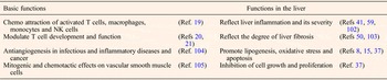
The precise biological functions of CXCL10 are yet to be completely identified. It is a pleiotropic molecule capable of exerting various biological functions by promoting the chemotactic activity of CXCR3+ cells (Ref. Reference Erhardt and Tiegs19). CXCL10 can modulate T cell development and function (Refs Reference Dufour20, Reference Jaruga21), as well as mitogenic and chemotactic effect on vascular smooth muscle cells (Ref. Reference Wallace22). It can also induce apoptosis and cell migration and regulate cell proliferation and angiogenesis in infectious and inflammatory diseases and cancer (Table 1).
Functions of CXCL10

Source and regulation of CXCL10
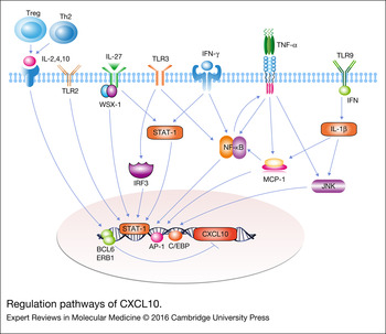
CXCL10 was initially identified as a chemokine that is induced by IFN-γ and secreted by both immune cells (monocytes, neutrophils and dendritic cells) (Refs Reference Dufour20, Reference Ciesielski23, Reference Ge24), and non-immune cells (endothelial cells, keratinocytes, fibroblasts, mesenchymal cells, astrocytes and hepatocytes) (Refs Reference Teijaro25, Reference Shields26, Reference Treacy27, Reference Hassanshahi28, Reference Kanda29, Reference Fukui30, Reference Phares31). CXCL10 induction is regulated by multiple mechanisms (Fig. 1): (1) regulatory T cells (Treg)-derived antiinflammatory cytokines (IL-10, IL-2) transmit intracellular signals to suppress CXCL10 induction through the inhibition of tumour necrosis factor (TNF-α) and IFN-γ directly or indirectly (Ref. Reference Sarfo32); (2) T helper cells 2 (Th2)-derived cytokines, or those that promote differentiation of naive T-cells into Th2 cells (e.g. IL-4), can suppress CXCL10 production and reduce inflammation at the site of infection (Ref. Reference Albanesi33); (3) in human liver progenitor cells, CXCL10 can be produced by Toll like receptor 3 (TLR3) and retinoic acid-inducible gene 1 (RIG-1), whereas the other mediators such as interferon regulatory factor 3 (IRF-3), p38 mitogen-activated protein kinases (p38 MAPK), and extracellular signal-regulated kinases (ERK) are partially involved (Ref. Reference Maire34); (4) in hepatitis C virus infection, CXCL10 is induced by endogenous expression of hyaluronan through an engagement of toll like receptor 2 (TLR2) and CD44 in hepatocytes (Ref. Reference Abe35); (5) in acute hepatitis, CXCL10 can be induced by Interleukin 27 (IL-27) in IFN-γ-dependent manner in hepatic cells, suggesting a complementary role of IL-27, WSX-1 (a subunit of IL-27 receptor) and IFN-γ in moderating liver inflammation by the regulation of CXCL10 (Ref. Reference Basset36); (6) in primary human hepatocytes, CXCL10 can be induced by interferon lambda 4 (IFN-λ4), which is a novel type-III interferon (Ref. Reference Onabajo37); (7) TNF-α and IFN-γ employ STAT1α and NF-κB, respectively, for CXCL10 expression in human fibroblast cell lines and LPS-stimulated KCs (Ref. Reference Kopydlowski38) (Fig. 1). Synergy was observed between TNF-α and IFN-γ regarding the induction of CXCL10 expression by different cell systems such as keratinocytes and hepatocytes. This synergistic effect is facilitated by NF-κB and interferon-sensitive response element (ISRE) in the promoter of CXCL10 (Ref. Reference Vazirinejad39). However, interferons are not necessarily required for CXCL10 induction. NF-κB and IRF3 can activate the CXCL10 promoter independent of type I or type III IFN signalling (Ref. Reference Brownell40) (Fig. 1).
Regulation pathways of CXCL10. (a) Treg-derived anti-inflammatory cytokines could transmit intracellular signals to suppress CXCL10 induction; (b) CXCL10 could be induced by endogenous expression of hyaluronan through an engagement of TLR2 and IL-27; (c) TLR3 through NF-κB signalling pathway; (d) IRF-3 and JNK/AP-1 pathway; (e) TNF-α and IFN-γ employ STAT1α and NF-κB for CXCL10 expression; (f) TLR9 activates IFN-γ regulatory factors that induce production of IL-1β, leading to production of CXCL10. AP-1, Activator protein-1; IFN-γ, Interferon gamma; IRF3, interferon regulatory factor 3; IL-1β, Interleukin 1β; JNK, Jun N-terminal kinase; NF-κB, nuclear factor kappaB; STAT1, signal transducer and activator of transcription 1; TLR, toll like receptor; Treg, regulatory T cells.

Role of CXCL10 in the liver
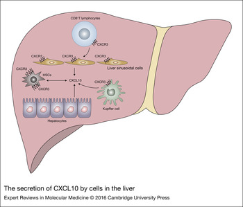
Hepatocytes, which conduct the metabolic, biosynthetic and biliary secretory functions in the liver, are important elements of liver inflammation in NASH. In the liver, CXCL10 is mainly secreted by hepatocytes located in areas of lobular inflammation, however CD8+ T lymphocytes can migrate towards it (Refs Reference Zeremski41, Reference Nishioji42) (Fig. 2). It has been shown to aggravate hepatic inflammatory conditions in patients with liver diseases such as hepatitis B, hepatitis C, autoimmune hepatitis and T cell hepatitis induced by Con A (Refs Reference Jaruga21, Reference Deng43, Reference Lagging44). Isolated hepatocytes can also produce CXCL10 after injection of αCD40 and IL-2 under interferon- γ (IFN- γ) induction (Ref. Reference Kimura45).
The secretion of CXCL10 by cells in the liver. CXCR3-expressing CD8+ T cells, hepatocytes, HSCs, Kupffer cells and liver sinusoidal cells that are recruited to the site of infection contribute to CXCL10 secretion. HSCs, hepatic stellate cells.

Hepatic stellate cells (HSCs) express functional chemokines and receptors, including CXCR3, CCL2, CCL3, CCL5, CXCL1, CXCL8, CXCL9 and CXCL10 (Refs Reference Wasmuth46, Reference Holt47, Reference Bonacchi48). Stimulation of CXCR3 expressing HSCs enhances their migration through the phosphorylation of ERK (Ref. Reference Holt47). In addition, a study using CXCL10-deficient mice revealed that CXCR3 expressing HSCs can both secrete and be activated by CXCL10 when stimulated with IFN- γ (Ref. Reference Hintermann49) (Fig. 2).
Liver sinusoidal endothelium is another important source of CXCL10 in the liver. Immunohistochemistry analysis in HCV infected tissue samples showed that CXCL10 is expressed in sinusoidal endothelial cells. CXCL10 expression strongly correlates with the number of transcripts for IFN- γ and is selectively upregulated in patients with chronic hepatitis B (CHB) (Ref. Reference Harvey50).
CXCL10 can also be produced by macrophages, which are a group of mononuclear cells that participate in the innate immune cell response. Kupffer cells (KCs), the intrahepatic form of macrophages, are a primary source of hepatic pro-inflammatory cytokines. Secretion of IL-6, IL-1β and CXCL10 by KCs from high fat diet (HFD)-fed mice is significantly higher than that from mice fed with normal diet after lipopolysaccharide (LPS) challenge (Ref. Reference Leroux51). KCs are essential for the early phase of NASH development as indicated by the ameliorated lipid accumulation and insulin resistance by KCs depletion in high-fat high-sucrose diet-fed mice (Refs Reference Tosello-Trampont52, Reference Huang53). Based on the presence of specific receptors and on the cytokines and chemokines they produce, macrophages are classified as M1 macrophage (pro-inflammatory) and M2 macrophage (anti-inflammatory). CXCL10 and other pro-inflammatory cytokines are mainly induced by M1 macrophages, the increase of which plays an important role in NASH pathogenesis (Refs Reference Maina54, Reference Odegaard55).
CXCL10 and NASH
CXCL10 plays a key role in liver inflammation
Inflammation may result from oxidative stress and pro-inflammatory chemokines and cytokines, which perpetuate liver injury and lead to fibrosis (Ref. Reference Farrell56). Inflammation can also promote insulin resistance, which is a feature of NASH, and is therefore thought to be the driving force for NASH progression (Refs Reference Larter and Farrell57, Reference Harmon, Tiniakos and Argo58). It has been reported that CXCL10 levels are correlated with the degree of inflammation in chronic hepatitis C infection (Refs Reference Zeremski41, Reference Mihm, Schweyer and Ramadori59).
Liver innate immune cells, including macrophages, neutrophils, dendritic cells and natural killer cells (NK cells), are involved in NASH progression. CXCL10-deficient mice showed less B- and T lymphocyte, and dendritic cell infiltrations. Neutrophils may induce ongoing inflammation in hepatocytes and oxidative stress, thereby promoting NASH development. The neutrophil-to-lymphocyte ratio is higher in NASH patients and it is related to the severity of NASH (Ref. Reference Alkhouri60). Neutralization of CXCL10 reduces the recruitment of NK cells to the liver after Hepatitis B virus infection (Ref. Reference Kakimi61). Dendritic cells also play a regulatory role in NASH (Ref. Reference Alkhouri60). Treg cells and Th17 cells are associated with NASH progression (Ref. Reference Ganz and Szabo62).
We have found that CXCL10 acts as a key pro-inflammatory cytokine through amplifying the effects of other pro-inflammatory cytokines, including TNF-α, Interleukin 6 (IL-6), Interleukin 1β (IL-1β), monocyte chemoattractant protein 1 (MCP-1), interferon gamma (IFN-γ) and toll like receptor 9 (TLR9). The active role of these cytokines is crucial in the progression of NAFLD not only by inducing hepatic inflammation but also by promoting hepatocyte apoptosis, necrosis and fibrosis (Refs Reference Tosello-Trampont52, Reference Marra and Tacke63). TNF-α produced by intrahepatic KCs can promote NASH development in both human NASH and experimental steatohepatitis by activating neutrophils and causing insulin resistance (Refs Reference Harvey50, Reference Romero64). Blockade of TNF-α might represents a novel therapeutic target in NASH with the potential to limit tissue injury and possibly prevent the progression to severe liver disease (Ref. Reference Tosello-Trampont52). Similar to TNF-α, IL-1β can activate neutrophils, promote fibrosis and cause insulin resistance. TNF-α and IL-1β are able to induce MCP-1 in vitro (Ref. Reference Mühlbauer65). MCP-1 is also an important molecule in NASH as it may bridge inflammatory responses with the induction of insulin resistance (Ref. Reference Kassel66). Moreover, MCP-1 can stimulate lipogenesis to promote steatosis in the liver, allowing inflammation to exacerbate steatosis (Ref. Reference Rull67). IFN-γ is a major inducer of CXCL10 related to NASH pathogenesis. TLR9 activates IFN-γ regulatory factors that induce production of IL-1β, leading to NASH development in mouse model. In addition to IFN-γ and TLR9, a close correlation between CXCL10 and TNF-α, MCP-1, IL-6 has been well documented (Refs Reference Wong68, Reference Tamimi69). This suggests that CXCL10 induces cytokine expression, leading to liver inflammation.
Most of the above mentioned cytokines and inflammatory factors are regulated by NF-κB signalling pathway, which is a key regulator of early hepatic inflammation and liver injury in NASH (Ref. Reference Leite70). NF-κB activation in macrophages initiates the inflammatory cascade and regulates inflammation through cytokine-directed cell differentiation (Ref. Reference Baker, Hayden and Ghosh71). NF-κB is a transcription factor that is highly expressed in human NASH patients and animal steatohepatitis models. Its activation occurs early in steatohepatitis and is essential for inflammatory cell recruitment in NASH progression. We found that CXCL10 could induce NF-κB activation in experimental steatohepatitis. Another main inflammatory pathway involved in NASH pathogenesis is Jun N-terminal kinase (JNK) signalling (Ref. Reference Farrell56), which can be activated in human and experimental NASH. Its activation is related to hepatocellular injury, macrophage activation, hepatocyte apoptosis and insulin resistance. JNK1 knockout ameliorated MCD-induced inflammation and lipid accumulation, suggesting the essential role of JNK in the development of steatohepatitis. It has been reported that CXCL10 induces sustained JNK activation (Ref. Reference Bertola72). CXCL10, NF-κB and JNK have a collaborative interaction in promoting inflammation and liver injury in hepatitis C infection.
CXCL10 contributes to hepatic steatosis and oxidative stress
Liver inflammation in obese mice results from lymphocyte hyper-responsiveness to chemotactic agents and also from steatosis (Ref. Reference Bigorgne73). A growing body of evidence suggests that lipid gathering in hepatocytes triggers the production of pro-inflammatory factors (Refs Reference Zhang8, Reference Tosello-Trampont52, Reference Haukeland74). Steatosis induces an increase of KC's lipid content, which is associated with a disturbance of lipid metabolism encompassing lipid synthesis, oxidation, uptake and secretion in KCs. Fat-laden KCs display a pro-inflammatory phenotype (Ref. Reference Leroux51). CXCL10 promotes steatosis by stimulating lipogenesis in experimental steatohepatitis models (Ref. Reference Zhang8).
Oxidative stress includes lipid peroxidation, Cytochrome P450 2E1 (CYP2E1), mitochondrial dysfunction, cytokine induction etc. CYP2E1 is a member of the oxido-reductase cytochrome family. It is upregulated in both human and experimental steatohepatitis (Ref. Reference Surapaneni, Priya and Mallika75). CYP2E1 may accelerate NASH through inflammation and insulin resistance. It has also reported to be the major enzyme responsible for microsomal lipid peroxidation in NASH, thus strengthening the mechanistic link between CYP2E1 and steatohepatitis (Refs Reference Abdelmegeed76, Reference Leclercq77). We have shown that CXCL10 contributes to oxidative stress through CYP2E1 and C/EBPβ (Ref. Reference Zhang8). Plasma CXCL10 level was found to be positively correlated with plasma MDA (another biomarker of oxidative stress) levels in patients with nonalcoholic fatty liver disease (Ref. Reference Chang78). These findings suggest that CXCL10 could be a pivotal molecule that facilitates transition from benign steatosis to hepatocellular damage and inflammation in steatohepatitis.
CXCL10 promotes hepatocyte apoptosis
Hepatocyte apoptosis is a critical event that responds to liver injury in NASH. The elevation of apoptotic cell death is closely associated with the severity of NASH (Ref. Reference Feldstein79). CXCL10 expression is positively correlated with liver cell apoptosis in human and mice. Neutralization of CXCL10 ameliorated conA–induced tissue injury in vivo, which is strongly associated with reduced liver cell apoptosis (Ref. Reference Sahin80). In IFN-λ4-expressing HepG2 cells, CXCL10 led to decreased proliferation and increased cell death (Ref. Reference Onabajo37). Apoptosis is apparent at the onset of liver injury in a mouse model of NASH and is temporally associated with the elevated serum ALT level and the severity of liver damage. There is reduced apoptosis in CXCL10−/− mice when compared with WT mice in steatohepatitis models thus elucidating the role of CXCL10 in regulating hepatic apoptosis. Protein expression of the active form of the apoptosis regulator caspase-3 is also significantly downregulated, consistent with the impaired apoptosis (Ref. Reference Zhang8). Strategies that function against the increased apoptosis may be a novel therapeutic modality to slow down NASH progression.
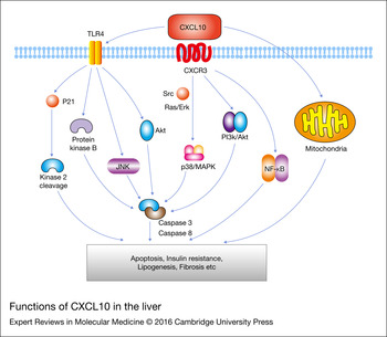
CXCL10–CXCR3 interaction induces both apoptotic, and survival signalling in T-lymphocytes through p38/MAPK, and PI3K/Akt activation pathways, respectively (Ref. Reference Sidahmed81). CXCL10-CXCR3 mediated cytoplasmic signalling triggers effector functions like cell migration and proliferation that are mediated through Src, Ras/ERK and PI3 K/Akt pathways (Ref. Reference Bonacchi48) (Fig. 3). IFN-γ contributes to apoptosis by signalling through CXCL10/CXCR3, which results in a dose-dependent increase in cell death and evokes a mitochondrial-mediated apoptotic pathway in cultured oligodendrocyte progenitor cells (Refs Reference Tirotta, Ransohoff and Lane82, Reference Tirotta83).
Functions of CXCL10 in the liver. (a) CXCL10-CXCR3 mediated cytoplasmic signalling triggers effector functions like cell migration, and proliferation that are mediated through Ras/ERK, and PI3 K/Akt pathways; (b) CXCL10/TLR4 induced long-term protein kinase B, JNK and Akt activation promotes hepatocyte apoptosis by caspase-8, caspase-3 and p21-activated kinase 2 cleavage; (c) CXCL10 evokes a mitochondrial-mediated apoptotic pathway. ERK, extracellular signal-regulated kinases; JNK, Jun N-terminal kinase.

CXCL10 and its noncognate receptor, TLR4, function as a pro-apoptotic signalling cascade during liver injury. CXCL10/TLR4 acts as a noncognate chemokine pathway specifically involved in hepatocyte apoptosis (Fig. 3). Through TLR4, but not its cognate receptor (CXCR3), CXCL10 induces liver cell apoptosis in HCV infection. CXCL10-activated protein kinase B, JNK and Akt can lead to caspase-8 and caspase-3 activation and p21-activated kinase 2 cleavage, thereby prompting hepatocyte apoptosis (Ref. Reference Sahin80) (Fig. 3). These data suggest that CXCL10 contributes to apoptosis in steatohepatitis. CXCL10 neutralization or therapeutical modulation of the CXCL10/TLR4 pathway might be a promising target for liver diseases associated with augmented hepatocyte apoptosis.
CXCL10 promotes liver fibrosis
Activated inflammation may further induce hepatic fibrosis associated with progressive steatohepatitis (Ref. Reference Brunt84). Hepatic fibrosis is a scarring response common in most types of chronic liver injury, which can be caused by pathogens, alcohol abuse, chemical toxins, autoimmune liver disorders, or as a side effect of the metabolic syndrome (Ref. Reference Ramón Bataller85). Liver fibrosis is characterised by an accumulation of extracellular matrix (ECM) proteins and a change in the type of ECM from the normal basement membrane-like matrix to a matrix rich in fibrils collagens. An abnormal ECM composition jeopardises the differentiated functions of all resident liver cells and results in cirrhosis, portal hypertension and ultimately in liver failure (Ref. Reference Gressner and Weiskirchen86). Elevated serum and hepatic CXCL10 levels are both associated with high activity grade and severity of fibrosis in liver biopsies (Ref. Reference Harvey50). CXCL10 might attract HSCs to site of damaged hepatocytes (Ref. Reference Holt47). CXCL10 plays an important role in chemically induced liver fibrosis, which participates in a crosstalk between hepatocytes, HSCs and immune cells. NK cells seem to play an important role in controlling HSC activity and fibrosis (Ref. Reference Hintermann49). Although the receptor is the same, stimulation of stellate cells with CXCL9 provides different signals to cells and leads to the repression of collagen expression (Refs Reference Wasmuth46, Reference Berres, Nellen and Wasmuth87). These data suggest that CXCL10 functions as a pro-fibrotic factor in steatohepatitis, and CXCL10 blockade may constitute a possible therapeutic target for hepatic fibrosis. Among liver-resident cells, hepatic stellate cells are considered a crucial cell type in chronic liver diseases and contribute to the development of fibrosis by producing large amounts of extracellular matrix proteins (Ref. Reference Lee and Friedman88).
The role of CXCR3 in NASH
CXCR3, the cognate receptor of CXCL10, also plays an important role in the development of steatohepatitis (Refs Reference Zhang9, Reference Tomita89). Enhanced CXCR3 expression can be detected in both human NAFLD liver tissues and in experimental steatohepatitis models (Ref. Reference Zhang9). By using CXCR3−/− mice, we have reported that CXCR3 can induce lipid accumulation, inflammatory cell infiltration and ER stress in the development of steatohepatitis (Ref. Reference Zhang9). Similarly, Tomita et al reported the protective effect of CXCR3 genetic deletion against high fat, fructose and cholesterol diet-induced steatohepatitis (Ref. Reference Tomita89). Moreover, CXCR3 levels were significantly reduced in the liver tissues of CXCL10 deficient mice, suggesting that CXCL10 deficiency can result in reduced trafficking of CXCR3 positive immune cells to the liver, thereby ameliorating steatohepatitis (Ref. Reference Tomita89). As an important immune mediator, CXCR3 can induce macrophage infiltration as well as Th1 and Th17 immune response in steatohepatitis (Refs Reference Zhang9, Reference Berres, Nellen and Wasmuth87). CXCR3 blockade by CXCR3 antagonists can reverse the established steatohepatitis (Ref. Reference Zhang9), suggesting that the small molecule CXCR3 inhibitors may be novel therapeutic options for NASH.
The utility of CXCL10 as a non-invasive diagnostic marker
The gold standard of the diagnosis of NAFLD remains to be liver biopsy, a procedure that is invasive, expensive and may cause life-threatening complications. Additionally, it has several limitations as a surrogate marker of clinical outcomes. For instance, it only allows an evaluation of a small sample of the liver parenchyma, which may not be representative of the pathology in the rest of the liver tissue. Moreover, it is not appropriate to do several liver biopsies on the same patient just to monitor the progress of the disease.
Despite these limitations, liver biopsy rates are increasing, possibly because of the changing epidemiology and management of common liver diseases (Ref. Reference Tomita89). However, non-invasive methods are needed on account of the risk of complications of biopsy and the inconvenience of monitoring disease progression. Multiple biomarkers are being evaluated for the non-invasive diagnosis of NASH, such as fibroblast growth factor 21 (FGF21), cytokeratin-18 (CK18) and the fatty liver index (FLI). According to a recent review, measurement of CK-18 levels appears to be the most accurate among serologic markers that have been evaluated for the detection of NAFLD (Ref. Reference Myers, Fong and Shaheen90). CK18 is the major intermediate filament protein in the liver and plasma levels of caspase-generated CK18 fragments reflects hepatocellular apoptosis, which is implicated in the pathogenesis of NAFLD (Ref. Reference Papagianni, Sofogianni and Tziomalos91). However, in contrast with its satisfactory performance in detecting steatosis (AUROC = 0.90), CK18 fragments have moderate accuracy area under the receiver-operating characteristics curve (AUROC) (AUROC = 0.70–0.83) in the diagnosis of NASH (Ref. Reference Myers, Fong and Shaheen90).
CXCL10 is upregulated in NASH patients (Ref. Reference Shen92) and positively correlated with the incidence of obesity and type 2 diabetes (Refs Reference Bertola72, Reference Schulthess93). The AUROC of CXCL10 indicated an overall accuracy of 0.81 to diagnose NAFLD and an accuracy of 0.77 to diagnose NASH. CXCL10 is also an independent risk factor for NASH patients, according to our data (Ref. Reference Zhang8). CXCL10 levels were positively correlated with ALT, HOMA-IR, hs-CRP, MDA, MCP-1 and TNF-α in NAFLD patients. CXCL10 was a very strong and independent factor associated with outcome of NAFLD progression [OR 3.05 (95% CI 1.86–5.00), P < 0.0001]. The AUROC for diagnosing incident diabetes was 0.74 (95% CI 0.65–0.83, P < 0.0001) (Ref. Reference Chang78).
Altogether, CXCL10 has a moderate specificity and sensitivity compared with now existing methods, thus is a non-invasive biomarker of NASH. However, available data are relatively limited and it should be validated in large studies in different populations. Further studies are needed to explore the combined accuracy with other predicting factors mentioned above.
CXCL10 as a therapeutic target for NASH
Our data showed that administration of the anti-CXCL10 mAb could prevent MCD-induced steatohepatitis both in vitro and in vivo as indicated by reduced lipid accumulation and inflammatory cell infiltration in MCD-fed mice administrated with anti-CXCL10mAb (Ref. Reference Zhang8). Moreover, neutralizing anti-CXCL10mAb treatment has been successful in reducing hepatic fibrosis in CCL4-induced liver fibrosis model (Ref. Reference Hintermann49). Thus, the development of reagents to manipulate the CXCL10/CXCR3 trafficking pathway may be a useful therapeutic strategy to decrease liver fibrosis. However, CXCR3–/– mice develop more severe liver damage after conA challenge than wild-type mice (Ref. Reference Zaldivar94). Since CXCL9 plays an anti-fibrotic effect, and it may no longer be able to recruit NK cells in the absence of CXCR3, as a consequence activated HSCs would further promote liver fibrosis (Ref. Reference Wasmuth46). The beneficial effects of CXCR3 in T lymphocytes-mediated hepatitis should be considered when introducing CXCR3 antagonists into clinical trials as these agents may be associated with increased liver injury under certain circumstances. Thus, reagents need to be highly specific for the CXCL10 pathway. Therefore, CXCL10 is a potential therapeutic target for NASH and fibrosing steatohepatitis.
CXCL10 in other inflammatory diseases
As an important pro-inflammatory cytokine, CXCL10 is also involved in many other inflammatory diseases, such as type 1 diabetes (Refs Reference Morimoto95, Reference Christen96), multiple sclerosis (Refs Reference Franciotta97, Reference Martire98, Reference Ko99), autoimmune Addison's Disease (Ref. Reference Edvardsen100) and allograft rejection (Ref. Reference Jackson101). High serum CXCL10 levels have been detected in type 1 diabetes patients. CXCL10 neutralisation by anti-CXCL10 antibodies abrogates type 1 diabetes in transgenic rat insulin promoter-lymphocytic choriomeningitis virus (RIP-LCMV) mice (Ref. Reference Christen96). The role of CXCL10 in multiple sclerosis has also been documented (Refs Reference Franciotta97, Reference Martire98, Reference Ko99). Serum and cerebrospinal fluid (CSF) CXCL10 levels were significantly increased in acute multiple sclerosis patients (Ref. Reference Franciotta97). CXCL10 can act as a biomarker to predict the interferon beta treatment response in multiple sclerosis patients (Ref. Reference Martire98). Astroglial CXCL10 knockout can delay the clinical onset of multiple sclerosis in a murine autoimmune multiple sclerosis model (Ref. Reference Ko99). Elevated serum CXCL10 levels can also be detected in patients with autoimmune Addison's Disease (Ref. Reference Edvardsen100), suggesting the involvement of CXCL10 in the progression of autoimmune Addison's Disease. Urinary CXCL10 has reported to be a noninvasive biomarker of renal allograft rejection (Ref. Reference Jackson101).
Conclusion and prospects
We and others have demonstrated CXCL10 plays a key inflammatory role in NAFLD development. CXCL10 is correlated with inflammation, oxidative stress, fibrosis and apoptosis in the progression of steatohepatitis. Recent studies also suggest that CXCL10 exerts its harmful effects in the progression of NASH by inducing cellular damage. These suggest that CXCL10 plays an important role in almost every phase of NASH development. By recruiting CXCR3+ T lymphocytes to sites of liver inflammatory areas, CXCL10 plays a key role as an inflammatory mediator in NASH development. CXCL10 is a non-invasive biomarker of NASH, and it might be a potential therapeutic target of NASH. Further studies in large human cohorts should be used in the future to test the clinical utility of plasma CXCL10 as a non-invasive biomarker for NASH. All in all, these diagnostic and therapeutic strategies provide a basis for the amelioration of NASH and related metabolic disorders.
Acknowledgements
The project was supported by research funds from RGC-GRF Hong Kong (1406415); National Basic Research Program of China (973 Program, 2013CB531401); Theme-based Research Scheme of the Hong Kong Research Grants Council (T12-403-11).
Conflict of interest
None.