Introduction
Allergic fungal sinusitis is a relatively recently identified but well described clinical entity which may result in expansion and erosion of the bony walls of the paranasal sinuses.Reference Marple1 The current literature describes the characteristics of allergic fungal sinusitis related bone erosion, which most commonly occurs in the ethmoid sinuses, with particular involvement of the lamina papyracea.Reference Nussenbaum, Marple and Schwade2 Such expansion and erosion can lead to ocular manifestations, including proptosis,Reference Carter, Graham and Carpenter3 telecanthus and visual loss.Reference Marple, Gibbs, Newcomer and Mabry4 This report highlights an atypical presentation of allergic fungal sinusitis and considers its importance, both in the diagnosis of patients exhibiting ophthalmological symptoms and in the endoscopic surgical treatment of allergic fungal sinusitis.
Case report
A 43-year-old man presented to the senior author (JBJ) complaining of increased nasal congestion, and progressive diplopia over the course of approximately four months. He had a history of non-insulin dependent diabetes mellitus and chronic allergic rhinosinusitis. He denied having other visual complaints, headache or rhinorrhoea.
Physical examination demonstrated a right lateral gaze deficit consistent with an abducens nerve palsy. Results for visual acuity and visual field testing were within normal limits.
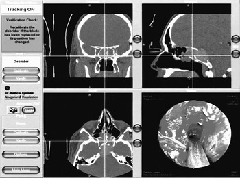
Magnetic resonance imaging revealed a complete, hypointense opacification of the right sphenoid sinus with expansion into the clivus, scattered inflammatory sinus mucosal disease within the ethmoid sinuses bilaterally, and near-complete opacification of the right frontal sinus (Figure 1). The brain, orbits and cavernous sinuses were unremarkable.
Example of the intra-operative computed tomography navigation system, showing the probe in the right ethmoid sinus. The associated crosshairs show the location of the probe in three different planes (coronal, sagittal and axial). Note the soft tissue mass in the right posterior ethmoid and sphenoid sinuses, with bony erosion of the posterior and inferior walls (sagittal section).

In order to better delineate the bony margins of the right sphenoid sinus, and to assist in planning image-guided surgery, a non-contrast paranasal sinus computed tomography scan was obtained. This study demonstrated a 3.0 cm, hyperdense, soft tissue mass of the right sphenoid sinus, sella and clivus. There was suprasellar extension abutting the optic chiasm. Polypoid mucosal disease was present in the middle and posterior ethmoid air cells.
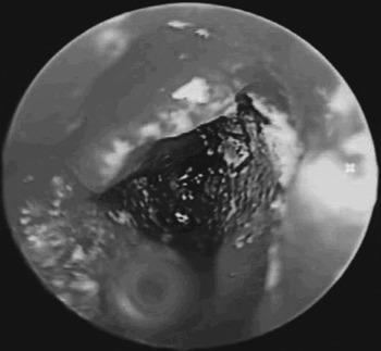
The patient was scheduled for a transnasal endoscopic sphenoidotomy with intra-operative image guidance. The surgical procedure involved an endoscopic transethmoid–sphenoid evacuation of a fungus ball and an endoscopic image-guided approach to the post-sphenoid posterior cranial fossa region, with evacuation of an inspissated collection of fungal debris (Figure 1). The sphenoid sinus contents were consistent with allergic fungal mucin (Figure 2). The latter part of this procedure utilised image guidance and endoscopic visualisation of the dura as well as the cavernous sinus. As the procedure was completed, the surrounding brain was seen to expand to a more normal position behind the sphenoid sinus.
Intra-operative endoscopic image demonstrating the black, ‘molasses-like’ material of the fungus ball.

Histopathological analysis of the surgical specimen demonstrated fungal organisms with mucus and inflammatory cells, consistent with a diagnosis of allergic fungal sinusitis.
In the recovery room, the patient's diplopia resolved completely. Post-operatively, the patient received aggressive medical treatment with antibiotics, systemic oral steroids and antifungals. Clinic follow up continued to demonstrate that the surgical site was mucosalised and clean, although endoscopically the brain could be seen pulsating behind the sphenoid sinus. At the time of writing, the patient was without sinus or visual complaints.
Discussion
In the presented case, transnasal endoscopy was both a diagnostic and a therapeutic procedure. Our patient's differential diagnosis prior to surgery included allergic fungal sinusitis as well as a primary sellar or clival lesion. In addition to eroding into the posterior cranial fossa, the expansion of the sphenoid sinus had resulted in a VIth cranial nerve palsy. As the abducens cranial nerves each rest lateral to the clivus, it was proposed that there was a neoplastic process, mass effect or inflammatory effect causing the patient's diplopia. Histopathological analysis confirmed the diagnosis of allergic fungal sinusitis.
Although allergic fungal sinusitis causes expansion and bone erosion, the mucoperiosteum remains intact, acting as a barrier to the spread of fungus. Bone erosion has been presumed to occur due to pressure atrophy from expansile growth. An alternative hypothesis includes inflammatory mediators in association with the allergic mucin. To date, there has not been direct evidence to support either hypothesis.Reference Nussenbaum, Marple and Schwade2 In the present case, after removal of the mass, the brain moved to rest in its normal position in the posterior cranial fossa, being separated from the sinus only by mucosa.
• This paper describes a case of allergic fungal sinusitis causing bone erosion and diplopia
• Surgical treatment involved nasal polypectomy, wide marsupialisation of the sphenoid sinus and removal of the extensive allergic fungal mucin
• Otolaryngologists should be aware that approximately 20 per cent of patients with allergic fungal sinusitis demonstrate paranasal sinus expansion and bone erosion involving surrounding anatomical structures
• The use of image-guided surgical navigation systems can assist treatment in such cases
Earlier aggressive surgical postures, adopted due to the perceived risk of fungal invasion, were subsequently followed by reports of successful surgical treatment of allergic fungal sinusitis with bone erosion using endoscopic approaches.Reference Marple1, Reference Marple5 Endoscopic techniques, used with image guidance, represent a minimally invasive approach to the diagnosis and management of this disease process. More recently, the use of intra-operative image guidance has allowed endoscopic sinus surgeons to work more safely outside the confines of the paranasal sinuses. In the present case, the fungal disease extended into the clivus and was able to be extirpated via an endoscopic approach. In the immediate post-operative period, there was complete resolution of the patient's diplopia.