Fetal valproate syndrome has wide spectrum of clinical features, including neural tube defects, extremity-facial anomalies, heart defects, genitourinary anomalies, developmental retardation, and autism. Reference Mohd and Green1,Reference Bromley, Mawer and Briggs2 It was first described in 1984 by DiLiberti et al due to teratogenic effects of valproic acid in pregnancy. Reference Diliberti, Farndon, Dennis and Curry3 Interestingly, our case with coronary arteriovenous fistula firstly reported in the literature with fetal valproate syndrome.
Case
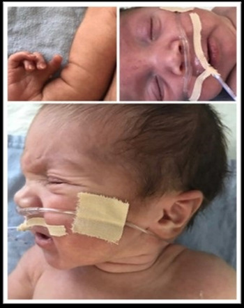
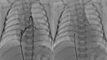
A 5-day-old male weighing 3250 g, who had been intubated due to respiratory distress, was transferred to the neonatal ICU. Before pregnancy, mother was using valproic acid for the treatment of epilepsy, and she continued to use during pregnancy with the advice of her physician. History revealed that her first-born child had autism. There was no consanguinity between the parents. On follow-up, the patient was extubated and tachypnea was observed. In physical examination, 1x1.5 cm diameter capillary haemangioma on forehead, frontal bossing and broad forehead, hypertelorism, posteriorly rotated low set ears, long philtrum, micrognathia, anteverted nares, pectus excavatum deformity, right thumb agenesis, contractures in the wrists, clinodactyly, lumbosacral hypertrichosis, and sacral dimple were observed (Fig 1). On auscultation, 2/6 systolic murmur was heard on the left sternal border. Other systems examination and laboratory findings suggested no abnormalities. In ECG, sinus rhythm and significant p waves were observed. X-ray showed prominent pulmonary vascular shadows, cardiomegaly, absence of radius bone in the right forearm and presence of spina bifida. Echocardiographic evaluation revealed an 8-mm atrial septal defect, and the right atrium and ventricle were dilated. Left ventricular functions were normal. During antibiotic therapy, respiratory distress persisted and X-ray showed no parenchymal lung abnormality. On the control echocardiography, the left main coronary artery was enlarged and there was a marked colour Doppler flow from the left coronary region to the right atrium. In angiography, when radio-opaque material was administered to the left coronary artery orifice, it showed a coronary AV fistula that is draining from the left coronary artery to the right atrium. The coronary AV fistula was closed with the ADO-II AS (AMPLATZER Duct Occluder II Additional Sizes) device through the aorta and left coronary artery (Fig 2). Peripheral blood genetic analysis did not reveal chromosomal abnormalities in favour of trisomy disorders. Also, clinical exome sequencing test performed and there was no mutation related to Holt–Oram syndrome and Fanconi anaemia. After transcatheter closure, the patient’s symptoms improved significantly and he was discharged.
Clinical findings of the patient.

A coronary AV fistula that draining from the left coronary artery to the right atrium (on the left). The coronary AV fistula was closed with the ADO-II AS device through the aorta (on the right).

Discussion
CHDs have been reported in several patients with fetal valproat syndrome, and the incidence of CHD is estimated to be around four times that seen in the general population. Reference Clayton-Smith and Donnai4 In some case series, septal defects and valvular diseases have been reported with extremity anomalies. Some features of these anomalies, including radius aplasia, clinodactyly, and contracture of wrists have been reported similar to Holt–Oram syndrome, Fanconia anaemia, and thrombocytopenia absent radii in fetal valproat syndrome. Reference Mohd and Green1,Reference Mutlu-Albayrak, Bulut and Çaksen5,Reference Shimamura and Alter6 The facial findings of fetal valproat syndrome include hypertelorism, frontal bossing, flat nasal bridge, broad forehead, anteverted nares, midface hypoplasia, bitemporal narrowing, posteriorly rotated ears, thin lips, and long philtrum. Reference Williams, King, Cunningham, Stephan, Kerr and Hersh7 Holt–Oram syndrome is an autosomal dominant disease characterised by various cardiac anomalies, including clinodactyly, absence of thumb, hypoplasia or agenesis of the radius, ulna hypoplasia, carpal bone anomalies, and atrial septal defect. Reference Mori and Bruneau8 In addition, Fanconi anaemia has similar clinical skeletal and cardiac abnormalities, but it is autosomal recessive disease and skin findings including hyper- or hypopigmentation or café-au-lait spots reported to be frequent. Eye malformations including strabismus and hypo/hypertelorism also reported and individuals do not develop bone marrow failure early in their course. Patients with thrombocytopenia absent radii present as newborns with thrombocytopenia and characteristic physical findings of bilateral absent radii (unilateral in about 2% of cases) with thumbs present albeit abnormal in appearance. Reference Shimamura and Alter6 In our case, clinical features of extremity and cardiac findings were observed similar to Holt–Oram syndrome and Fanconia anaemia, but there was no family history of disease and consanguinity between the parents. Patient’s whole blood count was normal that ruled out thrombocytopenia absent radii. Skin manifestations including hyper- or hypopigmentation or café-au-lait spots were not seen in our patient contrary to frequently seen in Fanconia patients. Also, any mutation was not found in favour of Holt–Oram syndrome and Fanconi anaemia in clinical exome sequencing test. Chromosome analysis excluded trisomy disorders. In addition, the presence of typical facial anomalies, sacral hypertrichosis, spina bifida, exposure to valproic acid in both pregnancies and the history of autism in her sibling suggest that diagnose is fetal valproate syndrome. Our case is the first case in the literature in which fetal valproate syndrome is accompanied by a coronary AV fistula. Coronary AV fistula before reported in Down syndrome and Klinefelter syndrome but it is firstly reported in our case with teratogenicity. Reference La Placa, Pinello, Schierz, Giuffrè and Corsello9,Reference Yoshida, Ryu and Ogata10 The coronary AV fistula of the patient was treated with percutaneous transcatheter closure and he was discharged from our centre.
Conclusion
Fetal valproate syndrome can be prevented by discontinue of the valproic acid, especially during first trimester of pregnancy. Close monitoring of pregnant women exposed to valproic acid and the detailed evaluation of babies born from these pregnancies can be life-saving.
Acknowledgements
None.
Financial support
This research received no specific grant from any funding agency, commercial or not-for-profit sectors.
Conflicts of interest
None.
Ethical standards
This report does not include any human or animal experimentation.