Introduction
Successful genetic selection programmes over the past 30 to 40 years have resulted in a modern dairy cow, biologically efficient at producing large volumes of milk but with an associated decreased level of fertility. Currently, the average embryonic and foetal loss rate in high-producing dairy cows is close to 60%. It has been established that 70% to 80% of this loss occurs during the period of rapid embryo growth, i.e. between days 8 and 16 after insemination (Diskin and Sreenan, Reference Diskin and Sreenan1980). Coinciding with the period of greatest embryo loss is the period of greatest change in embryo growth and development, characterized by an exponential increase in size and protein content and an apparent peak in relative synthetic activity. It is also characterized by critical changes in embryo morphology, in particular the three phases of compaction, blastocyst formation and elongation. It is reasonable to speculate that, as the embryo is completely dependent on its uterine environment and the composition of uterine fluid during these critical phases before implantation, a uterine environment that is optimal for normal embryo development can exist. There is increasing evidence of an association between the early luteal phase concentration of systemic progesterone and embryo loss; however, little is known about the mechanisms surrounding this loss and how progesterone affects uterine function, particularly during the first few days after artificial insemination (AI). The objective of this paper is to review the current knowledge on embryo growth, development and survival in the cow and how these may be influenced by changes in uterine function and the concentration of systemic progesterone.
Degree and timing of embryo loss
Early embryo loss
Embryo mortality refers to embryo losses that occur during the period from fertilization to completion of differentiation, which occurs at approximately day 45 in the cow (Committee on Bovine Reproductive Nomenclature, 1972). However, there are many published reports of fertilization rates in heifers and moderate-yielding dairy cows with estimates varying from 90% to 100% (see review by Diskin and Sreenan, Reference Diskin and Sreenan1980). Data relating to the high-producing dairy cows are rather limited, with only three published reports (Wiebold, Reference Wiebold1988; Ryan et al., Reference Ryan, Prichard, Kopel and Godke1993; Sartori et al., Reference Sartori, Sartor-Bergfelt, Mertens, Guenther, Parrish and Wiltbank2002). These latter reports indicate that at least under temperate ambient temperatures, fertilization rates are most likely similar for moderate and high-producing dairy cows. Yet, the calving rate to first service is significantly lower in high-producing cows compared to low-producing cows or heifers. The extent and pattern of early embryo loss have been reasonably well characterized and reviewed by Sreenan and Diskin (Reference Sreenan and Diskin1986), Diskin et al. (Reference Diskin, Murphy and Sreenan2006) and Peters (Reference Peters1996). Sreenan and Diskin (Reference Sreenan and Diskin1986) have reported that embryonic mortality is about 40% for heifers and moderate-producing dairy cows, with most of this (70% to 80%) occurring between days 8 and 16 after insemination. There are little data on the extent or timing of early embryo loss in high-producing dairy cows. However, based on morphological criteria, three studies (Wiebold, Reference Wiebold1988; Ryan et al., Reference Ryan, Prichard, Kopel and Godke1993; Sartori et al., Reference Sartori, Sartor-Bergfelt, Mertens, Guenther, Parrish and Wiltbank2002) have shown that a high proportion (41% to 67%) of embryos recovered on days 6 to 7 from moderate- to high-producing dairy cows had lower cell numbers or were of a lower quality grade than those from heifers or non-lactating cows.
Late embryo loss
There is some evidence that in high-producing dairy cows a significant proportion of embryo loss can be classified as late embryo loss or that deemed to occur after 24 days of gestation. Vasconcelos et al. (Reference Vasconcelos, Silcox, Lacerda, Pursley and Wiltbank1997) reported that intensively fed cows yielding 11 000 to 12 000 kg of milk per cow sustained a 20% embryo loss rate between 28 and 98 days of gestation. In our own laboratory, Silke et al. (Reference Silke, Diskin, Kenny, Boland, Dillon, Mee and Sreenan2002) reported a 7.2% embryo loss rate between days 28 and 84 in 1046 lactating dairy cows, yielding on average 7247 kg of milk per cow per lactation, which was similar to heifers at 6.1%. The pattern of loss was also similar between cows and heifers, with almost half of the late embryo loss occurring between 28 and 42 days of gestation. In another large single-herd study consisting of 1442 pregnant dairy cows yielding between 9200 and 10 600 kg of milk per cow, López-Gatius et al. (Reference López-Gatius, Santolaria, Yániz, Garbayo and Hunter2004) reported a 9.6% loss rate between days 36 and 90, with 75% of the losses occurring between days 45 and 60 of gestation. Horan et al. (Reference Horan, Mee, Rath, O’Connor and Dillon2004) have also recently reported a similar loss rate of 7.5% between days 30 and 67 of gestation in dairy cows managed under pastoral conditions indicating that, at least for moderate-yielding dairy cows, early embryo loss is the greatest factor contributing to overall embryo loss.
Embryo development
Embryo growth
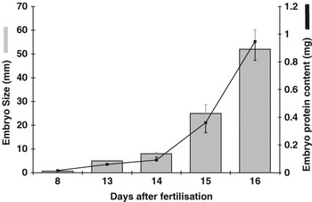
Following fertilization in the oviduct and a number of early cleavage stages, the embryo moves to the uterus at the 8 to 16-cell stage at around days 3 to 4 after fertilization. At around days 5 to 6 when the embryo, still within the zona pellucida, has reached the 16 to 32-cell stage, tight junctions are formed between the individual blastomeres and the embryo begins to compact to form the morula stage. This is the first critical stage of embryo development and is an essential step for differentiation and blastocyst formation (Larue et al., Reference Larue, Ohsugi, Hirchenhain and Kemler1994). Day 8 signals the formation of a blastocoele cavity and the differentiation of the embryonic cells now numbering about 120 into an inner cell mass and trophectoderm, which will ultimately give rise to the foetus and placental tissues, respectively. The blastocyst now expands to approximately 200 μm, and between days 9 and 10 it hatches from the zona pellucida. From fertilization until blastocyst formation and while still confined within the zona pellucida, the size of the embryo remains relatively unchanged and spherical in nature. Following blastocyst formation and hatching, however, the embryo continues to expand and change from being spherical to ovoid and finally elongated in shape by days 13 to 16 (Grealy et al., Reference Grealy, Diskin and Sreenan1996; Morris et al., Reference Morris, Diskin and Sreenan2000). Embryo diameter and length increase from around 0.9 and 5.3 mm on day 13 to about 1.8 and 52 mm on day 16 (Grealy et al., Reference Grealy, Diskin and Sreenan1996). Overall, the embryo increases approximately 300-fold in size from the morula to the elongated stage on day 16 (Figure 1). From about day 19, the process of implantation begins with coruncular–cotyledons attachment points detectable from day 21 and is completed by day 42 (Hunter, Reference Hunter1980).
Cattle embryos undergo an exponential increase in size and protein content from day 8 through to day 16 after fertilization.

Embryo protein content and protein synthesis
Embryo protein content can also be used as an additional parameter to understand the nature of embryo growth and is necessary for the objective characterization and understanding of embryo metabolism. Protein synthesis and phosphorylation are inextricably linked, with phosphorylation itself a consequence of signal transduction and protein synthesis as well as being a post-translational control mechanism. There is a strong positive correlation between embryo size and protein content (Grealy et al., Reference Grealy, Diskin and Sreenan1996). The protein content changes very little from 132 ng at the 2-cell to 183 ng at the compact morula stage. It increases significantly and in an exponential fashion, however, to 367 ng by the expanded blastocyst stage, to 73 μg by the ovoid stage on day 13 and to 947 μg by the elongated stage on day 16. Overall, from the compact morula stage to the elongated stage on day 16, the protein content of the embryo increases over 5000-fold (Grealy et al., Reference Grealy, Diskin and Sreenan1996) (Figure 1). The synthetic activity is relatively quiescent up until activation of the embryonic genome around the time of compaction and subsequent blastocyst formation on day 8 (Frei et al., Reference Frei, Schultz and Church1989). Concomitant with an increase in protein content is an increase in protein synthetic rate and protein phosphorylation measured by the incorporation of radiolabeled 35S methionine and 32P phosphate, respectively (Morris et al., Reference Morris, Diskin and Sreenan2000). While the absolute rates of protein synthesis and phosphorylation increased with day and degree of elongation between days 13 and 15, when expressed per unit of embryonic protein the relative rates were found to actually decrease from a much higher level from around the time the blastocyst began to elongate.
Embryo metabolism
Commensurate also with this rapid development, the embryo has an increased requirement for energy substrates such as glucose and for amino acids, the building blocks of proteins and nucleic acids. Beginning with blastocyst expansion, the energy requirement of the bovine embryo increases dramatically (Donnay and Leese, Reference Donnay and Leese1999) and glucose becomes a major energy source (Rieger et al., Reference Rieger, Loskutoff and Betteridge1992). Glucose utilization and lactate production increase through blastocyst elongation (Morris et al., Reference Morris, Humpherson, Leese and Sreenan2001); however, when expressed on a per unit of protein basis earlier stage day 14 embryos tend to have a higher metabolic rate than the later day 15 or day 16 embryos. Similarly, amino acid metabolism per unit of protein differs with stage of embryo development with alanine production and arginine depletion tending to be greater in earlier day 14 than in elongated day 16 embryos (Morris et al., Reference Morris, Humpherson, Leese and Sreenan2002).
Uterine fluid composition
Until implantation, the bovine embryo is dependent on the oviduct and uterine environment for the provision of an adequate supply of nutrients including amino acids, ions and growth factors. Using short-term collection (under 3 h), we have recently been able for the first time, however, to collect both oviduct and uterine fluids under physiological conditions (Kenny et al., Reference Kenny, Humpherson, Leese, Morris, Tomos, Diskin and Sreenan2002). This has enabled the objective measurement of the concentrations a range of amino acids (Hugentobler et al., Reference Hugentobler, Diskin, Leese, Humpherson, Watson, Sreenan and Morris2007a), ions (Hugentobler et al., Reference Hugentobler, Morris, Sreenan and Diskin2007b) and energy substrates (Hugentobler et al., Reference Hugentobler, Humpherson, Leese, Sreenan and Morris2007c) in oviduct and uterine fluid throughout the oestrous cycle. In these studies, the secretion rate of uterine fluid tended to decrease from 1.83 μl on day 6 to 1.04 μl on day 14; however, this decrease was not significant. A number of authors (Leese et al., Reference Leese, Tay, Reischl and Downing2001; Salleh et al., Reference Salleh, Baines, Naftalin and Milligan2005) have suggested that reproductive tract fluids change throughout the cycle, increasing in response to oestradiol and declining with increasing progesterone concentrations. We were unable, however, to find a significant association between the concentration of systemic progesterone and the secretion rate of uterine fluid or the concentrations of amino acids, ions, or glucose, lactate and pyruvate in uterine fluid nor was there any differences between days 6, 8 and 14 (Hugentobler et al., Reference Hugentobler, Diskin, Leese, Humpherson, Watson, Sreenan and Morris2007a, Reference Hugentobler, Morris, Sreenan and Diskin2007b and Reference Hugentobler, Humpherson, Leese, Sreenan and Morris2007c).
Progesterone and embryo loss
Many factors have been implicated in embryo and foetal loss, including those of genetic, physiological, endocrine and environmental origin. There is now increasing evidence of an association between systemic concentrations of progesterone both in the cycle preceding ovulation and in the early luteal phase of the cycle following insemination with embryo survival. Folman et al. (Reference Folman, Rosenberg, Herz and Davidson1973) and Meisterling and Dailey (Reference Meisterling and Dailey1987) reported reduced fertility in dairy cows associated with low concentrations of progesterone during the cycle preceding insemination. Diskin et al. (2002) reported a positive linear association between the concentration of progesterone on the day of PGF2α-induced luteolysis and subsequent embryo survival rate and have suggested that this is most likely due to premature oocyte maturation, which compromises subsequent normal embryo development (Diskin et al., Reference Diskin, Murphy and Sreenan2006).
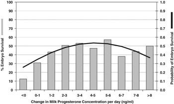
Many more studies have looked at the association between the concentration of systemic progesterone in the cycle following insemination and embryo loss; however, the findings have been equivocal with some studies reporting a positive association (Ahmad et al., Reference Ahmad, Beam, Butler, Deaver, Duby, Elder, Fortune, Griel, Jones, Milvae, Pate, Revah, Schreiber, Townson, Tsang and Inkeep1996; Starbuck et al., Reference Starbuck, Darwash, Mann and Lamming2001) while others reported no association (Bulman and Lamming, Reference Bulman and Lamming1978; Lamming et al., Reference Lamming, Darwash and Back1989). The inconsistencies in some of the earlier studies may have arisen because invariably they compared the mean concentration of progesterone between cows that conceived and those that did not. More recently, Stronge et al. (Reference Stronge, Sreenan, Diskin, Mee, Kenny and Morris2005) and McNeill et al. (Reference McNeill, Diskin, Sreenan and Morris2006a) have chosen to model the relationship between the concentration of milk progesterone (a continuous variable) and embryo survival (a binomially distribute variable) using logistic regression. Stronge et al. (Reference Stronge, Sreenan, Diskin, Mee, Kenny and Morris2005), following 871 inseminations across six herds in spring-calving dairy cows, found a linear and quadratic relationship between the concentration of milk progesterone on days 5, 6 and 7 following insemination and also between the rate of increase in progesterone between days 4 and 7 and embryo survival (Figure 2). Based on derivative analysis of the logistic regression curves, an optimum embryo survival rate was associated with milk progesterone concentrations of 7.4, 13.2 and 16.8 ng/ml on days 5, 6 and 7, respectively, while the optimum rate of increase in milk progesterone between days 4 and 7 was found to be 4.7 ng/ml per day. Based on these values, it was calculated that 60%, 80% and 75% of animals had progesterone concentrations that were sub-optimal whereas 85% of animal had a rate of increase that was below the optimum. McNeill et al. (Reference McNeill, Diskin, Sreenan and Morris2006a), in a smaller study, also found a similar linear and quadratic relationship between the concentration of milk progesterone and embryo survival on days 4 to 6 after ovulation and embryo survival. They also found that progesterone concentration on days 4 and 5 were highly predictive of concentrations on days 7 and 6, respectively. The quadratic component of the relationship in these studies indicates an increased probability of embryo survival with increasing concentrations of progesterone, but at a diminishing rate. Furthermore, they also suggest the existence of an optimal value beyond which embryo survival decreases. These data are consistent with that of Starbuck et al. (Reference Starbuck, Darwash, Mann and Lamming2001) who reported increased embryo survival at progesterone concentration above 3 ng/ml, declining at concentrations greater than 9 ng/ml, indicating that low as well as high concentrations of progesterone are both associated with a low embryo survival rate. Given these findings, it is not surprising that earlier studies failed to find an association between mean progesterone concentrations and embryo survival.
Relationship between the rate of change in milk progesterone between days 4 and 7 after artificial insemination and the probability of embryo survival.

A reduction in embryo survival rate has also been associated with high energy intake and this has been explained on the basis of an inverse relationship between feed intake and systemic progesterone concentration in sheep (Parr et al., Reference Parr, Davis, Fairclough and Miles1987), pigs (Jindal et al., Reference Jindal, Cosgrove, Aherne and Foxcroft1996) and cows (Sangsritavong et al., Reference Sangsritavong, Combs, Sartori and Wiltbank2002). Increased feeding is associated with elevated liver blood flow and increased steroid metabolism in lactating cows. Sangsritavong et al. (Reference Sangsritavong, Combs, Sartori and Wiltbank2002) showed that high feed intake increased liver blood flow by 58% by 4 hours after feeding, while cows left unfed during the same period showed an 83% decrease in liver blood flow. They also found that the baseline liver blood flow in lactating cows was over twice that of non-lactating cows, and that lactating cows had lower serum progesterone and oestrogen levels than non-lactating cows.
Progesterone supplementation
The importance of adequate systemic concentrations of progesterone during the early embryo development is supported by a number of studies involving supplementation with exogenous progesterone. Again, the results of these studies are often inconsistent. In an early review, Diskin and Sreenan (Reference Diskin and Sreenan1980) highlighted the differential effects of progesterone supplementation in cows with either low or normal pregnancy rates and the effect of the timing of administration of progesterone on the outcome of supplementation in terms of pregnancy rate. They summarized the earlier work of Herrick (Reference Herrick1953), Dawson (Reference Dawson1954) and Wiltbank et al. (Reference Wiltbank, Hawk, Kidder, Black, Ulberg and Casdia1956), who studied the effects of intra-muscular progesterone supplementation in cows with low pregnancy rates. Each study showed a consistent increase in pregnancy rate following progesterone supplementation, although this increase was not significant in any single study. When these data were collated and analysed, there was a significant increase in pregnancy rate from 23% in control animals to 42.5% in the treated groups.
Effect of progesterone supplementation during the early to mid-luteal phase
Robinson et al., (Reference Robinson, Leslie and Walton1989) used progesterone-releasing internal devices (PRIDs) to treat lactating cows from days 5 to 12 after insemination. Control cows had a 30% pregnancy rate compared to 60% for treated cows. In another study (Johnson et al., Reference Johnson, Ross and Fourt1958), cows administered 100 mg of repositol progesterone over 7 days from days 2 to 9, or 500 mg of progesterone on day 2 showed an increase in pregnancy rate to 70% (combined data) from 42% in controls. The insertion of controlled internal drug release devices (CIDRs), containing 1.9 g progesterone, on days 6, 7 or 8 was associated with a significant increase in pregnancy rates from about 66% for control animals to about 79% in progesterone-supplemented animals (Macmillan et al., Reference Macmillan, Taufa, Day and Peterson1991).
Effect of progesterone supplementation during the late luteal phase
Dairy cows with a pregnancy rate of 63% showed no increase in pregnancy rate after progesterone supplementation (100 mg i.m.) from days 10 to 20 (Diskin and Sreenan, Reference Diskin and Sreenan1980). Similarly, there was no improvement in pregnancy rates when CIDRs were inserted on days 14 to 17 with removal on day 21, or when CIDRs were inserted on days 10, 12, 14 or 16 for 6 days (Macmillan et al., Reference Macmillan, Taufa, Day and Peterson1991). Mann et al. (Reference Mann, Merson, Fray and Lamming2001) found no significant increase in conception rate in cows supplemented with progesterone from days 10 to 17 and concluded that supplementation during the second half of the luteal phase to be of no benefit in treating early embryo loss in dairy cows. Collectively, these data indicate the importance of the timing of initiation of progesterone treatment in affecting pregnancy rate, and show early supplementation to be more effective in increasing pregnancy rates.
Effect of progesterone supplementation to cows with low systemic progesterone
When progesterone was supplemented to cows with low systemic progesterone concentrations (1 to 2 ng/ml) on day 5, the pregnancy rate was doubled from about 29% to 58% (Starbuck et al., Reference Starbuck, Darwash, Mann and Lamming2001). However, supplementation had little or no effect on pregnancy rate when administered to cows with day 5 values of less than 1 ng/ml, and the authors suggested that late ovulation might have compromised embryo survival prior to day 5 in these animals. Starbuck et al. (Reference Starbuck, Darwash, Mann and Lamming2001) also showed that supplementation had no effect on pregnancy rate when progesterone was administered to animals with day 5 plasma progesterone levels of 2 to 3 ng/ml. This finding may indicate a threshold value for progesterone concentration, above which animals do not benefit from progesterone supplementation. This theory is supported by a depression in pregnancy rate when progesterone concentrations exceeded 9 ng/ml on day 5 of the cycle (Starbuck et al., Reference Starbuck, Darwash, Mann and Lamming2001). The authors suggested that this represents an upper limit of progesterone, which, if exceeded either spontaneously or by progesterone supplementation, was actually detrimental to embryo survival and may have caused embryo mortality by excessive growth stimulation, leading to asynchrony between the embryo and its environment. For progesterone supplementation to be successful, in a therapeutic sense, it must be strategically targeted towards animals that show specific luteal progesterone concentrations within a range shown to benefit from supplementation.
Mechanisms of action of progesterone
Progesterone has been called the ‘hormone of reproduction’, acting through its receptor and several co-factors it orchestrates molecular, biochemical and physiological interactions in the uterus that affect embryo growth, development and viability (Spencer et al., Reference Spencer, Burghardt, Johnson and Bazer2004). Progesterone affects the volume of uterine secretions (Garrett et al., Reference Garrett, Geisert, Zavy and Morgan1988) and the rate of conceptus development (Garrett et al., Reference Garrett, Geisert, Zavy and Morgan1988; Green et al., Reference Green, Hunter and Mann2005) as well as the ability of embryos to produce the luteolysis inhibitor interferon-tau (IFN-τ) (Garrett et al., Reference Garrett, Geisert, Zavy and Morgan1988; Kerbler et al., Reference Kerbler, Buhr, Jordan, Leslie and Walton1997; Mann et al., Reference Mann, Fray and Lamming2006) and the timing and strength of PGF2α (Mann and Lamming, Reference Mann and Lamming1995; Mann et al., Reference Mann, Lamming and Payne1998). The rationale for progesterone supplementation in animals with low post-ovulatory progesterone concentrations is to create an embryotrophic environment allowing the embryo to grow, develop and survive, until, for example, it is capable of preventing luteolysis by secretion of the luteotrophin, INF-τ (Mann and Lamming, Reference Mann and Lamming2001). Mann et al. (Reference Mann, Fray and Lamming2006) reported a 4-fold increase in trophoblast length together with a 6-fold increase in uterine concentrations of INF-τ in non-lactating cows supplemented with progesterone between days 5 and 9, while no effect was seen in cows supplemented between days 12 to 16. The actions of progesterone may be mediated either by a direct effect on the embryo or indirectly via the uterus or possibly both.
Direct effect
For a direct effect of progesterone on the embryo, progesterone receptors (PGRs) must be present on the embryo. Hou and Gorski (Reference Hou and Gorski1993) could not detect PGRs in mice embryos until the blastocyst stage. To our knowledge there are no reports of PGR expression in early-stage bovine embryos. Furthermore, there is little or no evidence of an effect of progesterone on in vitro-produced cattle embryos (Fukui and Ono, Reference Fukui and Ono1989). The most likely route by which progesterone affects embryo survival is through an indirect effect on the uterus.
Indirect effect
Progesterone may elicit indirect effects by binding to uterine stroma or endometrial PGRs and initiating a cascade of events including changes in gene expression (and ultimately protein expression) or changes in uterine permeability to ions, amino acids or metabolites from plasma or by non-genomic effects on the uterine endometrium (Revelli et al., Reference Revelli, Massobrio and Tesarik1998; Duras et al., Reference Duras, Mlynarczuk and Kotwica2005). The PGRs play a central role in modulating the uterine response to both progesterone and oestrogen during the oestrous cycle. Previous authors have examined the expression of the PGR transcript throughout the oestrous cycle and pregnancy in both cattle (Robinson et al., Reference Robinson, Mann, Lamming and Wathes1999 and Reference Robinson, Mann, Lamming and Wathes2001) and sheep (Spencer and Bazer, Reference Spencer and Bazer1995). However, there is a paucity of information regarding the effect of concentrations of progesterone on the expression of the PGR in the bovine endometrium. It is known that steroid hormones are involved in regulating the secretory activity of the oviduct and uterus (for review see Geisert et al., Reference Geisert, Morgan, Short and Zavy1992; Vallet et al., 2001), and some uterine progesterone-responsive genes have been identified (for reviews see Basha et al., Reference Basha, Bazer, Geisert and Roberts1980; and Spencer and Bazer, Reference Spencer and Bazer2002). However, the identities of progesterone-regulated uterine genes, in particular, those expressed during the early stages of gestation, are largely unknown.
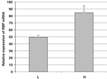
In a recent study, McNeill et al. (Reference McNeill, Cairns, Sreenan, Diskin, Fitzpatrick, Smith and Morris2006b) have shown that uterine expression of mRNA encoding the PGR, oestradiol receptor (ESR) and particularly the retinol binding protein (RBP) is sensitive to small changes in the systemic concentration of progesterone in the first few days after AI. A 1 ng/ml increase in systemic progesterone was associated with an approximate 70% increase in RBP mRNA expression (Figure 3). They also reported possible duration or cycle stage effects of progesterone supplementation on the expression of some of these genes. Furthermore, in a preliminary report, McNeill et al. (2004) showed that there was a positive linear relationship between the concentration of systemic progesterone and folate binding protein (FBP) mRNA expression on day 6 after ovulation. While RBP and FBP genes are likely to be important for early embryo development, in particular the transport of essential nutrients to the embryo, they have not previously been characterized in cow uterine endometrium at this early stage after ovulation or shown to be sensitive to changes in the concentration of systemic progesterone. Changes in mRNA are, however, often not accompanied by measurable or corresponding changes in protein expression (Greenbaum et al., Reference Greenbaum, Colangelo, Williams and Gerstein2003). In a recent report, Costello et al. (2006) have shown that uterine RBP concentrations are similar to plasma concentrations on days 3, 7 and 11 of the oestrous cycle. However, concentrations were 6- to 13-fold higher in uterine fluid on day 15 compared to all the other days and concentrations in the uterine horn ipsilateral to the corpus luteum were over 2-fold higher than those in the contralateral side. The concentration of progesterone is known to be higher in the tissue (Pope et al., Reference Pope, Maurer and Stormshank1982) and uterine horn ipsilateral to the corpus luteum in cattle while Weems et al. (Reference Weems, Lee, Weems and Vincent1988) have shown that the concentration of progesterone in the ipsilateral uterine vein is almost 2-fold higher than that in the contralateral vein, which was in turn similar to the concentration in jugular venous blood. Based on these data, it is tempting to conclude that the concentration of uterine RBP is sensitive to the concentrations of systemic progesterone. However, Costello et al. (2006) could find no association between the concentrations of systemic progesterone and uterine RBP concentrations.
A 1 ng/ml change in the concentration of blood plasma progesterone is associated with a 70% increase in retinol binding protein (RBP) gene expression on day 6 after artificial insemination.

Conclusions
There is a large body of evidence that early embryo loss is one of the greatest factors affecting conception rate in dairy cows. The timing of the greatest increment of loss is associated with the period of greatest change in embryo growth, morphological development and metabolism during which the embryo is completely dependent on its uterine environment. Despite the association between systemic progesterone and early embryo loss, there is an acute lack of information on uterine function in the cow during this time and how it is affected by progesterone. There is an urgent need to characterize the uterine environment, particularly in the high-producing cow, in order to understand embryo loss at a functional level and in order to devise strategies to minimize or reduce embryo loss.