Introduction
Western population demographics have shifted toward a higher median age,[1] attributable to advances in medical care and public health promotion. With this increase in life expectancy, age-related morbidity is rising. In particular, the musculoskeletal system is affected [e.g., osteoarthritis (OA) affects 33% of people over 45 years old[2]] and numerous tissue engineering approaches have been developed for the treatment of localized pathologies.
In treating the musculoskeletal system with tissue engineering strategies, mesenchymal stem cells (MSCs) are a key cell source (endogenous or exogenous) that can differentiate into osteogenic, chondrogenic, myocyte, and adipocyte lineages to facilitate repair.[Reference Jiang, Jahagirdar, Reinhardt, Schwartz, Keene, Ortiz-Gonzalez, Reyes, Lenvik, Lund, Blackstad, Du, Aldrich, Lisberg, Low, Largaespada and Verfaillie3] Their repair potential can be enhanced using biomaterial scaffolds, which are fabricated from biocompatible materials. These form a template for tissue regeneration, providing mechanical support, and protection to cells during proliferation and tissue formation. There are myriad of examples of materials used for scaffold formation, including natural materials such as collagen, alginate and chitosan, and synthetic materials such as polyethylene glycol, polystyrene, and poly-l-lactic acid.[Reference O'Brien4]
The extensive work on bioreactor (i.e., devices that mechanically stimulate cells within these biomaterial scaffolds, typically prior to implantation) and mechanical property effects on MSC differentiation demonstrates a clear link between the application of mechanical force or strain and MSC differentiation.[Reference Lutolf and Hubbell5–Reference Ivanovska, Shin, Swift and Discher11] Bioreactors can be used to apply mechanical strain to MSCs, resulting in upregulation of chondrogenic and osteogenic markers at the gene and protein level, forming, for example, articular cartilage in vitro that is biomechanically similar to its in vivo counterpart.[Reference Brady, Vaze, Amin, Overby and Ethier12, Reference Démarteau, Wendt, Braccini, Jakob, Schäfer, Heberer and Martin13]
Based on these phenomena, our objective is to develop an intrinsically responsive biomaterial(s) that can be used to provide dynamic mechanical cues to cells. An intrinsically responsive material could be stimulated in a non-contact manner prior to implantation and/or following implantation. One option to achieve this goal is to fabricate a magnetically active scaffold (MAS) such as those previously described, which can be used to release drugs (small molecules and proteins) and cells in response to magnetic stimulation.[Reference Cezar, Kennedy, Mehta, Weaver, Gu, Vandenburgh and Mooney14–Reference Zhao, Kim, Cezar, Huebsch, Lee, Bouhadir and Mooney16] More recently, the mechanical stimulation effect alone (i.e., no biologics) of these MASs demonstrated enhanced skeletal muscle regeneration over non-stimulated controls.[Reference Cezar, Roche, Vandenburgh, Duda, Walsh and Mooney17]
With this platform, we are focused on the retention and direct mechanical stimulation of cells attached to the scaffold. To achieve this, we modified two biomaterial systems that are known to facilitate the attachment, growth, and proliferation of cells to make them responsive to an externally applied magnetic field. First, we developed and characterized magnetically responsive, macroporous alginate- and collagen-based biomaterials that can deform in a magnetic field through the incorporation of iron-oxide microparticles. In addition to their ability to apply loads to cells in response to magnetic stimulation, these alginate- and collagen-based MASs have potential as an on-demand drug delivery system. Finally, we confirmed the ability to seed these scaffolds with rat MSCs and have investigated the effects of externally induced deformation via magnetic stimulation on cell survival.
Methods
Alginate MAS fabrication
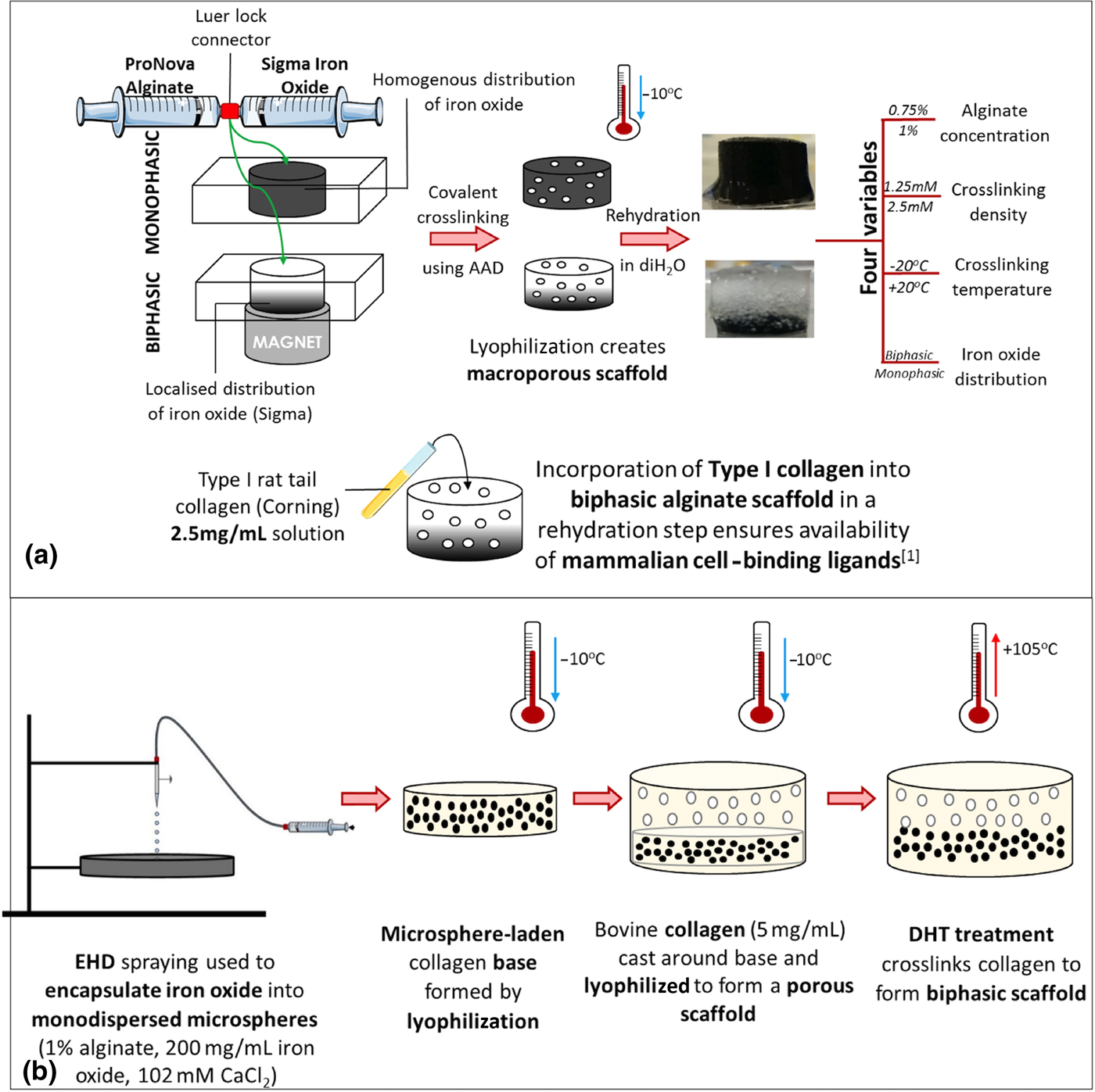
Alginate MAS were fabricated as previously described[Reference Cezar, Kennedy, Mehta, Weaver, Gu, Vandenburgh and Mooney14, Reference Zhao, Kim, Cezar, Huebsch, Lee, Bouhadir and Mooney16] [Fig. 1(a)]. Briefly, alginate (0.75–1 wt.%, high G-block containing alginate, MVG [M:G = 40:60 as specified by manufacturer; MW ~250 kDa), FMC Biopolymer, Norway] was resuspended in MES [2-(N-Morpholino)ethanesulfonic acid] buffer, mixed with 7 wt.% iron-oxide microparticles (Sigma-Aldrich) and crosslinked with adipic acid dihydrazide (AAD; 1.25–2.5 mM; Sigma-Aldrich) using carbodiimide chemistry. Biphasic MAS were fabricated through application of a non-uniform magnetic field,[Reference Cezar, Kennedy, Mehta, Weaver, Gu, Vandenburgh and Mooney14] at two crosslinking temperatures (+20 °C, −20 °C) and samples were underwent freeze-drying (Advantage EL, Virtis Co., SP Scientific, NY, USA) at −10 °C using optimized protocols previously described.[Reference O'Brien, Harley, Yannas and Gibson18]
Fabrication steps for alginate and collagen–microsphere MASs. (a) Alginate MAS: Iron oxide and alginate are mixed directly, crosslinked with AAD and lyophilized. A range of mechanical properties were achieved by adjusting four variables: alginate concentration, crosslinking density, temperature of crosslinking, and iron-oxide distribution. In a final step, the scaffold was soaked with type I collagen to incorporate cell-binding ligands. (b) Collagen–microsphere MAS: Iron oxide–alginate microspheres were fabricated using EHD spraying. Microspheres were mixed with collagen to create a base and lyophilized. Collagen was cast around the base and the entire construct lyophilized and crosslinked with DHT treatment to produce the final bilayered scaffold.

Cell binding was facilitated by soak-loading biphasic alginate MAS in collagen type I (2.5 mg/mL, Corning). Scaffolds were then freeze-dried a second time and subsequently dehydrothermal (DHT) treated.[Reference Haugh, Jaasma and O'Brien19] Specimens were placed in open heat-resistant plastic containers (Sarstedt) in a vacuum oven (Vacucell) at 0.05 Bar and 105 °C for 24 h.
Collagen–microsphere MAS fabrication
Collagen suspension
A solution of 5 mg/mL bovine collagen type I suspension was formulated as previously described.[Reference Haugh, Jaasma and O'Brien19] Briefly, microfibrillar bovine tendon collagen (Collagen Solutions) was blended together with 0.5 M glacial acetic acid using an overhead blender (Ultra Turrax T18 Overhead blender, IKA Works Inc., Wilmington, NC, USA) in a reaction vessel at 15,000 rpm for 90 min. Temperature was maintained at 4 °C using a circulation cooling system (WKL230, Lauda Brinkman, Westbury, CT, USA), to prevent collagen denaturation. The subsequent 5 mg/mL collagen suspension was degassed in a freeze-dryer (VirTis Genesis 25EL, SP Scientific, NY, USA) for 60 min and stored at 4 °C.
Electrohydrodynamic spraying
An electrohydrodynamic spraying (EHDS, Spraybase, Avectas, Ireland) technique was employed in order to encapsulate iron-oxide microparticles into alginate microspheres. Iron oxide (200 mg/mL) was incorporated into a solution of 1 wt.% alginate by direct mixing. This solution was placed in the syringe pump of the EHDS apparatus and sprayed through a 21 G needle into a 102 mM CaCl2 solution at a rate of 200 µL/min. When a consistent flow rate had been achieved, voltage was increased to 8 kV, to ensure Taylor cone formation. A working distance of 50 mm was maintained throughout the spraying process. Microspheres were rotated in 102 mM CaCl2 solution for 10 min to facilitate complete ionic crosslinking.
Scaffold fabrication
Roughly 350 mg of microspheres were mixed with 0.3 mL of 5 mg/mL bovine collagen slurry using Luer lock syringes [Fig. 1(b)]. This mixture was cast into a polyvinyl chloride (PVC) mold and freeze-dried as described above. A further 0.7 mL of collagen slurry was then poured around this collagen–microsphere base and the construct freeze-dried again. The resultant bilayered scaffolds (iron-oxide layer and collagen-only layer) were removed from the molds and subjected to DHT treatment, described above. Where noted, additional EDAC [1-ethyl-3-(3-dimethylaminopropyl)carbodiimide; Sigma-Aldrich] crosslinking was performed at 6 mM EDAC solution in the presence of N-hydroxysuccinimide (NHS, Sigma-Aldrich), a commonly used catalyst, for 2 h (molar ratio of 5 M EDAC:2 M NHS). Scaffolds were then rinsed twice in PBS for 30 min.
Scaffold characterization
Unconfined compression
Unconfined compression testing of alginate and collagen MAS, was conducted using a Zwick machine (Z005, Zwick-Roell). Samples were hydrated for 24 h pre-testing. Specimens in at least triplicate in a water bath were tested for three cycles in unconfined compression up to 10% strain and data recorded using TestXpert II.
Magnetic deformation
Deformation in a non-uniform magnetic field of 6.2 kG was measured by photographing the samples in their undeformed state, and then imaged following application of the magnet and the magnetically induced deformation % calculated as relative change in height using ImageJ software. The magnet used for these studies had a magnetic field of 6.2 kG (S-020-045-N, Neomagnete, Germany) and magnetic stimulation was performed manually, using a metronome where appropriate.
Pore characterization
Sputter-coated (gold/palladium) alginate and collagen–microsphere MAS were characterized using scanning electron microscopy (SEM) techniques. Specimens were imaged at 5 kV using a Zeiss Supra™ SEM (Carl Zeiss, Germany). Additionally, alginate and collagen MAS were fixed (4% paraformaldehyde), washed three times in phosphate-buffered saline (PBS), dehydrated in ethanol and paraffin embedded. Slides were prepared and stained with Alcian Blue (alginate samples) or Picrosirius Red (collagen–microsphere samples).
Platelet-derived growth factor (PDGF)-BB release study
Biphasic 0.75 wt.% alginate MAS (2.5 mM AAD, −20 °C, freeze-dried) were fabricated. Scaffolds were rehydrated in 500 µL of 400 ng/mL PDGF-BB solution, and incubated for 24 h at 4 °C. Scaffolds were washed for 3 min in release buffer [PBS, 2% fetal bovine serum (FBS)] and transferred to 12-well plates containing 4 mL release buffer. Scaffolds were stimulated (1 Hz, 1 min) at 2 h intervals over 4 h. Release buffer was sampled post-stimulation and stored at 4 °C. PDGF-BB content was analyzed using a standard commercial ELISA kit (R&D Systems).
Cell culture
Rat MSCs were cultured to passage 7 in expansion media [high-glucose Dulbecco's modified Eagle's medium (DMEM), 1% non-essential amino acids, 1% GlutaMax™ (both by Gibco® by LifeTechnologies), 10% FBS (BioSera), 1% L-glutamine, and 1% Penicillin–Streptomycin]. Freeze-dried, biphasic 0.75 wt.% alginate MAS (2.5 mM AAD, −20 °C), soak-loaded with type I collagen, and collagen–microsphere MAS were used in this study. Scaffolds were sterilized by DHT treatment. Scaffolds were placed in individual wells of a 12-well plate (Corning), and rehydrated over 10 min with 500 µL of a 1 × 10⁶/mL cell suspension and incubated for 30 min to facilitate cell attachment. 3 mL of fresh media was added to each well, and the plate placed in at 37 °C and 5% CO2 for 7 days. Media was changed every 2 days.
Scaffolds were stimulated using a non-uniform magnetic field of 6.2 kG, at 1 Hz for 1 min/day, on days 4, 5, and 6. Following stimulation a sample of media was taken and released cells counted. On day 7 scaffolds were washed in PBS, halved, and incubated for 1 h in 3 mL of Live-Dead® solution (2 µM Calcein AM, 4 µM ethidium homodimer-1). Imaging was carried out using a Carl Zeiss LSM 710 confocal microscope and Zen® 2008 software. Live and dead cells were counted in five representative fields of view (1 mm2) per scaffold and average live/dead cell counts recorded. Following removal of the scaffold, the well plate was trypsinized and a cell count performed (i.e., to measure cells released from the scaffold and attached to the well plate).
Data analysis
All results are stated as mean±SD. Unpaired t-tests and two-way analysis of variances (ANOVAs), with Bonferroni correction, were carried out as stated; P < 0.05 was considered statistically significant.
Results
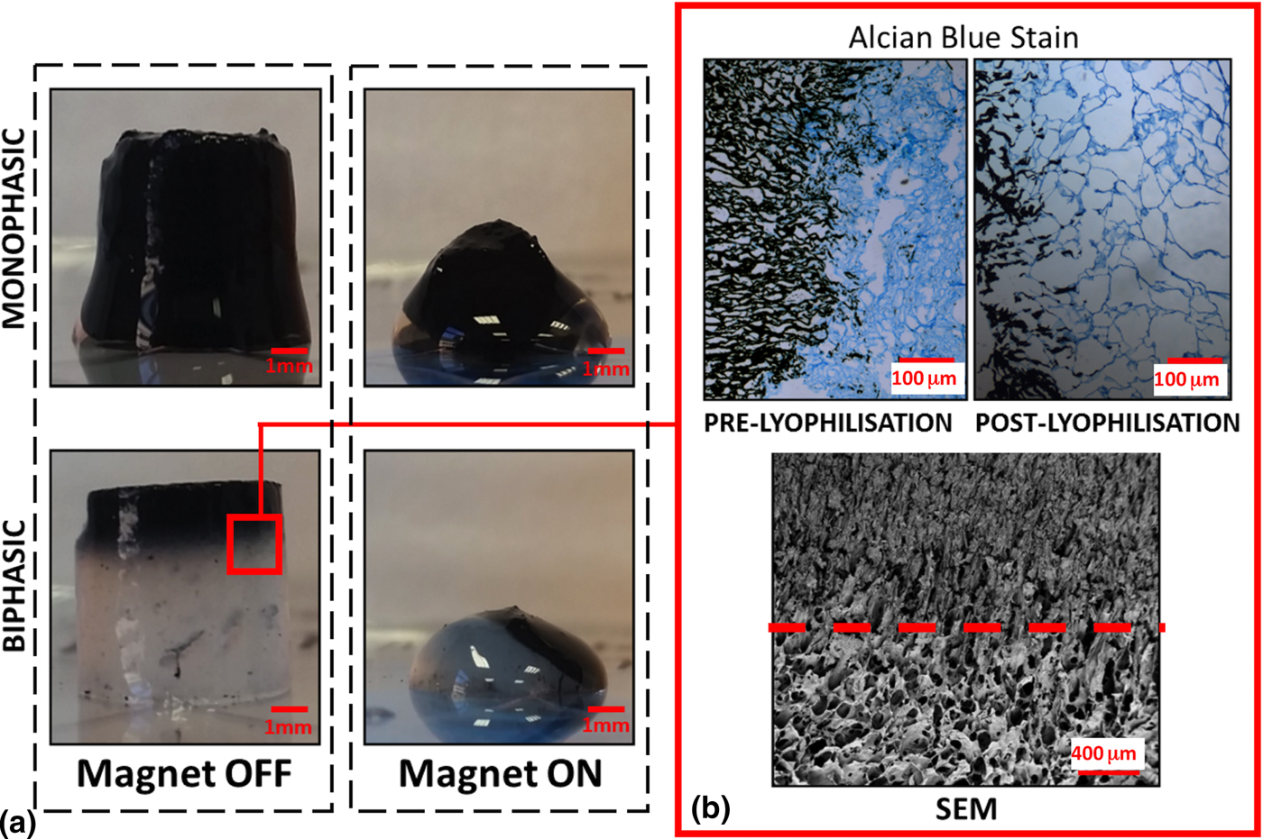
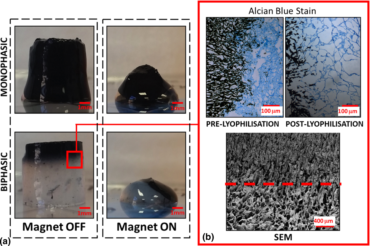
Alginate MAS and collagen–microsphere MAS were successfully fabricated, demonstrated good handling properties, and could be stored for several weeks in diH2O at room temperature without any negative effects on physical properties. The alginate MAS were capable of reversible deformation of up to 50% in response to a non-uniform magnetic field [Fig. 2(a)], with the biphasic scaffolds being more deformable, consistent with previous results.[Reference Cezar, Kennedy, Mehta, Weaver, Gu, Vandenburgh and Mooney14] Following lyophilization, these scaffolds demonstrated an interconnected, macroporous material, of pore diameter up to 200 µm, consistent with previous data for alginate materials [see Fig. 2(b)].[Reference Augst, Kong and Mooney20, Reference Lee and Mooney21] Imaging of the biphasic scaffolds demonstrated iron-oxide particles localized to discrete areas of the scaffold.
Imaging of alginate MAS scaffolds. (a) Macroscopic images of monophasic and biphasic alginate scaffolds with and without magnetic stimulation. (b) Alcian blue histological staining of biphasic alginate samples pre- and post-lyophilization. Images show clear demarcation of the iron and alginate phases, as well as the open porous structure. (c) SEM images of alginate scaffold shows interface between iron-oxide phase (superiorly) and alginate phase (inferiorly; dashed line indicates the interface).
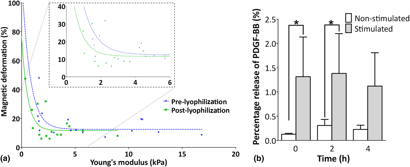
Modulation of the various fabrication parameters affected the mechanical and magnetically induced deformation properties of alginate MAS [see Fig. 3(a)]. Magnetic deformation ranged from 6% to 50%, while Young's modulus ranged from 0.3 to 16.8 kPa. The asymptotic relationship suggests that there is a threshold value of Young's modulus, below which deformation is observed. Exemplary results from the complete data set show that, in particular, AAD and monophasic versus biphasic distribution of iron oxide were found to have the most significant impact on the Young's modulus and deformation (P < 0.05; Supplemental Fig. S1). A twofold reduction in AAD resulted in a threefold decrease in Young's modulus, and a 1.5-fold increase in deformation. Similarly, scaffolds with identical processing parameters but mono- versus bi-phasic had a twofold change in Young's modulus, and a twofold increase in deformation (Supplemental Fig. S1).
Evaluation of the mechanical properties of the alginate MAS. (a) Relationship between Young's modulus (kPa) and magnetically-induced deformation pre- and post-lyophilization demonstrates an asymptotic relationship [pre-lyophilzation: f(x) = 118.8–12.52 e(−1.335x) + 12.52; R 2 = 0.32; post-lyophilization: f(x) = 76.7–11.68 e(−1.795x) + 11.68; R 2 = 0.58; n = 3–4]. (b) Release profile of PDGF-BB from biphasic alginate scaffolds at each stimulation cycle (three cycles total) (mean ± SD; n = 4–6; two-way ANOVA with Bonferroni correction, *P < 0.05).
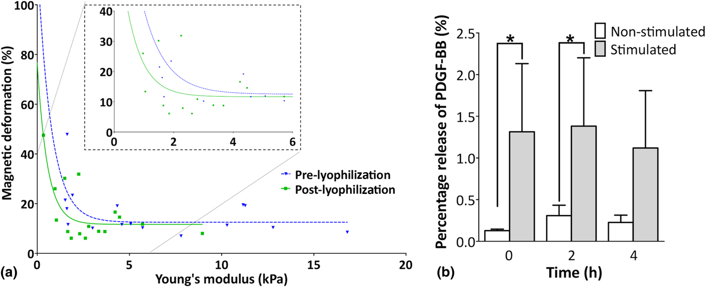
Although the primary goal of this study is to mechanically stimulate cells, it has been previously demonstrated that collapse of the scaffolds in a magnetic field can be used to control drug release. We confirmed the ability to release a model therapeutic protein, PDGF, from the biphasic alginate MAS [Fig. 3(b)]. Although the total amount of release is low—due to the high affinity alginate for PDGF (likely due to alginate's affinity for heparin-binding proteins[Reference Kearney and Mooney15])—the stimulated group shows a significantly (P < 0.05) higher protein release at timepoints 1 and 2, compared with the corresponding non-stimulated group.
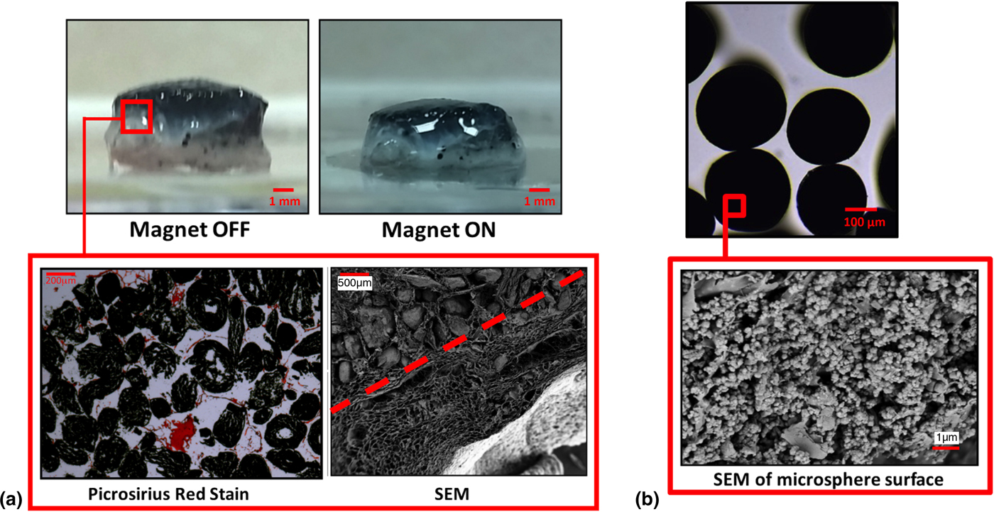
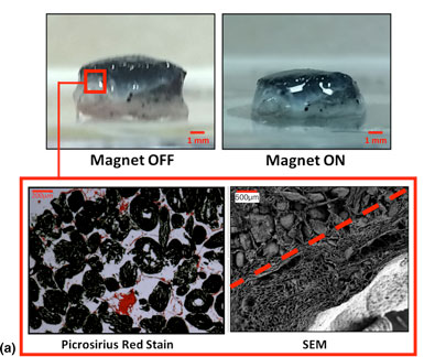
Collagen–microsphere MAS were successfully fabricated and deformed up to 30% in a non-uniform magnetic field [Fig. 4(a)]. Macroscopic, and SEM/light microscopy imaging of the collagen–microsphere MAS [Fig. 5(b)] show ferrous alginate microspheres of diameter 177 ± 2 µm, surrounded by a porous collagen matrix of pore size 100–200 µm. Iron-oxide particles were localized within the ferrous microspheres, with the collagen phase being iron-free.
Imaging of collagen–microsphere MAS. (a) Macroscopic images of the collagen scaffolds, pre- and post-deformation. Scale bar indicates 1 mm. Picrosirius red histological staining and SEM images of collagen scaffolds indicate the two layers and their interface. Dashed line on the SEM images delineates the ferrous microsphere and collagen phases. (b) Light microscopy of ferrous alginate microspheres reveals well-rounded and iron-oxide-loaded spheres. SEM imaging of ferrous alginate microsphere surface reveals the iron oxide–alginate interface.

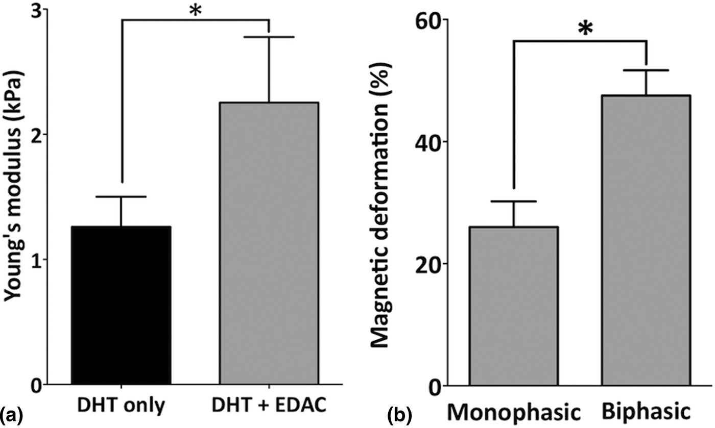
Mechanical properties of collagen–microsphere MAS. (a) Young's modulus and (b) magnetically-induced deformation of collagen–microsphere MAS crosslinked with DHT only and with DHT + EDAC treatment (mean ± SD; n = 4; t-test, *P < 0.05).

The effect of crosslinking parameters (EDAC ± DHT) on the Young's modulus and deformation of the collagen–microsphere MAS was also tested [Fig. 6(a)]. Scaffolds crosslinked using DHT + EDAC treatments were significantly (P < 0.05) stiffer, with a mean modulus of 2.3 ± 0.3 kPa when compared with non-DHT (1.3 ± 0.3 kPa). This resulted in an approximate halving of the magnetic-deformation responsiveness of the DHT + EDAC scaffold (26 ± 5% versus 18 ± 5%; non-significant). As these studies identified DHT only treatment as the scaffold with the highest magnetically-induced deformation, DHT only treatment was used for subsequent studies.
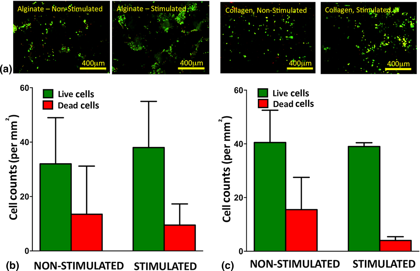
Cell survival study in alginate MAS and collagen–microsphere MAS, with and without magnetic stimulation (1 Hz for 1 min/day, for 7 days). (a) live/dead cell staining within cell-seeded scaffolds. Quantification of live/dead cell staining for (b) alginate MAS and (c) collagen–microsphere MAS scaffolds (mean ± SD, no significance between stimulated and unstimulated).

Finally, cells were incorporated in both systems and repeated cycles of stimulation in a magnetic field of 6.2 kG was applied to assess cell survival in alginate MAS and collagen–microsphere MAS (Fig. 6). Live/Dead® imaging demonstrated cell penetration (see Supplemental 2) into the scaffold and cell survival in both non-stimulated and stimulated MAS indicating suitable biocompatibility of these substrates. Cell counts of cells found in the media following magnetic stimulation were equivalent to non-stimulated controls at a maximum of 4% of the total amount of cells seeded on the scaffolds. Furthermore, on day 7, <1% of the total number of seeded cells were found attached to the well plates for both magnetically stimulated and non-stimulated scaffolds, suggesting cells adhered and remained in the scaffold throughout the culture period. There were no significant differences in cell survival in non-stimulated versus stimulated groups for either scaffold.
Discussion
The objective of this work was to develop an intrinsically responsive biomaterial(s) that can be used to provide dynamic mechanical cues to cells. Herein, we developed and characterized magnetically responsive, macroporous alginate-, and collagen-based biomaterials that could be manipulated in a magnetic field, through incorporation of iron-oxide microparticles. We then seeded these scaffolds with rat MSCs to begin to explore the constructs use for dynamic mechanical stimulation of cells.
Biphasic alginate MAS were successfully fabricated, discretely localizing iron-oxide microparticles and limiting the potential toxicity of iron oxide to cells seeded into the alginate phase. Lyophilization of alginate hydrogels resulted in the formation of a porous alginate MAS, due to formation of ice crystals during the freeze phase of the lyophilization process that were sublimated to create a porous architecture (as confirmed on SEM and light microscopy). A steady cooling rate with a final freezing temperature of −10 °C was utilized to maximize pore size and, therefore enhance magnetic deformation as previously shown.[Reference Zhao, Kim, Cezar, Huebsch, Lee, Bouhadir and Mooney16] The decrease in Young's modulus in response to the introduction of pores via freeze drying is believed to be the primary effect on Young's modulus; this relationship between porosity and Young's modulus is well established for porous materials,[Reference Gibson and Ashby22] collagen,[Reference Harley, Leung, Silva and Gibson23] and alginate.[Reference Zhao, Kim, Cezar, Huebsch, Lee, Bouhadir and Mooney16] However, as these are composite materials, the densification of the iron-oxide phase within the collagen/alginate and their interactions may result in other local/microscale effects that may have additional effects on the mechanical properties; these effects would be best understood by probing the microscale struts (e.g., using AFM techniques). A reduction in Young's modulus (e.g., by reducing the AAD crosslinker concentration) also increased magnetic deformation. In particular, localizing iron-oxide microparticles to a discrete phase of the biphasic scaffolds minimized their particle-reinforcement effect on the bulk of the scaffold, resulting in a sample with a lower Young's modulus that is more deformable in a non-uniform magnetic field.[Reference Cezar, Kennedy, Mehta, Weaver, Gu, Vandenburgh and Mooney14] Additionally, the higher density of magnetic microparticles found in the iron phase of the biphasic samples results in a greater force being applied to the alginate phase of biphasic samples upon application of a magnetic field. Stimulation of the hydrated biphasic alginate MAS with a non-uniform magnetic field results in deformation of the scaffold, and expulsion of fluid by forced convection. This flow can be exploited to accelerate the release of molecules from the structure via convection. The release study demonstrated that magnetic stimulation of PDGF-BB loaded biphasic alginate MAS released a significantly higher amount of PDGF-BB than non-stimulated scaffolds.
Graphing Young's modulus and magnetically induced deformation for all fabrication parameters tested, demonstrates an asymptotic relationship [Fig. 3(c)]. As Young's modulus is reduced below 2.5 kPa, there is an exponential increase in % deformation; lyophilization (i.e., the introduction of pores) shifts the curve closer to the origin. Deformations of up to 50% are possible with an appropriate combination of variables, and Young's modulus can range from 0.3 to 16.8 kPa.
Collagen-based scaffolds were made magnetically active by incorporation of ferrous alginate microspheres. We were unable to encapsulate iron-oxide microparticles directly into the collagen. This may have been due to the nature of DHT crosslinking—“zero length” crosslinks may be insufficient in entrapping the iron-oxide microparticles within the macroporous collagen network, whereas the nanopores of the alginate microspheres may be more suited to this purpose.[Reference Haugh, Jaasma and O'Brien19] This incorporation method was successful as demonstrated on histological staining and SEM imaging, showing a collagen network with pores of approximately 100 µm, consistent with previous work carried out in this area[Reference O'Brien4, Reference O'Brien, Harley, Yannas and Gibson18] surrounding ferrous alginate spheres of 100–200 µm diameter. EDAC crosslinking of DHT-crosslinked collagen–microsphere scaffolds was carried out to examine the effects on Young's modulus and magnetically induced deformation. The Young's modulus of these collagen scaffolds was higher than found in previous studies,[Reference Haugh, Jaasma and O'Brien19] and was significantly (P < 0.05) higher in the DHT + EDAC group compared with DHT only treatment. This is likely due to the presence of small, dense ferrous alginate microspheres in the scaffold structure, and the use of DHT treatment in addition to EDAC crosslinking. DHT + EDAC crosslinked scaffolds showed a non-significant reduction in % deformation compared to DHT only.
Ferrous alginate–collagen and collagen–microsphere MAS remained stable in media over 7 days. Both scaffold groups exhibited good cell penetration into the constructs and exhibited good cell survival. Incorporation of type I collagen into the alginate MAS adequately functionalized the scaffolds with cell-binding ligands as evidenced by the maintenance of cells within the scaffold. By contrast, Zhao et al. and Cezar et al. specifically designed their alginate materials with low cell-binding ligand density (covalently coupled RGD, i.e., the tripeptide sequence of L-arginine, glycine, and L-aspartic acid) to facilitate cell release.[Reference Cezar, Kennedy, Mehta, Weaver, Gu, Vandenburgh and Mooney14, Reference Zhao, Kim, Cezar, Huebsch, Lee, Bouhadir and Mooney16] This illustrates that the processing steps and addition of iron oxide does not affect the properties of cell interaction. It should be noted, however, that the alginate and collagen are not covalently linked herein and that the magnetic deformations may not be directly coupled.
Typically, bioreactors that provide continuous dynamic compression of MSCs around the 1 Hz frequency range (<1 up to 10 Hz) result in cartilage formation, whereas tensile loading of MSC constructs is preferred for osteogenesis.[Reference Delaine-Smith and Reilly24, Reference Schulz and Bader25] Our system was tested at 1 Hz in compression; however, before full validation of the technology, its ability to be loaded over a longer timescale (e.g, its fatigue properties) will need to be evaluated. Additionally, it is conceivable that the same principles applied here could be used to apply tension to the constructs (i.e., by fixing the base of the scaffold and using a magnet to apply tension to the upper surface); however, this was not examined here.
These results, taken together, demonstrate exciting, versatile MASs. In future work, we will investigate the potential of these scaffolds to affect cell behavior and explore their use in tissue engineering applications. The tunable properties of these materials makes them ideal for providing a variety of stimulation profiles to direct MSC differentiation and enhance tissue regeneration.
Supplementary material
The supplementary material for this article can be found at https://doi.org/10.1557/mrc.2017.41.
Acknowledgements
The authors acknowledge support from the TCD MSc program in Bioengineering and Science Foundation Ireland (AMBER, SFI/12/RC/2278). C.J.K. and F.J.O’.B. acknowledge RCSI's Office of Research and Innovation Seed Fund Award (Grant Number GR 14-0963) and the European Union for a Marie Curie European Reintegration Grant under H2020 (Project Reference 659715). V.N. and C.H. would like to thank the ERC (StG 2DNanocaps) and SFI (PIYRA and AMBER) for their support and the Advanced Microscopy Laboratory, TCD for the provision of their facilities.