Introduction
Despite recent therapeutic advances, severe secondary hyperparathyroidism induced by chronic renal failure is often refractory to medical treatment and necessitates parathyroidectomy. In this setting, parathyroidectomy has positive biochemical and symptomatic effects, and has been proven to be the most successful treatment for advanced secondary hyperparathyroidism.Reference Llach1, Reference Tominaga2
Subtotal parathyroidectomy (with complete removal of three parathyroid glands and subtotal removal of the fourth) and total parathyroidectomy with autotransplantation (i.e. re-implantation of a piece of parathyroid tissue in the sternocleidomastoid muscle or in the forearm) are considered to be equivalent procedures, both showing a low morbidityReference Cattan, Halimi, Aidan, Billotey, Tamas and Drüeke3, Reference Rothmund, Wagner and Schark4 and low recurrence or persistence rates.Reference Tominaga, Tanaka, Sato, Numano, Uchida and Falkmer5–Reference Walgenbach, Hommel and Junginger9 Nevertheless, the optimal surgical strategy for the treatment of secondary hyperparathyroidism is still controversial.Reference Cattan, Halimi, Aidan, Billotey, Tamas and Drüeke3, Reference Rothmund, Wagner and Schark4 Recurrent or persistent disease remains a problem in both techniques, as each includes only a reduction of hyperplastic parathyroid tissue. Those who prefer total parathyroidectomy plus autotransplantation argue that, in cases of recurrent disease due to a hyperplastic autograft, the graft can be easily excised under local anaesthesia.Reference Cattan, Halimi, Aidan, Billotey, Tamas and Drüeke3, Reference Rothmund, Wagner and Schark4, Reference Walgenbach, Hommel and Junginger9 Opponents, however, point to the difficulties in resecting a hyperplastic autograft;Reference Henry, Denizot, Audiffret and France7, Reference Walgenbach, Hommel and Junginger9, Reference Neonakis, Wheeler, Krishnan, Coles, Davies and Woodhead10 in addition, they hypothesise that the rate of severe hypoparathyroidism is higher following this procedure.Reference Cattan, Halimi, Aidan, Billotey, Tamas and Drüeke3, Reference Henry, Denizot, Audiffret and France7
The rate of recurrence or persistence after subtotal parathyroidectomy and total parathyroidectomy plus autotransplantation is reported to be 2.5–12 per cent.Reference Tominaga, Tanaka, Sato, Numano, Uchida and Falkmer5, Reference Henry, Denizot, Audiffret and France7–Reference Walgenbach, Hommel and Junginger9 Following the initial operation, the time taken to develop recurrent disease ranges from four to 216 months (mean: 54 ± 36 months).Reference Richards, Wormuth, Bingener and Sirinek11 Tominaga et al. Reference Tominaga, Katayama, Sato, Matsuoka, Goto and Haba12 identified persistent secondary hyperparathyroidism in 49 of 1156 patients (4.2 per cent) who had undergone total parathyroidectomy plus autotransplantation; these patients' mean age was 50 years ± a standard deviation (SD) of 5.5 years. Re-operation was required in 21/49 (2.8 per cent) cases.
The aim of the current study was to describe the surgical findings in a series of patients who had undergone re-intervention for secondary hyperparathyroidism, and especially to assess what extent these findings correlated with (Reference Llach1) the data of the initial operation, and (Reference Tominaga2) the results of localisation tests carried out before the re-intervention.
Methods
We reviewed the files of 18 patients operated upon for recurrent secondary hyperparathyroidism. They were all referred by our clinic's nephrology department or from other institutions. Most patients were treated with haemodialysis, although five had undergone a kidney transplantation. There were 11 women and seven men, aged from 18 to 78 years (median age: 44 years). The time interval between the first and second operation ranged from 12 months to 22.5 years.
Surgery was carried out, taking into account the available data for the initial parathyroidectomy and the results of the localisation studies performed before the re-intervention. Neck ultrasonography, Mibi (99mTc-sestamibi scintigraphy) and computed tomography (CT) had been carried out in a non-systematic way, because most patients had been referred for re-intervention from another clinic. The surgical files for the first intervention were studied, except in three patients for whom no reliable report of the first intervention could be obtained. During re-intervention, all enlarged parathyroid tissue was removed. Simultaneously, a partial or total thyroidectomy was performed in six cases and a superior sternotomy (manubriotomy) in three patients. The location of the recurrent hyperplasia was noticed: right or left; superior, inferior or thyroidal (when adjacent to the thyroid gland but not clearly superior or inferior); and superior mediastinum or sternocleidomastoid muscle.
Results
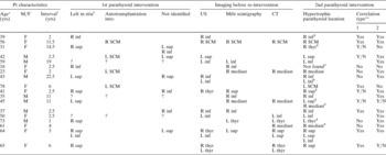
The individual data for the 18 patients operated upon for recurrent secondary hyperparathyroidism are summarised in Table I. In one patient, no parathyroid tissue was found, even after two re-interventions including sternotomy and complete thymectomy. In the 17 remaining patients, hyperplastic parathyroid tissue was found at one or more locations.
Patient characteristics

*At time of re-intervention; †between 1st and 2nd intervention; ‡i.e. subtotal resection (or biopsy). **1 = correlation between location of recurrence and data for first intervention; 2 = correlation between location of recurrence and pre-operative imaging; Y/N = partial correlation. §With unilateral or total thyroidectomy; #with partial sternotomy. Pt = patient; yrs = years; M = male; F = female; US = ultrasonography; Mibi = 99mTc-sestamibi scintigraphy; CT = computed tomography; R = right; L = left; inf = inferior parathyroid region; sup = superior parathryoid region; thyr = unclear if superior or inferior; SCM = sternocleidomastoid muscle; mediast = superior mediastinum; ? = file of first intervention unavailable
We studied the findings during re-intervention from two points of view, as follows.
Correlation with original surgery
The correlation between the location of recurrent parathyroid hyperplasia and the original surgical findings was assessed in 15 patients. At the initial parathyroidectomy, the following findings were noted. One or more glands had been partially left in situ in nine patients, because subtotal parathyroidectomy had been performed or, more rarely, a biopsy of an apparently normal gland had been performed. Total parathyroidectomy with autotransplantation in the sternocleidomastoid muscle had been performed in four patients. Less than four glands had been identified in six patients, i.e. at one location (right or left, superior or inferior) the exploration had remained negative. (The sum of findings was more than 15, as some patients had had a combination of the above findings).
As expected, recurrent hyperplasia was often present at sites where parathyroid tissue had either been left in situ, been re-implanted or had not previously been identified. However, there was no general rule. Hyperplasia was also found at other locations adjacent to the thyroid and in the superior mediastinum. On the other hand, sometimes no parathyroid tissue was identified at sites where parathyroid tissue had been retained. In six out of nine patients in whom parathyroid tissue had been left in situ, recurrent disease was seen at that location, while in three cases it was not. Four patients had undergone a re-implantation in the sternocleidomastoid muscle; in half of these, recurrence was found in the sternocleidomastoid muscle. Summarising the individual data of Table I, one sees that, in approximately one-third of patients, the location of the present parathyroid hyperplasia completely correlated with the data for the first intervention. In one-third, there was a partial correlation, and in one-third there was no correlation at all.
Correlation with pre-operative imaging
The correlation between the location of recurrent parathyroid hyperplasia and the pre-operative imaging (performed before the re-intervention) was assessed in 18 patients. Ultrasonography, Mibi scintigraphy and CT showed a lesion suspected of representing parathyroid hyperplasia in 10, 10 and seven patients, respectively (an example of a relevant CT scan is shown in Figure 1). These suspicions were confirmed during surgery in nine, nine and seven patients, respectively (giving positive predictive values of 90, 90 and 100 per cent, respectively). On the other hand, parathyroid hyperplasia was sometimes found in sites not predicted on pre-operative imaging. On the whole, the site of the recurrent disease was adequately predicted by one or more localisation tests in 13 patients. In two patients, there was a partial correlation between intra-operative findings and pre-operative imaging. In three cases there was no correlation at all, including the two cases in which the pre-operative tests were negative.
Axial computed tomography scan of the neck, showing on the left side a large thyroid cyst and, dorsal to this, between the thyroid, oesophagus and trachea, a partially cystic, hypertrophic parathyroid gland. Both were confirmed by surgery and pathological examination.

From these results, it would appear that imaging enabled better prediction of the location of recurrent disease than did the surgical record of the first intervention.
Discussion
Generally, the operative findings of primary surgery for secondary hyperparathyroidism show that, in the great majority of patients, four or more enlarged parathyroid glands are found. Renal failure is the stimulus for hyperplasia, and there is no reason why this should not affect all parathyroid tissue. However, it is known that the size and weight of the parathyroid glands in cases of secondary hyperparathyroidism can vary considerably within one patient;Reference Debruyne, Ostyn and Delaere13 some glands can be approximately normally sized, which can make their identification more difficult. In addition, the number of parathyroid glands can vary, as supernumerary glands are not uncommon. In a study of 570 patients,Reference Numano, Tominaga, Uchida, Orihara, Tanaka and Takagi6 the frequency of supernumerary glands was up to 16.5 per cent. The thymic tongue was the most frequent location of these supernumerary glands (51 per cent). In 1.1 per cent, supernumerary glands were located in the mediastinum.
Thus, there are several possible reasons for recurrent disease following parathyroidectomy.
Missed cervical glands: removal of less than four glands
To avoid persistent or recurrent disease, all parathyroid glands, including supernumerary ones, should be removed at the initial operation. The removal of only three or fewer glands leads to a persistence or recurrence rate of 34.7–73 per cent.Reference Henry, Denizot, Audiffret and France7 In a study of 822 patients,Reference Hibi, Tominaga, Uchida, Takagi, Imai and Funahashi14 fewer than four glands were removed in 16 patients (1.9 per cent); in 15 of these cases, three glands were extirpated, and in one case only two glands were detected. Remarkably, 12 out of these 16 patients did not develop persistent or recurrent disease. It was therefore concluded that subtotal removal or autotransplantation should be performed, even if fewer than four parathyroid glands are found.
Removal of fewer than four glands can be due to inadequate exploration, ectopic localisation, small gland size or multinodular goitre. Although it has been suggested that some individuals may indeed have fewer than four glands, in autopsy studies this is rather rare.Reference Pattou, Pellissier, Noel, Wambergue, Huglo and Proye15 In six of our patients, a gland had been missed during primary surgery; in four, hyperplasia was found at that location.
Missed glands: supernumerary glands
The incidence of supernumerary glands is reported to be up to 20 per cent.Reference Walgenbach, Hommel and Junginger9, Reference Richards, Wormuth, Bingener and Sirinek11, Reference Pattou, Pellissier, Noel, Wambergue, Huglo and Proye15 Pattou et al. Reference Sokol, Kavolius, Schaaf and D'Avis16 even reported that supernumerary glands were found in 30 per cent of 290 patients, and that such glands accounted for 32 per cent of persistent or recurrent disease. The majority of these glands is located in the thymus, while a lower percentage is found in the mediastinum. Therefore, routine removal of the thymic tongue and careful examination of the regions surrounding the (lower) poles of the thyroid are proven to be important steps in the surgical procedure. In our series, a hypertrophic parathyroid gland was found in the mediastinum in three patients.
Hyperplasia of parathyroid remnant
If the metabolic stimulus for hyperplasia persists after parathyroidectomy, the parathyroid remnant may become hypertrophic again. Cattan et al. Reference Cattan, Halimi, Aidan, Billotey, Tamas and Drüeke3 concluded that re-operation for recurrent secondary hyperparathyroidism is easier after subtotal parathyroidectomy than after total parathyroidectomy plus autotransplantation. Success rates of re-operation were significantly higher after the former procedure than after the latter (87 and 70 per cent, respectively; p = 0.02). Cattan et al. argued that recurrence after subtotal parathyroidectomy is usually located in the parathyroid remnant, which is easy to identify with the help of the initial operative report, while management of recurrence after total parathyroidectomy plus autotransplantation is often difficult – after this procedure, recurrence was located in the graft in only half the patients, while hyperplastic tissue was found in the neck or the mediastinum in the other half. In our series, parathyroid tissue had been left in situ in nine patients; hyperplasia of this remnant was found in six of them.
Autograft hyperplasia
A meta-analysis of 53 publicationsReference Richards, Wormuth, Bingener and Sirinek11 identified 501 patients who had undergone a re-operation for recurrent or persistent hyperparathyroidism after subtotal parathyroidectomy or total parathyroidectomy plus autotransplantation. Findings at re-operation included: autograft hyperplasia (49 per cent), supernumerary glands (20 per cent), remnant hyperplasia (17 per cent), a missed in situ gland (7 per cent) and a negative exploration (5 per cent). Although significantly more recurrences occurred in autografts than in remnants (p < 0.05), there was an equivalent incidence of negative exploration, missed in situ glands and supernumerary glands after both subtotal parathyroidectomy and total parathyroidectomy plus autotransplantation. Four of our patients underwent autotransplantation; in half of them, recurrence was found in the sternocleidomastoid muscle, which corresponds with the results of Cattan et al. Reference Cattan, Halimi, Aidan, Billotey, Tamas and Drüeke3
‘Neoplastic’ seeding
‘Neoplastic’ seeding or parathyromatosis can be a problem in secondary hyperparathyroidism. This is caused by hyperfunctioning parathyroid tissue scattered during the primary operation.Reference Sokol, Kavolius, Schaaf and D'Avis16–Reference Kollmorgen, Aust, Ferreiro, McCarthy and van Heerden18
It is agreed that re-operations for secondary hyperparathyroidism remain a challenge. Before surgery, one should try to obtain a reliable record of the initial intervention, because this is evidently a very useful source of information. In one-third of our patients, the initial operative record correctly predicted the location of the recurrent disease. However, in a substantial number of patients this correlation was weak or absent. This discrepancy can take various forms. No recurrent disease may be present at sites where a subtotal resection has been performed or a re-implantation carried out. The small remnant of parathyroid tissue left in situ may no longer be viable due to devascularisation during the dissection. This is also the case after re-implantation, when viability is a fortiori not guaranteed, because revascularisation of the implanted tissue is needed. On the other hand, hyperplasia was sometimes found in ‘new’ locations – adjacent to the thyroid, in the thyroid gland (intrathyroidal) or in the superior mediastinum – and this would seem to represent ‘supernumerary’ parathyroid tissue which had not been identified during the primary surgery.
• When facing a re-intervention for recurrent secondary hyperparathyroidism, the surgical files of the first intervention are a useful information source because they may indicate: the probable embryological origin of an eventually unidentified hyperfunctioning gland (i.e. right vs left; superior vs inferior); the location of subtotal resection; or the location of autotransplantation of parathyroid tissue
• However, in a substantial number of patients, the surgical findings during re-intervention do not correspond with such previously recorded data
• Imaging before re-operation (with ultrasonography, Mibi scintigraphy or computed tomography) is indispensable; it is not only complementary to the original operative record but also shows a higher degree of accuracy in predicting the location of recurrent hyperplasia
For these reasons, pre-operative imaging with neck ultrasonography, Mibi scintigraphy and/or CT is indispensable. Ultrasonography is the primary localisation procedure, as it is noninvasive and relatively inexpensive. It is appropriate in patients with a missed cervical gland, but it is not useful for mediastinal glands. Mibi scintigraphy shows the highest sensitivity for localising diseased glands in patients with primary hyperparathyroidism (91 per cent) or recurrent secondary hyperparathyroidism,Reference Clements and Kent19, Reference Takebayashi, Hidai and Chiba20 and should be used prior to magnetic resonance imaging or CT.Reference Ihibashi, Nishida and Hiromatsu21, Reference Shen, Duren and Morita22 It is an excellent method of detecting mediastinal parathyroid glands. Mibi scintigraphy should not be performed routinely before the first neck exploration for renal hyperparathyroidism, because it is not helpful in detecting all parathyroid glands in one patient.Reference Carneille, Oudar, Combemale, Huglo, Noël and Wambergue23, Reference Olaizola, Zingraff and Heuguerot24
In our patient group, the results of imaging were very useful. When imaging could show hyperplastic parathyroid tissue, the location appeared to be correct in almost all cases. It seems that the predictive value of imaging is complementary to, but also clearly superior to, that of information deduced from the initial surgical record.