


Introduction
Cells and tissues are highly complex and diverse in function, maintained by the coordinated action of a manifold of biomolecules such as proteins, transcripts, lipids, and metabolites. These biomolecules operate in a tightly regulated manner, and even subtle perturbations, whether upregulation or downregulation, can drive disease progression or lead to fatal outcomes. The need for the understanding of such molecular complexity has driven the advancement of the field of omics, in which studies like proteomics, transcriptomics, lipidomics, and metabolomics are dedicated to mapping the identity, quantity, and interaction of these molecular classes.
Mass spectrometry (MS) is a powerful analytical technique for detecting and quantifying a broad range of biomolecules. In particular, the multiplexing capability of MS makes it uniquely suited for omics studies, as it enables the concurrent probing of molecular complexity and heterogeneity within biological specimens. Given that biomolecular function is defined by its spatial context, where its location dictates its abundance and interactions, researchers are increasingly adopting omics approaches in a spatially resolved manner to analyze biomolecules within their native spatial context. To this end, MS provides a robust platform for spatial molecular analyses through two primary avenues: mass spectrometry imaging (MSI) (or imaging mass spectrometry, IMS), which enables in situ mapping of molecular distributions across intact cells and tissues, and analyte extraction, where specific regions of interest (ROIs) of the specimen are isolated for downstream MS analysis.
While MS- and MSI-based spatial analyses can offer label-free and highly multiplexed molecular detection, their spatial resolution has often trailed behind that of optical and electron microscopy. Consequently, substantial research efforts have been directed toward bridging this gap. For instance, recent advances in MS instruments and techniques, such as transmission-mode matrix-assisted laser desorption/ionization (MALDI)-2, secondary ion mass spectrometry (SIMS), and custom-built mass spectrometers, have achieved (sub)cellular spatial resolution (Kraft et al., Reference Kraft, Weber, Longo, Hutcheon and Boxer2006; Kompauer et al., Reference Kompauer, Heiles and Spengler2017; Niehaus et al., Reference Niehaus, Soltwisch, Belov and Dreisewerd2019). The latest developments in capillary microsampling techniques have allowed sample collection from single cells as well as subcellular compartments (Portero et al., Reference Portero, Pade, Li, Choi and Nemes2022). Additionally, the development of multimodal and computational frameworks has enabled high-spatial-resolution MSI for spatial molecular mapping of individual cells (Capolupo et al., Reference Capolupo, Khven, Lederer, Mazzeo, Glousker, Ho, Russo, Montoya, Bhandari, Bowman, Ellis, Guiet, Burri, Detzner, Muthing, Homicsko, Kuonen, Gilliet, Spengler, Heeren, Dotto, La Manno and D’Angelo2022).
In parallel, considerable efforts in the field have been focused on developing sample preparation workflows that enhance the final spatial resolution of MS and MSI techniques. For example, early work on sample stretching has demonstrated the potential to improve the spatial resolution of lipid MSI by tissue fragmentation (Monroe et al., Reference Monroe, Jurchen, Koszczuk, Losh, Rubakhin and Sweedler2006; Tucker et al., Reference Tucker, Lanni, Serebryannyy, Rubakhin and Sweedler2011). Furthermore, single-entity deposition techniques have overcome the conventional spatial resolution constraints by placing dispersed cells or vesicles onto a substrate for MSI analysis (Comi et al., Reference Comi, Do, Rubakhin and Sweedler2017; Zhang et al., Reference Zhang, Liu, Fields, Shi, Huang, Lu, Schneider, Tang, Puglielli, Welham and Li2023; Su et al., Reference Su, Hollas, Butun, Kanchustambham, Rubakhin, Ramani, Greer, Early, Fellers, Caldwell, Sweedler, Kafader and Kelleher2024).
Recently, the introduction of expansion microscopy (ExM) has enabled uniform sample stretching at the subcellular level (Chen et al., Reference Chen, Tillberg and Boyden2015). ExM is an emerging optical microscopy technique that physically magnifies biological specimens by embedding and stretching them in a superabsorbent hydrogel (Wang et al., Reference Wang, Chan, Kwon, Tandukar and Gao2022). In ExM, the expansion factor of the stretched samples can be defined either as a linear (1D) or a volumetric (3D) value, with the latter scaling as the cube of the former. The effective spatial resolution achievable by ExM can therefore be determined by dividing the actual imaging resolution by the linear expansion factor, which is defined as the ratio of the post- to the pre-expansion sample size. For consistency, we use the linear definition of expansion factor to describe hydrogel-enabled sample embedding and expansion throughout this article.
A typical ExM workflow involves sample anchoring, polymerization, homogenization, and expansion. Here, anchoring refers to the process of retaining biomolecules to the hydrogel, which is essential for the successful implementation of ExM. In standard ExM workflows, this anchoring step is often achieved using a bifunctional anchoring molecule (‘linker’) that contains an acryloyl group, which participates in hydrogel polymerization, and a reactive group that covalently conjugates biomolecules of interest. Depending on the target molecular classes, this conjugation chemistry can be tuned. For example, an NHS-ester group is commonly used to anchor proteins through the primary amine residues, whereas a nitrogen mustard group can be used for RNA anchoring (Wen et al., Reference Wen, Leen, Rohand, Sauer and Hofkens2023). More recently, universal anchoring strategies have been reported that enable covalent anchoring of multiple classes of biomolecules using the same linker molecule (Cui et al., Reference Cui, Yang, Goodwin, O’Flanagan, Sinha, Zhang, Kitko, Shin, Park, Aparicio, Consortium and Boyden2023; Klimas et al., Reference Klimas, Gallagher, Wijesekara, Fekir, DiBernardo, Cheng, Stolz, Cambi, Watkins, Brody, Horani, Barth, Moore, Ren and Zhao2023). In some studies, noncovalent anchoring has also been proposed or implied. For instance, lipids can be reversibly tethered to hydrogel via noncovalent interactions with membrane proteins that are themselves covalently anchored (Hung et al., Reference Hung, Xie, Wang, Diao, Li, Wang, Qiu, Fang and Cai2024; Chan et al., Reference Chan, Pathmasiri, Pierre-Jacques, Hibbard, Tao, Fischer, Yang, Cologna and Gao2024b; Zhang et al., Reference Zhang, Ding, Hu, Shi, Huang, Lu, Tillberg, Wang and Li2025).
Equally important, homogenization refers to the process that breaks down the structural integrity of the hydrogel-embedded specimen for uniform expansion while preserving its original spatial and molecular information. This homogenization step often includes a proteolytic digestion process or a denaturation step. After homogenization, the sample–hydrogel composite can be expanded, separating biomolecules in space while retaining their relative localization. This physical ‘decrowding’ effect not only enhances the spatial resolution during subsequent imaging, but also increases physical separations between the biomolecules. As a result, the ‘decrowded’ biomolecules become more chemically and physically accessible for downstream detection and analysis as previously demonstrated with antibody labeling (Sarkar et al., Reference Sarkar, Kang, Wassie, Schroeder, Peng, Tarr, Tang, Niederst, Young, Su, Park, Yin, Tsai, Blanpied and Boyden2022).
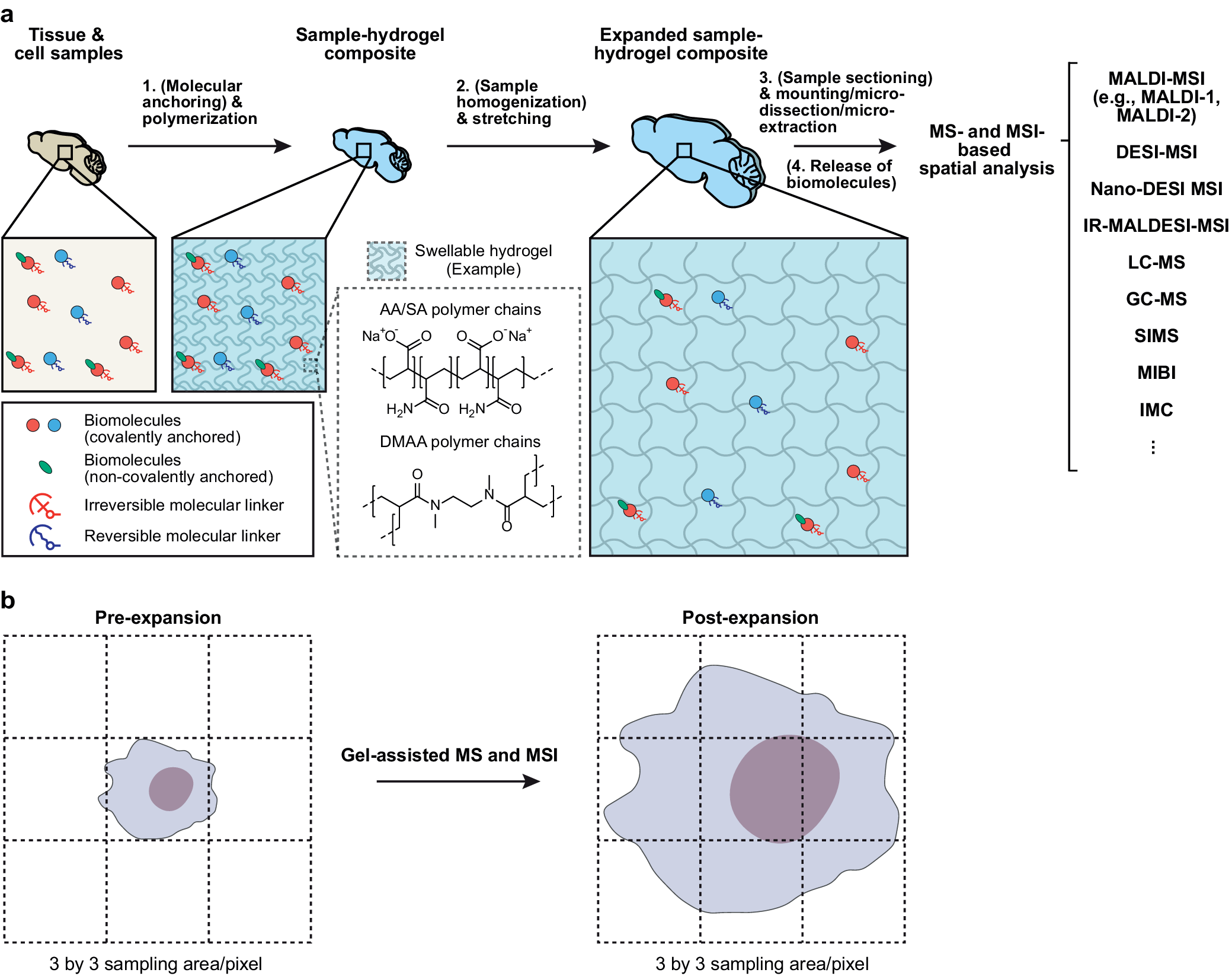
The recent combination of hydrogel-based sample embedding and expansion with MS- or MSI-based spatial analyses has thereby opened up new possibilities for single-cell and subcellular spatial omics investigations of intact cells and tissues (Figure 1). This emerging approach promises to transform spatial multiomics by providing the improved spatial resolution of MS and MSI for mapping the molecular organization of individual cells and subcellular compartments in complex tissues, such as the brain, kidney, pancreas, and tumors. From a methodological perspective, early developments have focused on optimizing the anchoring and homogenization steps, along with other critical steps for hydrogel embedding and expansion, to maximize biomolecule preservation and compatibility with MS and MSI techniques.
Principle and workflow of gel-assisted mass spectrometry (MS) and mass spectrometry imaging (MSI). (a) Schematics showing the principle and generalized workflow of hydrogel-based sample embedding and expansion for MS and MSI analyses. Intact cell and tissue samples undergo (1) treatment with a (reversible) small-molecule linker (optional) and polymerization to form a hydrogel composite, (2) homogenization (optional) and stretching, (3) sectioning (optional) and immobilization, microdissection, or microextraction, and (4) release of the tethered biomolecules (optional, e.g., via cleavage of a reversible molecular linker) for MS- and MSI-based spatial analyses (with exemplary modalities shown on the right). Biomolecules of interest (e.g., proteins and RNAs) (red and blue circles) can be covalently conjugated to the hydrogel through irreversible (red anchor) or reversible (blue anchor) molecular linkers. The reversible molecular linkers are especially useful for gel-assisted MS and MSI as covalently anchored molecules can be released for the downstream MS and MSI analyses. Some biomolecules (e.g., lipids) (green ovals) can be tethered to the hydrogel via noncovalent interactions with the hydrogel polymer chains or other biomolecules. The magnified schematics (solid squares) illustrate (left and middle) anchoring of biomolecules to the hydrogel, and (right) the anchored biomolecules in the expanded sample–hydrogel composite. The magnified schematic (dashed square) illustrates examples of commonly used swellable hydrogels, including acrylamide (AA)/sodium acrylate (SA) and N,N-dimethylacrylamide (DMAA) polymers. MALDI: matrix-assisted laser desorption ionization; DESI: desorption electrospray ionization; nano-DESI: nanospray desorption electrospray ionization; IR-MALDESI: infrared matrix-assisted laser desorption electrospray ionization; LC-MS: liquid chromatography mass spectrometry; GC-MS: gas chromatography mass spectrometry; SIMS: secondary ion mass spectrometry; MIBI: multiplexed ion beam imaging; IMC: imaging mass cytometry. (b) Schematics showing single-cell MS and MSI analyses enabled by gel-assisted MS and MSI. For example, expanding a cell 3-fold linearly results in a 9-fold increase in the areal pixel density.

Figure 1. Long description
Panel a shows a four-step workflow. Step 1 involves tissue and cell samples undergoing molecular anchoring and polymerization to create a sample-hydrogel composite. A magnified view shows red and blue circular biomolecules covalently anchored to a light blue wavy grid representing the hydrogel polymer network. Step 2 involves sample homogenization and stretching to create an expanded sample-hydrogel composite. A dashed box shows chemical structures for AA/SA and DMAA polymer chains. Step 3 involves sample sectioning, mounting, microdissection, or microextraction. Step 4 is the release of biomolecules for MS- and MSI-based spatial analysis. Listed MS and MSI modalities include MALDI-MSI, DESI-MSI, nano-DESI MSI, IR-MALDESI-MSI, LC-MS, GC-MS, SIMS, MIBI, and IMC. A legend identifies red and blue circles as covalently anchored biomolecules, green ovals as non-covalently anchored biomolecules, red anchors as irreversible linkers, and blue anchors as reversible linkers. Panel b compares pre-expansion and post-expansion states on a 3 by 3 sampling grid. At the pre-expansion state, a single cell occupies only the center pixel. At the post-expansion state, the cell has expanded to cover all 9 pixels of the grid, demonstrating increased areal pixel density.
In this article, we highlight the latest advances at this critical intersection of method development and discuss its implications for single-cell and subcellular spatial omics studies. To avoid potential confusion with specific methodologies developed, we have adopted the term ‘gel-assisted MS and MSI methods’ as a collective descriptor of this approach. This term encompasses techniques that utilize hydrogel sample embedding, often followed by sample stretching, to facilitate enhanced ionization, detection, and molecular analysis in MS and MSI.
Gel-assisted sample embedding and expansion for mass-spectrometry-based spatial lipidomic/metabolomic analyses
Lipids are an essential component of life, containing rich information about cellular processes, including cell signaling, membrane formation, and energy storage. Disruption in lipid metabolism is closely linked to pathological conditions across a broad range of organ systems, such as the brain, liver, and spleen (Vanier, Reference Vanier1983; Beltroy et al., Reference Beltroy, Richardson, Horton, Turley and Dietschy2005; Di Paolo and Kim, Reference Di Paolo and Kim2011; Tobias et al., Reference Tobias, Pathmasiri and Cologna2019; Xicoy et al., Reference Xicoy, Wieringa and Martens2019). MS has long been an indispensable tool in lipidomics studies, often applied to bulk samples such as tissue homogenates and dissociated cells. While these approaches provide valuable insights into the sample lipidome, the spatial organization of detected lipids is often lost during sample preparation. This loss of spatial context obscures how lipids are distributed across different cell types, tissue compartments, or microenvironments, which are important to unravel lipid functions and the regulation of downstream metabolic pathways. To address these methodological needs, MSI-based spatial analysis has been optimized to study the spatial organization of lipids and metabolites in intact cells and tissues.
Given its soft ionization capability and high imaging throughput, MALDI mass spectrometry imaging (MALDI-MSI) is a widely used MSI technique for lipidomic and metabolomic studies. In a standard MALDI-MSI workflow, tissue sections, often fresh-frozen, are mounted on a MALDI substrate such as a stainless steel plate or an indium tin oxide (ITO)-coated glass slide. A matrix layer composed of UV-absorbing organic molecules is then deposited on the sample surface, enabling efficient desorption and ionization of lipids during laser irradiation.
With its standard workflow, the spatial resolution of MALDI-MSI is constrained by both fundamental and practical factors. For instance, the crystal size of the deposited matrix sets an inherent limit to its spatial resolution, while the laser spot diameter and the precision of the sample stage, among other factors, introduce an instrumental limit to the imaging pixel size. In most commercially available MALDI time-of-flight (MALDI-TOF) mass spectrometers, the smallest raster distance or pixel size ranges from 5 to 10s of micrometers. While this is sufficient for resolving tissue-level spatial organization of target molecules, higher spatial resolution is needed for single-cell and subcellular applications.
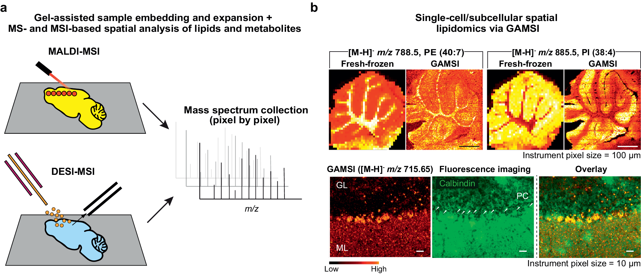
To address this limitation, recent studies have demonstrated that integrating gel-assisted sample embedding and expansion with MALDI-MSI offers a viable strategy for enhancing the spatial resolution of lipid MSI without modifying the existing hardware or analysis pipeline (Chan et al., Reference Chan, Pathmasiri, Pierre-Jacques, Cologna and Gao2023, Reference Chan, Maddison and Gao2024a, Reference Chan, Pathmasiri, Pierre-Jacques, Hibbard, Tao, Fischer, Yang, Cologna and Gao2024b; Hung et al., Reference Hung, Xie, Wang, Diao, Li, Wang, Qiu, Fang and Cai2024; Xie et al., Reference Xie, Wang, Diao, Guo, Lam, Li, Chen, Zhang, Wang, Fang and Cai2024a, Reference Xie, Wang, Diao, Wang and Cai2024b; Samuel et al., Reference Samuel, Baby, Mayo, Yan, Liang and Prentice2025; Zhang et al., Reference Zhang, Ding, Hu, Shi, Huang, Lu, Tillberg, Wang and Li2025; Mayo et al., Reference Mayo, Samuel, Guo, Ciccone, Liang and Prentice2026) (Figure 2). In this approach, intact cells and tissues are embedded and expanded in a swellable hydrogel and subsequently mounted on a MALDI substrate for MSI-based spatial lipidomic analyses. By tuning the polymer chemistry of the embedding hydrogel, the effective spatial resolution of MALDI-MSI can be enhanced by ~3- to 10-fold and potentially more. Of all the published protocols, the majority use a small-molecule reagent (‘linker’) to anchor biomolecules, polymerize and embed the sample in a swellable superabsorbent hydrogel, and mechanically homogenize and expand the sample. Most of these protocols are designed to maximize the retention of target analytes, including the lipids, within the original sample.
Gel-assisted MS and MSI for spatial lipidomic and metabolomic analysis. (a) (Top) Schematics showing the workflow of gel-assisted MALDI-MSI. Hydrogel-embedded and expanded samples are coated with a matrix and subsequently analyzed via laser desorption ionization. The red circles indicate the laser ablation spots, each corresponding to a single mass spectrum. (Bottom) Schematics showing the workflow of gel-assisted DESI-MSI. Hydrogel-embedded and expanded samples are analyzed using desorption electrospray ionization. In this process, a capillary releases charged solvent droplets (orange circles) onto the tissue, which desorbs and ionizes the analytes. The MS inlet then collects these charged analytes for the MS analysis, generating a single mass spectrum at each location. (b) Gel-assisted mass spectrometry imaging (GAMSI) enables sub-micrometer-pixel spatial lipidomic analysis of intact tissue samples with commercial mass spectrometers. (Top) Spatial distributions of selected lipids in (left) fresh-frozen and (right) GAMSI-processed (~3-fold expanded) mouse cerebellum sections. The instrument (AB SCIEX 4800) pixel size was set at 100 μm. Scale bars: 1 mm (3 mm). Here and after, scale bars are provided at the pre-expansion scale (with the corresponding postexpansion size indicated in the brackets). (Bottom) A GAMSI image of a ~4-fold expanded mouse cerebellum section, a fluorescence image of the same tissue section immunostained against calbindin (a Purkinje cell marker), and an overlay of the GAMSI and fluorescence images. The arrows in the fluorescence image indicate individual Purkinje cells. Scale bars: 25 μm (100 μm). The instrument (Bruker rapifleX) pixel size was set at 10 μm. The m/z (mass-to-charge) ratio values are provided for the singly charged deprotonated ions [M-H]−. The instrument pixel size refers to the physical size of a pixel as determined by the instrument specifications. The color scale bars for the MS images represent the relative intensity of the signals. GL, granular layer; ML, molecular layer; PC, Purkinje cell. Panels in (b) reprinted/adapted by permission from Springer Nature: (Chan et al., Reference Chan, Pathmasiri, Pierre-Jacques, Hibbard, Tao, Fischer, Yang, Cologna and Gao2024b), copyright 2024.

Figure 2. Long description
Panel a contains two schematics. The top schematic for MALDI-MSI shows a laser firing onto a yellow tissue sample with red ablation spots on a gray slide. The bottom schematic for DESI-MSI shows a capillary releasing orange charged droplets onto a blue tissue sample, with an inlet collecting analytes. Both workflows point to a central graph of mass spectrum collection (pixel by pixel) with intensity peaks plotted against m over z.
Panel b shows two rows of comparative imaging. The top row compares the spatial distribution of two lipids for fresh-frozen and GAMSI-processed mouse cerebellum sections: PE (40:7) at m over z of 788.5 and PI (38:4) at m over z of 885.5. The GAMSI images show significantly higher spatial resolution and detail than the pixelated fresh-frozen images .
The bottom row of panel b shows three high-resolution images of a mouse cerebellum section. The first is a GAMSI image at m over z of 715.65 showing the GL (granular layer) and ML (molecular layer) in a red hot color scheme. The second is a green fluorescence image panel for calbindin, with white arrows pointing to PCs (Purkinje cells). The third is an overlay of the two, showing the co-localization of the lipid signal with individual Purkinje cells. The color scale at the bottom indicates signal intensity from low (black) to high (yellow-white) for the red hot color scheme.
Gel-assisted mass spectrometry imaging (GAMSI) is a gel-assisted MSI method for single-cell spatial lipidomic and proteomic analyses (Chan et al., Reference Chan, Pathmasiri, Pierre-Jacques, Cologna and Gao2023, Reference Chan, Maddison and Gao2024a, Reference Chan, Pathmasiri, Pierre-Jacques, Hibbard, Tao, Fischer, Yang, Cologna and Gao2024b) (Figure 2b). As with other expansion-coupled MSI techniques, the sample homogenization step of GAMSI is crucial for breaking down the structural integrity of the gelled sample while preserving its spatial and molecular information. However, common homogenization conditions used for ExM, such as strong proteolysis and surfactant treatment, can cause substantial lipid loss. To address this issue, GAMSI makes use of trypsin-based sample digestion, which has been shown to provide better lipid retention compared to, for example, proteinase K (ProK)-based digestion.
With standard GAMSI protocol, tissue samples can be expanded ~3- to 6-fold, which corresponds to a ~3- to 6-fold increase in the effective spatial resolution for lipid MSI. GAMSI was initially validated on multiple commercially available MALDI-TOF mass spectrometers, which demonstrated its applicability and versatility across the MS instruments (Chan et al., Reference Chan, Pathmasiri, Pierre-Jacques, Cologna and Gao2023, Reference Chan, Pathmasiri, Pierre-Jacques, Hibbard, Tao, Fischer, Yang, Cologna and Gao2024b). Additional validations showed that the assigned lipid compositions remained largely comparable between GAMSI samples and the fresh-frozen controls. With ~4-fold expansion and ~2.5 μm effective pixel size, GAMSI allowed spatial lipidomic analysis of individual Purkinje cells in intact mouse cerebellum sections (Figure 2b). With ~6-fold expansion and ~0.88 μm effective pixel size, GAMSI resolved the lipidomic profiles of subcellular structures such as the glial and neuronal nuclei in the cerebellar white matter (Chan et al., Reference Chan, Pathmasiri, Pierre-Jacques, Hibbard, Tao, Fischer, Yang, Cologna and Gao2024b). Collectively, these results show that GAMSI provides a robust workflow for (sub)cellular MSI. Although current applications of GAMSI have focused on lipids (and proteins), expanding the technique to small-molecule imaging remains an active area for future development.
Tissue expansion mass spectrometry imaging (TEMI) utilizes a gel-assisted sample expansion process that does not involve a homogenization step (Zhang et al., Reference Zhang, Ding, Hu, Shi, Huang, Lu, Tillberg, Wang and Li2025). In TEMI, tissue samples are embedded in a swellable hydrogel along with a small-molecule linker, and expanded with a moderate expansion factor (e.g., ~1.5-fold). After repeating this gel-(re)embedding and expansion multiple times, the sample is sequentially swollen to the target expansion factor (e.g., ~3.5-fold after three iterations). Since TEMI expansion only relies on gradual swelling of the sample, it does not require a homogenization step to achieve crack/distortion-free tissue expansion. This mechanism significantly contributes to the enhanced preservation of native molecules, including lipids. When TEMI was applied to mouse cerebellum sections, for example, the molecular layer, granular layer, and white matter were more distinctly visualized compared to the unexpanded controls. TEMI further allowed imaging of individual Purkinje cells in expanded mouse cerebellum sections, revealing cell-specific lipid expression. Beyond lipidomics, the TEMI workflow is highly versatile, facilitating the spatial mapping of proteomic, glycomic, and metabolomic profiles. Future TEMI developments include maximizing hydrogel expansion and designing new anchoring chemistries to capture a broader molecular landscape.
Expansion mass spectrometry imaging (Ex-MSI) improves the spatial resolution of MALDI-MSI through a ~4.5-fold expansion process (Hung et al., Reference Hung, Xie, Wang, Diao, Li, Wang, Qiu, Fang and Cai2024). This method uses a hydrogel recipe similar to that introduced in the original protein retention ExM (ProExM) with ProK sample digestion (Tillberg et al., Reference Tillberg, Chen, Piatkevich, Zhao, Yu, English, Gao, Martorell, Suk, Yoshida, Degennaro, Roossien, Gong, Seneviratne, Tannenbaum, Desimone, Cai and Boyden2016). Briefly, the Ex-MSI protocol follows a general workflow of small-molecule anchoring, sample embedding in an expandable hydrogel, proteolytic digestion, and expansion. When applied to mouse brain sections, Ex-MSI samples exhibited more clearly defined features compared to the unexpanded samples across different acquisition pixel sizes, ranging from 50 μm (equivalent to a ~11 μm effective pixel size), 20 μm (~4.4 μm), 10 μm (~2.2 μm), to 5 μm (~1.1 μm). With an effective pixel size of ~2.2 μm, distinct patterns of lipid species such as phosphatidylethanolamine (PEs) and sulfatides (SHexCers) were revealed in the brain section, with the fibrous structure in the lateral geniculate complex and the nucleus of the pyramidal cells clearly resolved. In short, the Ex-MSI protocol leverages the standard proExM workflow, ensuring an easy adoption for laboratories with prior experience with ExM. A critical step of Ex-MSI involves the high-temperature protease treatment (with surfactants), which needs to be carefully optimized to balance uniform tissue expansion and lipid retention.
Expansion imaging mass spectrometry (ExIMS) has been developed to investigate the lipid retention of expanded tissues by testing different gel compositions, analyzing serial brain tissue sections, and utilizing computational techniques to assess the data (Samuel et al., Reference Samuel, Baby, Mayo, Yan, Liang and Prentice2025). Following the widely used ExM protocol of sample anchoring, gelation, homogenization, and expansion, lipid retention was analyzed in both positive and negative ion modes. When comparing cerebral serial tissue sections, ~4.5-fold expanded tissues exhibited clear images of Purkinje cells via positive ion mode MSI, which was confirmed by registering the MS images with fluorescent images. The cell bodies within the granular layer were further resolved in the expanded tissues for lipids such as phosphatidylcholine (PC) 34:2 and phosphatidylethanolamine (PE) 40:7. In total, 131 lipids were mapped in the negative ion mode, and 60 lipids were visualized in the positive ion mode using ExIMS. Notably, the lipid profiles of expanded and unexpanded tissues remained highly consistent in the negative ion mode. In contrast, the positive ion mode revealed some compositional discrepancies between the two groups, likely attributable to the tissue washing steps in the protocol. Despite these variations, ExIMS demonstrated a high degree of molecular preservation, with 75% of the lipid species identified in the unexpanded samples successfully retained after the expansion process. Overall, with protease treatment performed at low concentration and temperature, higher lipid retention is expected for ExIMS. Importantly, the hydrogel formulation, polymerization, and fixation conditions of ExIMS can be further adjusted for better retention of specific lipid classes. Future directions for ExIMS include expanding its use to other MS modalities and tailoring sample preparation protocols for tissues beyond the mouse brain.
Continuing efforts are made to enhance the spatial resolution of gel-assisted lipid MSI. 10xMSI, for example, employs a highly swellable polymer chemistry that takes advantage of self-crosslinked N,N-dimethylacrylamide (DMAA) polymers (Klimas et al., Reference Klimas, Gallagher, Wijesekara, Fekir, DiBernardo, Cheng, Stolz, Cambi, Watkins, Brody, Horani, Barth, Moore, Ren and Zhao2023; Xie et al., Reference Xie, Wang, Diao, Guo, Lam, Li, Chen, Zhang, Wang, Fang and Cai2024a). The 10xMSI workflow uses methacrolein as a small-molecule linker, which is introduced during the gelation step to eliminate the need for a separate anchoring step. After ProK digestion at a controlled temperature, the 10xMSI sample can be expanded ~10-fold over ice-cold water. With ~10-fold expansion, an effective pixel size of ~500 nm can be achieved using a 5-μm instrument pixel size. At this spatial resolution, 10xMSI could resolve subcellular structures such as the nucleus and dendrites of Purkinje cells in mouse cerebellum sections. In brief, 10xMSI represents a compelling approach for lipid MSI experiments due to its exceptional spatial resolution. However, as with other gel-assisted MS and MSI methods, the gains in resolution must be weighed against the dilution of target analytes that occurs as the expansion factor increases.
10x-DESI ExMSI utilizes a ~10-fold expansion workflow combined with desorption electrospray ionization (DESI)-MSI (Xie et al., Reference Xie, Wang, Diao, Wang and Cai2024b). This method uses a home-built DESI probe combined with an Orbitrap mass spectrometer, achieving an effective spatial resolution of 5 μm capable of resolving single cells. With this technique, the granule cell layer of the dentate gyrus in the hippocampus was clearly resolved. Furthermore, the white matter, granular, and molecular layers were resolved in mouse cerebellum tissues using a 40-μm step size (equivalent to a 4-μm effective step size), further revealing detailed structures of fibrous myelin within the granular layer. Utilizing this approach is particularly advantageous as the samples can be analyzed under ambient ionization environments. However, given that standard DESI imaging is generally restricted to tens of micrometers in step size, achieving (sub)cellular MSI requires a high expansion factor, such as the 10-fold expansion demonstrated here. More recently, ~10-fold sample expansion was combined with MALDI-MSI in ExFusion to achieve lipidomic mapping at the subcellular level (Mayo et al., Reference Mayo, Samuel, Guo, Ciccone, Liang and Prentice2026).
Gel-assisted sample embedding and expansion for mass-spectrometry-based spatial proteomic analyses
Proteomics refers to the comprehensive study of proteins, which includes their expression, function, and degradation within biological systems. In the central nervous system, proteins are fundamental to maintaining both homeostatic cellular functions and structural integrity. Disruptions and changes in protein expression and abundance are closely linked to neurodegenerative diseases (Taylor et al., Reference Taylor, Hardy and Fischbeck2002). This protein-driven complexity extends to the tumor microenvironment (TME). Pancreatic ductal adenocarcinoma (PDAC), for example, exhibits a highly heterogeneous proteomic landscape, and recent studies show its progression is mediated by signaling between the malignant cells and stromal cells (Huang et al., Reference Huang, Gao, Fu, Wang, Li, Chu, He, Li, Deng, Zhang, Kong, Yuan, Wang, Shi, Gao, Qin, Hunter and Tian2025). Proteomic understanding of complex cell signaling pathways is therefore essential for the discovery of new biomarkers and therapeutic strategies.
The human proteome, as characterized by the Human Proteome Project, is currently estimated to comprise approximately 20,000 proteins, a figure derived from the total number of protein-coding genes (Thul et al., Reference Thul, Åkesson, Wiking, Mahdessian, Geladaki, Ait Blal, Alm, Asplund, Björk, Breckels, Bäckström, Danielsson, Fagerberg, Fall, Gatto, Gnann, Hober, Hjelmare, Johansson, Lee, Lindskog, Mulder, Mulvey, Nilsson, Oksvold, Rockberg, Schutten, Schwenk, Sivertsson, Sjöstedt, Skogs, Stadler, Sullivan, Tegel, Winsnes, Zhang, Zwahlen, Mardinoglu, Pontén, von Feilitzen, Lilley, Uhlén and Lundberg2017). However, the true diversity of proteins likely extends far beyond this number. For instance, different forms of protein, known as proteoforms, are produced from the same gene through mechanisms such as alternative splicing and post-translational modifications (PTMs) (Smith and Kelleher, Reference Smith and Kelleher2013, Reference Smith and Kelleher2018). Although the exact number of proteoforms in the human body is unknown, it is estimated to be around 50 million, far exceeding the number of genes (Smith et al., Reference Smith, Agar, Chamot-Rooke, Danis, Ge, Loo, Paša-Tolić, Tsybin and Kelleher2021). To further compound proteomic studies, protein functions are highly dependent on their cellular and subcellular locations. In particular, the chemical environment, including local pH and redox conditions, and the availability of interaction partners and substrates can vary substantially across cellular and subcellular compartments. This variability creates a highly heterogeneous biochemical landscape that directly influences protein function, or the lack thereof. Therefore, spatial proteomics has garnered increasing interest in the identification and quantification of protein localizations and their possible functions (Lundberg and Borner, Reference Lundberg and Borner2019).
Fluorescence microscopy (FM), a widely used tool for spatial proteomics studies, provides superb spatial resolution and high sensitivity at the cellular and subcellular levels. In contrast, MS offers a distinctively powerful approach to spatial proteomics, characterized by deep molecular coverage, label-free detection, and high multiplexing capabilities in a single experiment. MS-based proteomics is primarily divided into top-down and bottom-up approaches (Aebersold and Mann, Reference Aebersold and Mann2016). In top-down proteomics, intact proteins are directly analyzed, enabling the characterization of their structures and PTMs. In bottom-up proteomics, proteins are first enzymatically digested into peptides, which are then analyzed and computationally assembled to reconstruct the parent proteins. Top-down approaches provide more direct structural information, while bottom-up workflows offer flexibility and scalability. Both strategies are widely employed depending on the experimental goals and sample requirements.
A typical MS-based spatial proteomics workflow begins with a microsampling method, such as laser capture microdissection (LCM) and liquid microjunction (LMJ) microextraction, to isolate ROIs within the sample (Mund et al., Reference Mund, Coscia, Kriston, Hollandi, Kovács, Brunner, Migh, Schweizer, Santos, Bzorek, Naimy, Rahbek-Gjerdrum, Dyring-Andersen, Bulkescher, Lukas, Eckert, Lengyel, Gnann, Lundberg, Horvath and Mann2022). Proteins are then extracted, fractionated, and digested from the sampled regions, and subsequently analyzed by LC-MS/MS when a bottom-up proteomics pipeline is used. In this workflow, the protein identities are reconstructed from the detected peptides, providing a simultaneous, label-free readout of multiple proteins. Since MS-based spatial proteomics relies on detection of intrinsic molecular structure rather than fluorescence, background signals from tissue autofluorescence do not interfere with the protein detection. Therefore, MS-based spatial proteomics is capable of detecting low-abundance proteins, facilitating deep proteomic coverage within heterogeneous biological samples.
MSI-based approaches are also commonly used for spatial proteomics studies, providing high-throughput profiling of protein distributions across intact tissue surfaces. MSI experiments typically involve either a top-down approach on tissue sections or a bottom-up approach combined with in situ enzymatic digestion (Groseclose et al., Reference Groseclose, Andersson, Hardesty and Caprioli2007). While MSI-based spatial proteomics may not achieve the same proteome coverage as the MS-based methods, its strength lies in its ability to map protein distributions within the broader tissue and organ context with high throughput.
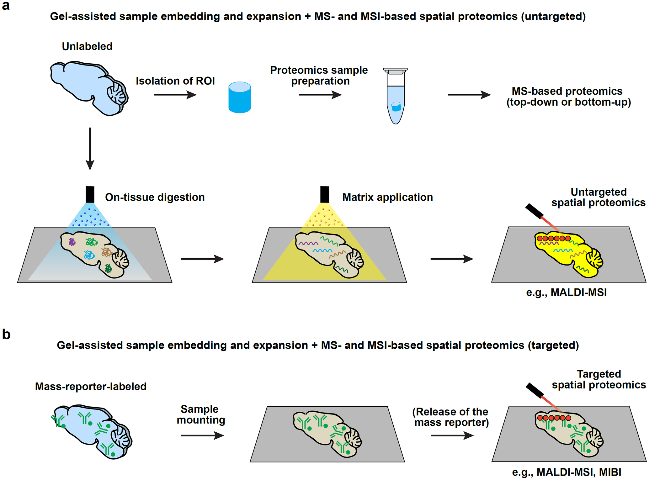
Despite these advances, achieving cellular and subcellular spatial resolution has remained a challenge for MS- and MSI-based spatial proteomics, primarily owing to the existing constraints in the accuracy and resolution of microsampling methods, as well as the limitations in the MSI spatial resolution. To overcome these constraints, gel-assisted sample embedding and expansion has been successfully integrated with MS- and MSI-based spatial proteomics pipelines, paralleling their application to lipid MSI (Drelich et al., Reference Drelich, Aboulouard, Franck, Salzet, Fournier and Wisztorski2021; Li et al., Reference Li, Sun, Sun, Dong, Wu, Sun, Zhang, Jiang, Zhou, Cen, Cai, Xia, Zhu, Guo and Piatkevich2022; Bai et al., Reference Bai, Zhu, Oliveria, Cannon, Feyaerts, Bosse, Vijayaragavan, Greenwald, Phillips, Schürch, Naik, Ganio, Gaudilliere, Rodig, Miller, Angelo, Bendall, Rovira-Clavé, Nolan and Jiang2023; Chan et al., Reference Chan, Pathmasiri, Pierre-Jacques, Cologna and Gao2023, Reference Chan, Pathmasiri, Pierre-Jacques, Hibbard, Tao, Fischer, Yang, Cologna and Gao2024b; Dong et al., Reference Dong, Jiang, Wu, Chen, Chen, Ding, Zheng, Piatkevich, Zhu and Guo2024, Reference Dong, Wu, Chen, Jiang, Piatkevich, Zhu and Guo2025; Zhang et al., Reference Zhang, Ding, Hu, Shi, Huang, Lu, Tillberg, Wang and Li2025; Franziscus et al., Reference Franziscus, Ferrand, Biehlmaier, Schmidt and Spang2026) (Figure 3).
Gel-assisted MS and MSI for spatial proteomic analysis. (a) Schematics showing the workflow of gel-assisted MS and MSI methods for untargeted spatial proteomics. (Top) For MS-based untargeted spatial proteomics, a sample ROI can be first isolated from the expanded sample-hydrogel composite using, for example, manual dissection or LCM. The isolated ROI is then subject to sample preparation for either top-down or bottom-up MS-based proteomics. (Bottom) For MSI-based untargeted spatial proteomics, after mounting the expanded sample-hydrogel composite on the MSI substrate, an in situ digestion step can be performed to release the peptides for the subsequent MSI workflow (e.g., MALDI-MSI). (b) Schematics showing the workflow of gel-assisted MS and MSI methods for targeted spatial proteomics. For MSI-based targeted spatial proteomics, the expanded sample-hydrogel composite can be first labeled with antibodies conjugated with a mass reporter (e.g., a photocleavable mass reporter or a metal isotope). After mounting the sample-hydrogel composite and (optionally) releasing the mass reporters (e.g., via photocleavage), the sample is subject to a standard MSI workflow (e.g., MALDI-MSI, MIBI).

Figure 3. Long description
Panel a shows two pathways for gel-assisted MS- and MSI-based untargeted spatial proteomics starting from an unlabeled blue tissue sample.
1. The top horizontal path shows isolation of a sample ROI, resulting in a blue cylindrical sample, followed by proteomics sample preparation in a microcentrifuge tube, leading to MS-based top-down or bottom-up proteomics.
2. The bottom path begins with on-tissue digestion. A blue spray is applied to the tissue on a gray substrate. This is followed by matrix application, where a yellow spray is applied. The final step shows untargeted spatial proteomics via a red laser beam scanning the tissue, using e.g., MALDI-MSI.
Panel b shows a single horizontal workflow for gel-assisted MS- and MSI-based targeted spatial proteomics.
1. The process starts with a mass-reporter-labeled tissue sample containing green antibodies.
2. The next step is sample mounting onto a gray substrate.
3. The final step, following an optional release of the mass reporter, shows targeted spatial proteomics analysis where a red laser scans the tissue to detect the mass reporters, via e.g., MALDI-MSI and MIBI.
By utilizing swellable polyacrylamide/sodium polyacrylate hydrogels for sample embedding and stretching, enhanced spatial resolution can be achieved for the subsequent untargeted MS proteomics analyses (Drelich et al., Reference Drelich, Aboulouard, Franck, Salzet, Fournier and Wisztorski2021). In this approach, tissue samples are denatured to allow uniform gel-assisted expansion while preserving protein integrity for bottom-up MS analysis. This workflow bypasses traditional proteolysis for sample-hydrogel composite homogenization, which can compromise protein sequence information and limit the depth of the identifiable proteome. After expansion, ROIs within the tissue are dissected with a scalpel blade and subsequently analyzed by LC-MS/MS for untargeted bottom-up proteomics. In another MS-based untargeted spatial proteomics workflow, expansion proteomics (ProteomEx) achieves ~6-fold expansion while utilizing biopsy punches to dissect out ROIs in the expanded tissue (Li et al., Reference Li, Sun, Sun, Dong, Wu, Sun, Zhang, Jiang, Zhou, Cen, Cai, Xia, Zhu, Guo and Piatkevich2022). With ProteomEx, high-spatial-resolution quantitative proteome profiling can be achieved in different tissue types, including the brain, liver, and breast cancer tissues. The ProteomEx workflow has also been applied to Alzheimer’s disease (AD) mouse tissues, identifying dysregulated proteins within specific brain regions. Lately, ~10-fold gel-assisted sample expansion has been integrated with LCM and downstream LC-MS/MS to achieve organelle-level spatial proteomics (Franziscus et al., Reference Franziscus, Ferrand, Biehlmaier, Schmidt and Spang2026).
Despite the enhanced spatial resolution, throughput remains a major challenge for these methods as manual handling often precludes parallel and large-scale processing. To overcome this limitation, filter-aided expansion proteomics (FAXP) integrates robotic automation with the ProteomEx workflow (Dong et al., Reference Dong, Jiang, Wu, Chen, Chen, Ding, Zheng, Piatkevich, Zhu and Guo2024, Reference Dong, Wu, Chen, Jiang, Piatkevich, Zhu and Guo2025). This robotics adaptation, along with additional modifications, can shorten the processing time by nearly 50% while drastically enhancing experimental reproducibility. FAXP is also compatible with LCM, enabling isolation of nuclei from the expanded tissue and the subsequent proteomic profiling. Together, FAXP and ProteomEx leverage the synergistic strengths of deep MS-based proteomic profiling and the high spatial resolution enabled by gel-assisted sample expansion. However, as with other MS-based proteomics approaches, throughput remains a bottleneck for whole-mount tissue investigations, and the implementation of automated workflow requires access to specialized hardware. Future developments include but are not limited to expanding the applicability of these approaches to a broader range of tissue types as well as clinical specimens.
For MSI-based untargeted spatial proteomics, the TEMI workflow supports on-sample digestion and MALDI-MSI-based spatial proteomic analyses (Zhang et al., Reference Zhang, Ding, Hu, Shi, Huang, Lu, Tillberg, Wang and Li2025). The resulting protocol enabled untargeted identification of over 50 tryptic peptides within the 3.5-fold expanded mouse cerebellum section. In another example, the in situ imaging proteomics via expansion (iPEX) workflow combines gel-assisted tissue expansion with MALDI-MSI for untargeted proteomic imaging at the micrometer scale (Wang et al., Reference Wang, Sun, Wu, Fu, Fan, Zhao, Huang, Pan, Lu, Han, Jia, Zeng, Zhang, Chen, An, Meng, Guo, Li, Lian, Sun, Hu, Yang, Feng, Li, Du, Liu, Piatkevich and Zou2026). The iPEX workflow begins with N-succinimidyl acrylate (NSA) modification and gelation, followed by SDS/boric acid homogenization, DL-dithiothreitol (DTT) reduction, and iodoacetamide alkylation. Unlike other gel-assisted MS and MSI methods, iPEX utilizes a sodium-free, ammonium bicarbonate buffer to achieve 3- to 7-fold expansion. After expansion, tissues are mounted on ITO slides for on-sample trypsin/Lys-C digestion and CHCA matrix deposition. By applying the method to the mouse retinal tissue, iPEX detected 1827 peptides that could not be detected on unexpanded tissues. The broad applicability of iPEX was further demonstrated across diverse tissue types by detecting hundreds to thousands of proteins at micrometer resolution in the brain, liver, intestine, and organoids. When applied to 5xFAD AD mouse tissues, iPEX identified early-onset biomarkers, including the downregulation of the peroxisomal enzyme ACAA1A in 2-month-old mice relative to the wild-type controls, which highlights a potential link between this enzyme and early-stage neurodegeneration. Overall, iPEX offers a versatile MSI-based untargeted proteomic profiling method with micrometer spatial resolution and high proteome coverage. Future iPEX developments include extending its applicability to formalin-fixed paraffin-embedded (FFPE) samples and further enhancing its proteomic coverage.
Gel-assisted tissue embedding and expansion has also been combined with MSI-based targeted spatial proteomics. One approach for protein labeling is to employ antibodies conjugated with a mass reporter, such as peptide-based photocleavable mass-tag (PC-MT) that can be ionized and detected in a highly multiplexed manner using a standard MALDI-MSI workflow (Yagnik et al., Reference Yagnik, Liu, Rothschild and Lim2021). This labeling strategy has been integrated with, for example, GAMSI, where the antibody-conjugated PC-MT is introduced to the expanded tissue samples for MALDI-MSI analysis (Chan et al., Reference Chan, Pathmasiri, Pierre-Jacques, Cologna and Gao2023, Reference Chan, Pathmasiri, Pierre-Jacques, Hibbard, Tao, Fischer, Yang, Cologna and Gao2024b). While the standard GAMSI workflow uses post-expansion labeling, it is also compatible with pre-expansion PC-MT labeling. This alternative, however, requires replacing the proteolytic digestion step with a denaturation step to ensure retention of the peptide mass reporters.
For MSI-based targeted spatial proteomics, Expand and comPRESS hydrOgels (ExPRESSO) integrates tissue expansion with multiplexed ion beam imaging (MIBI), a secondary ion MSI technique (Bai et al., Reference Bai, Zhu, Oliveria, Cannon, Feyaerts, Bosse, Vijayaragavan, Greenwald, Phillips, Schürch, Naik, Ganio, Gaudilliere, Rodig, Miller, Angelo, Bendall, Rovira-Clavé, Nolan and Jiang2023). In ExPRESSO, proteins are labeled with metal-isotope-tagged antibodies through a chelating linker. Under the high-energy ion beam of MIBI, the chelator-metal complex is disrupted, releasing isotope signals that correspond to the antibody-targeted proteins. When combined with ~3.7-fold tissue expansion, ExPRESSO achieves a spatial resolution of ~40 nm, providing subcellular spatial resolution. The ExPRESSO method was applied to human brain tissue to study protein distribution at subcellular resolution. Using a 21-marker panel, the technique successfully resolved astrocyte morphology and delineated fine subcellular structures. Furthermore, the method was applied to AD patient tissue to map proteins associated with blood–brain barrier homeostasis, tau tangles, and amyloid-β plaques. Combined, by leveraging the spatial resolution and high-plex capabilities of MIBI, ExPRESSO achieves unparalleled spatial resolution for multiplexed protein imaging at the subcellular scale. Future developments include broadening the scope of detectable molecules, as well as optimizing the technique’s imaging depth to enable 3D imaging through repetitive scanning.
Gel-assisted MS and MSI for spatial multiomics analyses
Multiplexed imaging enables spatial mapping of diverse biomarkers, often of different molecular classes and entities, on the same sample. MSI, for example, is suitable for multiplexed molecular imaging of intact cells and tissues due to its capability to generate mass spectra of virtually infinite numbers of molecules and their fragments within a single imaging modality. Enhancing the spatial resolution of MS- and MSI-based multiplexed imaging thus holds great potential for single-cell and subcellular spatial multiomics applications.
Leveraging the multiplexing capability of MSI, the previously discussed PC-MTs utilize the unique masses of an array of photocleavable peptide-based mass reporters, which are conjugated to antibodies to target specific proteins (Yagnik et al., Reference Yagnik, Liu, Rothschild and Lim2021). When the GAMSI workflow is combined with PC-MT-based protein labeling, multiplexed spatial lipidomic and proteomic information can be obtained from the same tissue section by performing sequential MSI (Chan et al., Reference Chan, Pathmasiri, Pierre-Jacques, Cologna and Gao2023, Reference Chan, Pathmasiri, Pierre-Jacques, Hibbard, Tao, Fischer, Yang, Cologna and Gao2024b). In TEMI, in addition to the PC-MT-based multiplexed imaging, N-glycan or tryptic peptide MSI can be performed following lipidomic or metabolomic MSI on the same tissue section (Zhang et al., Reference Zhang, Ding, Hu, Shi, Huang, Lu, Tillberg, Wang and Li2025). With this approach, up to 29 N-glycans and 57 (peptide) features were detected for the same tissue section on top of its rich lipidomic or metabolomic features.
In addition to single-modality imaging, multimodal and correlative imaging provides another effective option for spatial multiomics studies. In particular, the multiplexity of gel-assisted MS and MSI techniques can be enhanced by coupling them with an orthogonal technique. For example, both GAMSI and TEMI have demonstrated that MSI and fluorescence images can be overlaid for multimodal molecular mapping within the same tissue section (Chan et al., Reference Chan, Pathmasiri, Pierre-Jacques, Cologna and Gao2023, Reference Chan, Pathmasiri, Pierre-Jacques, Hibbard, Tao, Fischer, Yang, Cologna and Gao2024b; Zhang et al., Reference Zhang, Ding, Hu, Shi, Huang, Lu, Tillberg, Wang and Li2025). In GAMSI, the lipid MS images were overlaid with calbindin immunofluorescence images to identify individual Purkinje cells in a mouse cerebellum section. In TEMI, an expanded mouse cerebellum was stained with DAPI and/or an NHS-ester-based fluorescent dye for pan-protein labeling. These fluorescence labels provided a structure reference for the MS images and revealed the anatomical heterogeneity in lipid distribution within each cerebellar layer. The recently reported ExFusion further extended the utility of multimodal imaging by refining the ExIMS images using the fluorescently labeled morphological information (Mayo et al., Reference Mayo, Samuel, Guo, Ciccone, Liang and Prentice2026).
Current challenges in gel-assisted MS and MSI
While the emerging gel-assisted MS and MSI methods have significantly enhanced the spatial resolution of MS and MSI for single-cell and subcellular spatial omics studies, several challenges remain. These limitations generally fall into two categories: those stemming from the hydrogel-embedding workflow and those inherent to the MS and MSI processes.
First, gel-assisted MS and MSI methods rely on a sequence of chemical treatments, typically involving fixation, anchoring, and gelation, often followed by homogenization and physical expansion. These steps may introduce chemical and physical changes to the sample, such as alteration of its molecular profile and delocalization of the analyte. For instance, aldehyde-based fixatives are commonly used in gel-assisted MS and MSI protocols to stabilize the tissue architecture for subsequent gelation. However, these cross-linking agents may inadvertently mask key analytes by hindering their efficient desorption and ionization during MS and MSI analysis. The enzymatic treatment or denaturing process introduced in the homogenization step may also alter the molecular profile within the sample.
In response to these concerns, recent studies have characterized, for instance, the lipidomic coverage of gel-assisted MS and MSI techniques, comparing the detected molecular profiles against the traditionally used fresh-frozen samples. These comparative analyses showed that the overall lipidomic landscape is preserved without substantial loss or selective depletion, confirming reduced bias across lipid classes found using gel-assisted lipidomic MS and MSI methods (Chan et al., Reference Chan, Pathmasiri, Pierre-Jacques, Cologna and Gao2023, Reference Chan, Pathmasiri, Pierre-Jacques, Hibbard, Tao, Fischer, Yang, Cologna and Gao2024b; Samuel et al., Reference Samuel, Baby, Mayo, Yan, Liang and Prentice2025; Zhang et al., Reference Zhang, Ding, Hu, Shi, Huang, Lu, Tillberg, Wang and Li2025). For proteomic studies, iPEX demonstrated a marked increase in protein identifications via MALDI-MSI following gel-assisted expansion compared to traditional fresh-frozen tissues (Wang et al., Reference Wang, Sun, Wu, Fu, Fan, Zhao, Huang, Pan, Lu, Han, Jia, Zeng, Zhang, Chen, An, Meng, Guo, Li, Lian, Sun, Hu, Yang, Feng, Li, Du, Liu, Piatkevich and Zou2026). Furthermore, the proteins identified by iPEX showed substantial overlap with LCM-proteomics performed on gel-expanded tissues, though iPEX detected fewer total proteins.
Despite these advances, adapting gel-assisted MS and MSI workflows to other widely used sample formats, such as formalin-fixed paraffin-embedded (FFPE) tissues, remains an important challenge (Lee et al., Reference Lee, Rubakhin, Kusmartseva, Wasserfall, Atkinson and Sweedler2020; Hiratsuka and Tsuruyama, Reference Hiratsuka and Tsuruyama2024). In FFPE sample preparation, native biomolecules can be chemically modified (e.g., via lipid oxidation) or washed out during fixation or paraffin embedding/removal (Dannhorn et al., Reference Dannhorn, Swales, Hamm, Strittmatter, Kudo, Maglennon, Goodwin and Takats2022). These steps may also disrupt the native distributions of biomolecules, thus compromising the accuracy of their native spatial contexts. Moreover, the FFPE sample format can complicate the downstream MS and MSI analysis, with the lower m/z ranges (~100–500) exhibiting potential disruptions from formaldehyde oligomers as well as reduced lipid and metabolite signals (Dannhorn et al., Reference Dannhorn, Swales, Hamm, Strittmatter, Kudo, Maglennon, Goodwin and Takats2022). In the context of sample preparation for gel-assisted MS and MSI, these effects can be retained or further amplified to affect the final results.
Analyte delocalization represents another technical challenge for gel-assisted MS and MSI techniques. Enzymatic digestion, for example, could in principle allow analytes of interest to diffuse away from their original locations, while precisely quantifying such effects remains challenging. One such characterization has been provided by iPEX, which introduced a benchmarking strategy based on pan-protein fluorescent labeling. In this approach, proteins were labeled with a fluorescence marker prior to expansion (Wang et al., Reference Wang, Sun, Wu, Fu, Fan, Zhao, Huang, Pan, Lu, Han, Jia, Zeng, Zhang, Chen, An, Meng, Guo, Li, Lian, Sun, Hu, Yang, Feng, Li, Du, Liu, Piatkevich and Zou2026). This enabled direct comparison of protein distributions before and after the gel-assisted sample expansion process. As a result, the displacement was found to be less than 9%.
For gel-assisted sample expansion, free-radical chain-growth polymerization is commonly used to form a dense hydrogel polymer network across the sample. The free radicals generated in this process often constrain the selection of fluorescent labels for multimodal imaging, as certain fluorophores undergo quenching or structural damage when exposed to these reactive species. For instance, cyanine dyes, including Alexa Fluor 647 and Cy5, have been reported to lose their fluorescence during polymerization (Asano et al., Reference Asano, Gao, Wassie, Tillberg, Chen and Boyden2018). A workaround is to use alternative fluorophores that are compatible with free-radical polymerization, such as ATTO 647N.
Notably, the decrowding effect inherent to sample expansion leads to a decrease in analyte concentration per pixel (or per ROI) as the expansion factor increases, an effect commonly referred to as ‘analyte dilution.’ The areal density of analytes near the sample surface are thought to decrease in proportion to the square of the sample expansion factor: a 4-fold linear expansion, for example, reduces this density by 16-fold. As a result, the MS instrument’s sensitivity quickly becomes a limiting factor that constrains the achievable effective spatial resolution with adequate signal counts.
Interestingly, recent experimental benchmarks, particularly those using MALDI-MSI, suggest that the observed signal decrease can be less severe than this prediction (Chan et al., Reference Chan, Pathmasiri, Pierre-Jacques, Cologna and Gao2023, Reference Chan, Pathmasiri, Pierre-Jacques, Hibbard, Tao, Fischer, Yang, Cologna and Gao2024b; Samuel et al., Reference Samuel, Baby, Mayo, Yan, Liang and Prentice2025). This suggests that additional factors are likely compensating for the analyte dilution effect and contributing to signal enhancement for gel-assisted MS and MSI. One possible explanation is that expansion dilutes both the target analytes and the nonanalyte matrix components. This can enhance signal quality by reducing the matrix effect, which hampers analyte ionization in complex biological samples.
Consistent with this explanation, gel-assisted MSI protocols with 10-fold linear expansion have reported acceptable signal-to-noise ratios. We speculate that even higher expansion factors (e.g., 20-fold or larger) are feasible with additional innovations in the sample preparation, including better retention of the analytes and additional signal enhancement. Equally important are the continued advances in MS hardware that offer enhanced detection sensitivity. Recent advancements in laser-induced postionization (e.g., MALDI-2), for example, may provide substantial signal enhancement, potentially offsetting the effects of analyte dilution at higher sample expansion. Combined, the parallel refinement of sample preparation and analytical instrumentation is expected to push the spatial resolution limits of gel-assisted MS and MSI even further.
In addition to instrument sensitivity, a fundamental tradeoff between the throughput and spatial resolution exists. For example, imaging an expanded tissue section takes significantly longer than its non-expanded counterpart across the same biological area. An additional challenge, which is inherent to all MSI methods, arises from run-to-run variations. Since commercial instruments often require each expanded sample to be analyzed and processed sequentially on its own imaging plate, proper standardization is needed to ensure coherent characterization across the samples.
Another potential challenge for gel-assisted MSI techniques, as with all MSI-based approaches, is their limited quantitative robustness. While MSI can be semiquantitative, absolute spatial quantification of protein abundance within a sample is challenging (Tobias and Hummon, Reference Tobias and Hummon2020). In particular, matrix effects and differences in sample preparation can diminish attempts at absolute quantification, as any heterogeneity in crystallization of matrix deposition on a sample can affect ion extraction efficiency and cause signal suppression (Bouschen and Spengler, Reference Bouschen and Spengler2007). To address potential intensity differences in analyte extraction efficiency or ion suppression, internal standards can be used. However, such standards still do not guarantee absolute quantification, as normalizing to a standard does not compensate for all ion suppression and matrix effects (Hansen and Janfelt, Reference Hansen and Janfelt2016).
As with other MS and MSI techniques, gel-assisted MS and MSI methods are intrinsically destructive. Sequential and multiplexed imaging or measurement are typically limited by the loss of analytes under these techniques. However, better retention of target molecules can be achieved by optimizing the order and timing of each round of imaging and measurement, as well as tuning the laser power of the MS instrument (Denti et al., Reference Denti, Capitoli, Piga, Clerici, Pagani, Criscuolo, Bindi, Principi, Chinello, Paglia, Magni and Smith2022; Zhang et al., Reference Zhang, Liu, Fields, Shi, Huang, Lu, Schneider, Tang, Puglielli, Welham and Li2023; Seeley, Reference Seeley2025).
Finally, the vast scale of resulting datasets presents a critical bottleneck for gel-assisted MS and MSI workflows. The increased imaging area as well as number of sample runs drastically increases the final data size, while combining it with other imaging modalities will only increase the data. Therefore, a robust and scalable computational pipeline to process, visualize, and analyze increasingly larger gel-assisted MS and MSI datasets is needed. For multimodal imaging, another key challenge is to perform coregistration of datasets acquired across different imaging rounds or modalities. This task is complicated by differences in the pixel sizes, dynamic intensity ranges, and inherent spatial features. Consequently, the incorporation of fiducial markers and common structural landmarks will be essential for ensuring accurate image alignment and data overlay in the subsequent computational workflow. Within this context, applying computational frameworks, such as MEISTER (Xie et al., Reference Xie, Castro, Rubakhin, Trinklein, Sweedler and Lam2024c) and ExFusion (Mayo et al., Reference Mayo, Samuel, Guo, Ciccone, Liang and Prentice2026), may enable automated multimodal image fusion, facilitate extraction of cell-specific molecular profiles, and thereby reduce MS and MSI acquisition times. Developing similar computational packages will ultimately increase the throughput and accessibility of large spatial multiomics datasets generated by gel-assisted MS and MSI methods.
Outlook
As MS and MSI become an increasingly important tool for spatial molecular analysis of biological specimens, their spatial resolution is also becoming a critical bottleneck for single-cell and subcellular spatial omics applications. To overcome the existing spatial resolution limitations, gel-assisted sample embedding and expansion provides an effective approach for enhancing spatial resolution without the need for instrument modifications or new computational pipelines. Given its accessibility and versatility, the development of gel-assisted MS and MSI methods is likely to accelerate, leading to its adoption for an even broader range of MS instruments and techniques, including nano-DESI MSI, DESI-MSI, IR-MALDESI-MSI, SIMS, and IMC (Figure 1). Furthermore, extending these approaches to spatial analyses of additional molecular classes (e.g., glycans and metabolites) will continue to advance high-plex, single-cell and subcellular multiomics studies.
Nevertheless, practical considerations remain when combining gel-assisted sample embedding and expansion with different MS and MSI platforms. Especially, the interaction between the hydrogel matrix and the selected MS and MSI techniques must be carefully examined during experimental design. For instance, applying SIMS to expanded samples may lead to more extensive hydrogel ablation compared to atmospheric or vacuum-based ionization techniques, such as MALDI and DESI. Optimizing parameters such as the laser power or dwell time is therefore essential to prevent signal loss and higher background while capitalizing on the decrowding effect of gel-assisted sample expansion.
While gel-assisted MS and MSI methods have been used extensively to map the molecular profile of brain tissues, continuing advances in sample preparation and MS instrumentation will enable a much broader range of specimens to be studied. For instance, iPEX successfully demonstrated proteomic profiling in the retina, intestines, and liver, while TEMI was employed to characterize the lipidomes of melanoma tumors, kidney, and pancreatic tissues. When combined with (serial) sectioning and 3D registration, gel-assisted MS and MSI techniques may further form the foundation for single-cell and subcellular multiomic mapping of whole organs of various types (Fusar Bassini et al., Reference Fusar Bassini, Schede, Capolupo, Haj Abdullah Alieh, Venturi, Valente, Droin, Trejo Banos, Khven, Asirim, Nasrallah, Kaysudu, Krymova, D’Angelo and La Manno2025; Lu et al., Reference Lu, Zhang, Yagnik, Lim, Rothschild, Li, Schneider, Puglielli and Li2025; Ma et al., Reference Ma, Shedlock, Medina, Ribas, Clarke, Hawkinson, Dande, Golamari, Wu, Ziani, Burke, Merritt, Vander Kooi, Gentry, Yadav, Chen and Sun2025). These developments will underscore the versatility of gel-assisted MS and MSI techniques for a broad spectrum of biomedical research.
To further advance gel-assisted MS and MSI, several critical technical challenges must be overcome. For example, to address the previously discussed issue of sensitivity, mass detection with high sensitivity, such as MALDI-2 and single-molecule MS, will become necessary to maintain higher spatial resolution without compromising molecular coverage (Niehaus et al., Reference Niehaus, Soltwisch, Belov and Dreisewerd2019; Deslignière et al., Reference Deslignière, Rolland, Ebberink, Yin and Heck2023). Also, as discussed, the substantial increase in the sample analysis time needs to be addressed. Due to actual limitations in instrument access and stability, it is challenging and often unfeasible to run samples for weeks and days instead of hours and minutes. Therefore, the development of high-throughput commercial and custom MS instruments is essential for making gel-assisted MS and MSI methods more accessible to the community (Jungmann et al., Reference Jungmann, MacAleese, Buijs, Giskes, de Snaijer, Visser, Visschers, Vrakking and Heeren2010).
As discussed, continued development of gel-assisted MS and MSI is crucial for achieving (sub)cellular resolution analyses of complex molecular landscapes within tissue architecture. One potential direction is developing and optimizing hydrogel recipes with high (e.g., >20-fold) expansion factors to enable subcellular spatial omics, as well as engineering low-background hydrogels to enhance the molecular coverage. Another direction can focus on developing MS- and MSI-optimized anchoring strategies for concurrent retention of multiple types of biomolecules. As MS and MSI techniques continue to evolve, in toto mapping of spatial biomolecular heterogeneity with complex tissue microenvironments is becoming a reality. Gel-assisted MS and MSI, in particular, will be instrumental in resolving the intricate molecular organization of these systems.
Open peer review
To view the open peer review materials for this article, please visit http://doi.org/10.1017/qrd.2026.10024.
Data availability statement
All relevant data are included in this publication or can be found in the cited literature.
Acknowledgments
We thank the members of the Cologna lab at the University of Illinois Chicago for their helpful discussions and support.
Author contribution
R.G. supervised the study. Y.H.C., M.C.H., H.P.D., and R.G. wrote the article.
Financial support
This work was supported by the National Institutes of Health (grant number: DP2MH136390); the Searle Scholars Program; and the McKnight Technological Innovations in Neuroscience Award.
Competing interests
R.G. is a co-inventor of multiple patents related to ExM. The other authors declare no competing financial interests.

 Open access


Comments
Dear Dr. Boyden and Dr. Phan,
Thank you again for your kind invitation to contribute to the special collection “Nanoscale Analysis of Brain Chemistry and Structure Collection” in QRB Discovery. We are excited to submit our Perspective article, “Mass-spectrometry-based single-cell and subcellular spatial omics via hydrogel-assisted sample embedding and expansion”, for your consideration. This article highlights the recent integration of hydrogel-assisted tissue expansion with mass spectrometry and mass spectrometry imaging, demonstrating its utility for single-cell/subcellular spatial molecular mapping of intact brain tissue. With a comprehensive discussion of the current limitations and prospective future opportunities, we believe this article will be highly relevant to the broad readership of QRB Discovery and this special collection.
Short Summary:
Mass spectrometry (MS) and mass spectrometry imaging (MSI) are powerful tools for molecular analysis of the brain, but their spatial resolution limits single-cell/subcellular applications. Hydrogel-assisted sample expansion overcomes this limit by physically magnifying specimens within a hydrogel, enabling (sub)cellular mapping of lipids and proteins without instrument modifications. In this article, we discuss this approach’s principles, applications, challenges, and future improvements for single-cell/subcellular spatial omics.
Thank you for your consideration.
Sincerely,
Ruixuan Gao