INTRODUCTION
The protozoan parasite Entamoeba histolytica is responsible for millions of cases of amoebic colitis and liver abscess in humans annually, resulting in up to 100 000 deaths (World Health Organization, 1997). Another intestinal-dwelling amoeba, E. dispar, is morphologically indistinguishable from E. histolytica but is non-pathogenic (Diamond and Clark, Reference Diamond and Clark1993). E. histolytica/E. dispar is also commonly found in the feces of non-human primates (Smith and Meerovitch, Reference Smith and Meerovitch1985; Muriuki et al. Reference Muriuki, Murugu, Munene, Karere and Chai1998; Tachibana et al. Reference Tachibana, Cheng, Kobayashi, Fujita and Udono2000; Verweij et al. Reference Verweij, Vermeer, Brienen, Blotkamp, Laeijendecker, van Lieshout and Polderman2003). E. histolytica infection in these primates is a serious problem for animal health and also has zoonotic potential, which makes it important to discriminate E. histolytica from other non-pathogenic amoebae.
In older literature, several E. histolytica-like amoebae in monkeys have been described. The name E. nuttalli Castellani, Reference Castellani1908 was proposed for an amoeba found in a liver abscess in a Macacus pileatus monkey in Colombo, Sri Lanka (Castellani, Reference Castellani1908). Thereafter, E. (Löschia) duboscqi Mathis, 1913; E. chattoni Swellengrebel, 1914; E. cercopitheci Macfie, 1918; and E. ateles Suldey, 1924 have been described as E. histolytica-like species from non-human primates (Wenyon, Reference Wenyon1965). However, except for E. chattoni, which has uninucleated cysts, these species are thought to be synonymous with E. histolytica (Hegner and Schumaker, Reference Hegner and Schumaker1928; Neal, Reference Neal1966).
We have recently isolated an E. histolytica-like amoeba (P19-061405 strain) from a rhesus monkey (Macaca mulatta) in Nepal (Tachibana et al. Reference Tachibana, Yanagi, Pandey, Cheng, Kobayashi, Sherchand and Kanbara2007). However, the rRNA gene sequence of the isolate is located phylogenetically between E. histolytica and E. dispar. Furthermore, inoculation of trophozoites of the P19-061405 strain into livers of hamsters causes amoebic liver abscesses, indicating that the strain is potentially virulent. Therefore, we have proposed the revival of the name E. nuttalli for the amoeba isolated from rhesus monkey. Two other E. histolytica-like amoebae, the EHMfas1 and JSK2004 strains, which are also located phylogenetically between E. histolytica and E. dispar, have been isolated from a cynomolgus monkey (M. fascicularis) and a De Brazza's guenon (Cercopithecus neglectus), respectively, although there are 1–3 nucleotide differences among the 18S-rRNA genes in these 3 isolates (Suzuki et al. Reference Suzuki, Kobayashi, Murata, Yanagawa and Takeuchi2007; Takano et al. Reference Takano, Narita, Tachibana, Terao and Fujimoto2007).
Japanese macaque (M. fuscata) is a monkey that is distributed widely in Japan. A high prevalence of E. dispar infection but not of E. histolytica has been found in this primate (Rivera and Kanbara, Reference Rivera and Kanbara1999; Tachibana et al. Reference Tachibana, Cheng, Kobayashi, Matsubayashi, Gotoh and Matsubayashi2001), which makes it of interest to examine whether E. nuttalli infections are found in Japanese macaques. In this study, we report a high prevalence of E. nuttalli infection in captive Japanese macaques, and we describe the characteristics of the amoeba after isolation in an axenic culture.
MATERIALS AND METHODS
Subjects and stool examination
Thirty stool samples from captive Japanese macaques were collected in an open-air corral located in a park in Nagasaki City, Japan, between December 2006 and March 2008. The samples were examined microscopically with iodine staining. Aliquots of each sample were suspended in 2% potassium dichromate solution and stored at room temperature.
Culture conditions
Fresh stool samples (within 6 h after collection) were suspended in water for 24 h and then cultured in modified Tanabe-Chiba medium at 37°C (Tachibana et al. Reference Tachibana, Yanagi, Pandey, Cheng, Kobayashi, Sherchand and Kanbara2007). Grown trophozoites were transferred to Robinson's medium (Robinson, Reference Robinson1968). After several passages, the trophozoites were treated with a cocktail of antibiotics and then cultured monoxenically with living Crithidia fasciculata in TYI-S-33 medium (Diamond et al. Reference Diamond, Harlow and Cunnick1978) supplemented with 15% adult bovine serum at 37°C. Finally, the trophozoites were cultured axenically in TYI-S-33 medium and then cloned by limiting dilution, followed by microscopic observation. Trophozoites of the E. nuttalli P19-061405 strain and the E. histolytica SAW1453 and HM-1:IMSS strains were also cultured axenically in TYI-S-33 medium. Trophozoites of E. dispar SAW1734RclAR were cultured monoxenically with autoclaved C. fasciculata in YIGADHA-S medium supplemented with 15% adult bovine serum at 37°C (Kobayashi et al. Reference Kobayashi, Imai, Haghighi, Khalifa, Tachibana and Takeuchi2005).
Sandwich ELISA
Fresh stool samples (within 6 h after collection) were processed using an E. histolytica II kit (TechLab) for detection of E. histolytica antigen. Approximately 104 trophozoites cultured axenically were also analysed using the same kit.
Determination of the trophozoite diameter
Diameters of trophozoites were measured using a method reported in the literature (López-Revilla and Gómez-Domínguez, Reference López-Revilla and Gómez-Domínguez1988). Briefly, cultured trophozoites in log growth phase were chilled on ice and then fixed with 2·5% glutaraldehyde in 0·1 m cacodylate buffer (pH 7·4). After washing with phosphate-buffered saline (PBS), the diameters of 100 round trophozoites were determined with an ocular micrometer. Statistical analysis was performed by Student's t-test.
Isolation of genomic DNA
Small aliquots of fecal samples in potassium dichromate solution were mixed with ether and centrifuged. Precipitates were washed 3 times with PBS and genomic DNA was then isolated using a DNeasy tissue kit (Qiagen). Genomic DNA was also isolated from cultured trophozoites using the same kit.
PCR amplification
For detection of various Entamoeba species, genomic DNA was subjected to 35 cycles of PCR amplification using Takara ExTaq DNA polymerase. Partial 18S rRNA genes of E. histolytica, E. dispar and E. nuttalli were amplified using primer sets described previously (Tachibana et al. Reference Tachibana, Yanagi, Pandey, Cheng, Kobayashi, Sherchand and Kanbara2007). Amplification of the partial 18S rRNA gene of E. coli was performed using the primers 5′-GAA TGT CAA AGC TAA TAC TTG ACG-3′ and 5′-GAT TTC TAC AAT TCT CTT GGC ATA-3′, which were designed based on sequences in the GenBank database (Accession numbers: AF149914 and AF149915). Detection of the E. chattoni partial 18S rRNA gene was performed using primers Echattoni1 and Echattoni2 (Verweij et al. Reference Verweij, Polderman and Clark2001). PCR conditions were as follows: denaturation at 94°C for 15 s (195 s in cycle 1), annealing at 55°C (for E. chattoni) or 60°C (for others) for 30 s, and extension at 72°C for 30 s (450 s in cycle 35). An approximately 2·4-kb region containing the 18S and 5·8S rRNA genes was amplified from genomic DNA of cultured trophozoites over 30 cycles using Takara ExTaq DNA polymerase (Tachibana et al. Reference Tachibana, Yanagi, Pandey, Cheng, Kobayashi, Sherchand and Kanbara2007), using the following PCR conditions: denaturation at 94°C for 15 s (195 s in cycle 1), annealing at 60°C for 30 s, and extension at 72°C for 180 s (600 s in cycle 30). Amplification of a serine-rich protein gene was performed essentially as reported by Ghosh et al. (Reference Ghosh, Frisardi, Ramirez-Avila, Descoteaux, Sturm-Ramirez, Newton-Sanchez, Santos-Preciado, Ganguly, Lohia, Reed and Samuelson2000). The genes encoding hexokinase (HXK) and glucose phosphate isomerase (GPI) were amplified in 30 cycles using PrimeSTAR HS DNA polymerase (Takara) with primers described previously (Tachibana et al. Reference Tachibana, Yanagi, Pandey, Cheng, Kobayashi, Sherchand and Kanbara2007). The phosphoglucomutase (PGM) gene was amplified using the primers 5′-TCG TTG AAC CAG ATC AGT GC-3′ and 5′-AAG CTT CTC TGG ATG GTG TTG-3′, as described in Takano et al. (Reference Takano, Tachibana, Kato, Narita, Yanagi, Yasutomi and Fujimoto2009). The PCR conditions using PrimeSTAR HS DNA polymerase were as follows: denaturation at 98°C for 10 s, annealing at 55°C (for HXK and GPI) or 60°C (for PGM) for 5 s, and extension at 72°C for 90 s.
Sequencing
PCR products of rRNA genes, serine-rich protein genes, and PGM genes were subjected to direct sequencing after purification using a QIAquick PCR purification kit (Qiagen). PCR products of the HXK and GPI genes were processed with a Zero Blunt TOPO PCR cloning kit for sequencing (Invitrogen). Six clones of each gene were sequenced using a BigDye Terminator v3.1 Cycle Sequencing kit (Applied Biosystems). The reactions were run on an ABI Prism 3100 Genetic Analyzer (Applied Biosystems).
Zymodeme analysis
Isoenzyme analysis of cultured trophozoites in starch gel was performed using a literature method (Sargeaunt, Reference Sargeaunt and Ravdin1988), with determination of the mobilities of 4 enzymes: HXK, GPI, PGM, and L-malate:NADP+ oxidoreductase (ME).
Hepatic inoculation
Five male Syrian hamsters (Mesocricetus auratus) weighing 95–110 g were purchased from Japan SLC, Inc. The hamsters were anaesthetized by intraperitoneal injection of pentobarbital and then 5×10⁵ trophozoites cultured axenically were inoculated into the left lobe of the liver. The hamsters were sacrificed 7 days after inoculation and formation of amoebic liver abscesses was examined. Representative liver samples including abscesses were fixed with 4% paraformaldehyde in phosphate buffer and embedded in paraffin. Sections were stained with haematoxylin and eosin (H&E) and PAS (periodic acid-Schiff), and examined by standard light microscopy (Olympus BX51).
Electron microscopy
Small pieces of liver close to an abscess were fixed in periodate/lysine/ paraformaldehyde (PLP) (McLean and Nakane, Reference McLean and Nakane1974) overnight at room temperature. The samples were washed with 0·1 M phosphate buffer (pH 7·4), post-fixed with 1% OsO4 in 50 mm phosphate buffer for 1 h at 4°C, and then dehydrated with a graded ethanol series and embedded in Quetol 812 (Nisshin EM). Ultrathin sections were stained with uranyl acetate and lead citrate and then examined using a JEOL JEM-1200 EX II transmission electron microscope.
RESULTS
Detection of amoebae by stool examination and PCR
The thirty stool samples were all formed, and not loose, watery or bloody. Cysts of E. histolytica/E. dispar/E. nuttalli, E. chattoni and E. coli were highly prevalent in microscopic observation. PCR amplification using primers specific for E. nuttalli yielded products from all samples, whereas no samples gave products using primers for E. histolytica and E. dispar. E. chattoni and E. coli were detected by PCR in all samples and the amplicons were confirmed to be from the expected species by sequencing. Examination of the 30 fecal samples using an E. histolytica antigen detection kit gave no positive results.
Isolation of E. nuttalli in culture
Growth of trophozoites in Tanabe-Chiba medium was observed in 4 samples. After several passages, E. nuttalli DNA was detected by PCR in these samples, but E. chattoni and E. coli DNA was not amplified. Three of the 4 samples were used as xenic strains and labelled as NASA821, NASA823 and NASA829 for subsequent experiments. One isolate, named the NASA6 strain, was axenized and then cloned. The living trophozoites in the E. nuttalli NASA6 strain had an elongated shape of length 18–60 μm. The average diameter of chilled trophozoites of the NASA6 strain (22·4±0·43 (mean ± s.e.) μm) was significantly smaller than that of the E. histolytica HM-1:IMSS strain (28·3±0·54 μm, P<0·0001), but larger than that of the E. nuttalli P19-061405 strain (20·7±0·29 μm, P=0·001). Trophozoites of the NASA6 strain cultured axenically gave a positive result with an E. histolytica II antigen detection kit.
Analysis of ribosomal RNA genes in the isolates
The 18S rRNA gene of a clone of the NASA6 strain (DDBJ, EMBL, and GenBank Accession number AB485592) showed 2 nucleotide differences compared with the P19-061405 strain (AB282657), but was identical to the EHMfas1 strain (AB197936). There were no differences in the 5·8S rRNA gene and the internal transcribed spacer (ITS) 1 and 2 regions among the 3 strains. The sequences of the rRNA genes from the 3 xenic isolates were also identical to that of the NASA6 strain.
Analysis of the serine-rich protein gene
The deduced amino acid sequence from the serine-rich protein gene of the NASA6 strain (AB485593) differed from those of the P19-061405 (AB282662) and EHMfas1 (AB197935) strains. In the NASA6 strain, the amino acid motifs EKASSSDKP(cca), EASSN(aat)DKP and EASSSDKS(tca) were present instead of EKASSSDKS(tca), EASSS(agt)DKP and EASSSDKP(cca), respectively, in the P19-061405 and EHMfas1 strains, resulting in single nucleotide substitutions. In addition, the ESSSN(aat)DKP motif was found in the NASA6 strain, for which ESSSS(agt)DKP is the closest sequence motif in human isolates. The sequence in the region with glutamic and aspartic acids in the NASA6 strain was similar to that in EHMfas1, but insertion of an additional ED was found in NASA6. The sequences of serine-rich protein genes in the 3 xenic strains were identical with that of the NASA6 strain, suggesting that only 1 type of E. nuttalli was prevalent in the monkeys in the corral.
Analyses of isoenzyme patterns
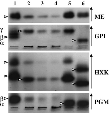
The electrophoretic patterns of 4 enzymes from the axenic strain (NASA6) and xenic strains (NASA821 and NASA829) of E. nuttalli isolated from Japanese macaques are shown in Fig. 1. The patterns for HXK and GPI were identical to those observed in the E. nuttalli P19-061405 strain and differed from those of E. histolytica and E. dispar. However, the mobility of the PGM band in the 3 NASA strains was identical with that in E. histolytica, whereas the mobility of this band in the P19-061405 strain was identical with that in E. dispar.
Isoenzyme patterns for ME, GPI, HXK and PGM of Entamoeba nuttalli strains obtained from Japanese macaques (lanes 2 to 4). Lane 1, E. histolytica SAW1453 (Zymodeme XIV); lane 2, E. nuttalli NASA6 (axenic); lane 3, E. nuttalli NASA821; lane 4, E. nuttalli NASA829; lane 5, E. nuttalli P19-061405; lane 6, E. dispar SAW1734RclAR (Zymodeme I). Vertical arrows indicate the direction of migration. Arrowheads indicate positions of bands.

Analyses of HXK, GPI and PGM genes
The calculated molecular mass and theoretical pI of HXK1 (AB485594) in the E. nuttalli NASA6 strain were 49·8 kDa and 5·38, respectively, and those for HXK2 (AB485595) were 49·3 kDa and 4·99, respectively, with both enzymes containing 445 amino acids. These pI values were identical to those for the E. nuttalli P19-061405 strain (AB282663 and AB282664), although HXK1 and HXK2 differed by 1 and 2 amino acids, respectively: Ala75 in both enzymes in NASA6 compared to Pro75 in P19-061405, and Ile232 in HXK2 in NASA6 compared to Val232 in P19-061405. The amino acid sequences of GPI1 (AB485596) and GPI2 (AB485597) in the E. nuttalli NASA6 strain were consistent with the calculated molecular mass of 61·4 kDa and theoretical pI of 6·6. These values were also identical to those of the P19-061405 strain (AB282665 and AB282666). Each protein had a single amino acid change from NASA6 to P19-061405: Ile232 to Val232 in GPI1, and Ala330 to Val330 in GPI2. PGM genes from E. nuttalli NASA6 and P19-061405 strains (AB485598 and AB485599) encoded proteins of 553 amino acids with calculated molecular masses of 60·8 kDa. Amino acid differences were detected in 2 positions: Asn416 and Asp441 in NASA6 compared to Ser416 and Asn441 in P19-061405. There were 4 differences in amino acids between NASA6 and E. histolytica (CAA74796), and 11 differences between NASA6 and E. dispar (CAA74797). The pI values of PGM from the NASA6 and P19-061405 strains, 5·87 and 5·99, were identical to those from E. histolytica and E. dispar, respectively.
Virulence in hamsters
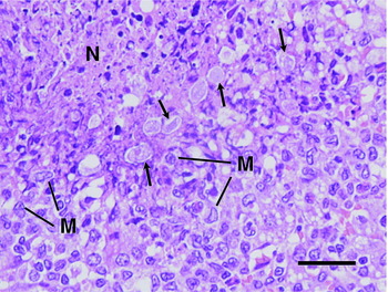
Of the 5 hamsters that received intrahepatic inoculation of trophozoites of the NASA6 strain, 1 died at 6 days after inoculation and another died at 7 days after inoculation. In these hamsters, the abscess weights as a percentage of the whole liver were 30% and 35%, respectively, and trophozoites were found in the abscesses in both animals. The other 3 hamsters were sacrificed at 7 days after inoculation and amoebic liver abscesses were also found in these animals. Histopathological analysis of the liver damage produced by E. nuttalli trophozoites showed large and irregular necrotic areas of liver parenchyma. These areas were peripherally limited by abundant live and pleomorphic trophozoites associated mainly with chronic inflammatory infiltrates (Fig. 2), forming palisade bands of epithelioid cells with evidence of a granulomatous reaction. The necrotic zones contained homogeneous and eosinophilic material with cell remnants and dense basophilic deposits.
Liver histology in hamsters inoculated with 5×10⁵ trophozoites of the Entamoeba nuttalli NASA6 strain. At 7 days after inoculation, areas of necrosis (N) appeared that were limited peripherally by multiple trophozoites (arrows) and mononuclear (M) inflammatory cells. (H&E stain, Scale bar=40 μm).

Electron microscopy of hamster liver inoculated with E. nuttalli trophozoites
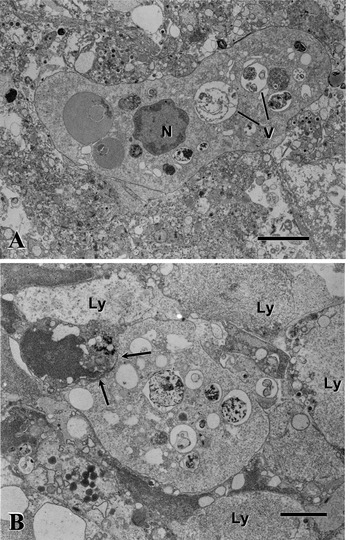
Electron microscopy of hepatic tissues taken from the vicinity of abscess walls at 7 days after inoculation showed the presence of elongated and pleomorphic trophozoites of E. nuttalli that were randomly spread at the outer limits of the abscess. The parasites were surrounded by abundant necrotic tissue formed by hepatic and inflammatory cell remnants composed of small vesicles and dense granules, membrane fragments and damaged cells (Fig. 3A). Some of these cells were partially damaged hepatocytes with cytoplasm containing multiple vesicles, lipid droplets and dilated organelles. Other damaged cells that were closely associated with the parasites and compatible with lysed inflammatory cells are shown in Fig. 3B. These irregularly outlined damaged cells surrounding the trophozoite contained only a fine granular material with a few dense granules distributed mainly at the periphery. Some trophozoites showed initial endocytic activity of damaged cells characterized by concavity at one pole of the parasite associated with an unrecognized damaged cell (Fig. 3B). The nuclei of the trophozoites were round but slightly irregular, and sometimes had an undulating profile (Fig. 3A). Dense chromatin was distributed at the periphery close to the nuclear membrane. The plasma membrane of each trophozoite was intact, thin and sharply defined. The cytoplasm included multiple phagocytic vacuoles of different sizes, with some containing membranous, amorphous or microvesicular material or other cell remnants (Fig. 3A and B). Geometrically arranged crystalloid structures were occasionally observed and glycogen particles were scarce.
Transmission electron micrographs of hamster liver at 7 days after inoculation of Entamoeba nuttalli NASA6 trophozoites. (A) The image shows an irregularly elongated trophozoite with a round nucleus (N) with peripherally distributed chromatin and a cytoplasm containing multiple vacuoles (V) enclosing membranous or amorphous material and granules of unknown origin. The thin, sharp and intact plasma membrane of the amoeba is apparent. The parasite is surrounded by abundant damaged host cells. (B) The image shows an ovoid trophozoite of E. nuttalli in the initial stage of the endocytic process (arrows) with a damaged cell contacting the left pole of the cell. The cytoplasm of the parasite contains vacuoles of different sizes containing unknown material. Multiple lysed (Ly) cells containing a fine granular material with small dense granules are seen surrounding the amoeba. (Scale bar=3 μm).

DISCUSSION
This study is the first report of isolation of E. nuttalli from Japanese macaques. Since E. nuttalli was detected in all fecal samples, Japanese macaques appear to be highly susceptible to E. nuttalli infection, as well as to E. coli and E. chattoni. Surprisingly, E. dispar was not detected, whereas our previous studies have demonstrated that E. dispar is prevalent in Japanese macaques (Rivera and Kanbara, Reference Rivera and Kanbara1999; Tachibana et al. Reference Tachibana, Cheng, Kobayashi, Matsubayashi, Gotoh and Matsubayashi2001). This suggests that the amoeba species prevalent in captive macaques may vary in each colony and may be different from that of wild macaques. The Japanese macaque (M. fuscata) is phylogenetically closer to rhesus monkey (M. mulatta) than cynomolgus monkey (M. fascicularis) (Hayasaka et al. Reference Hayasaka, Fujii and Horai1996; Chu et al. Reference Chu, Lin and Wu2007), but the sequence of the 18S rRNA gene of the NASA6 strain differed from that of the P19-061405 strain isolated from a rhesus monkey but was identical to that of the EHMfas1 strain isolated from a cynomolgus monkey. At present, the number of E. nuttalli isolates is insufficient to establish the relationship of evolution and dispersal in hosts and parasites, but the NASA6 and EHMfas1 strains clearly differ based on the serine-rich protein gene sequences and isoenzyme patterns.
In zymodeme analysis, the location of the slower running band for HXK and the appearance of a single γ band for GPI may be unique characteristics of E. nuttalli-type amoebae, compared with E. histolytica and E. dispar. In contrast, the pattern for PGM was distinctly different in NASA strains compared with other E. nuttalli-type strains in zymodeme analysis, with a β band in the NASA strains and an α band in the other strains. Sargeaunt has suggested that the absence of an α band and the presence of a β band for PGM are characteristics of E. histolytica that distinguish it from E. dispar, with the exception of zymodeme XIII (Sargeaunt, Reference Sargeaunt and Ravdin1988). NASA6 was the first strain showing E. histolytica-type mobility for PGM, while the P19-061405, EHMfas1 and JSK2004 strains showed E. dispar-type mobility (Tachibana et al. Reference Tachibana, Yanagi, Pandey, Cheng, Kobayashi, Sherchand and Kanbara2007; Takano et al. Reference Takano, Narita, Tachibana, Terao and Fujimoto2007; Suzuki et al. Reference Suzuki, Kobayashi, Murata, Yanagawa and Takeuchi2007). However, gene analyses have shown that there are fewer differences in the amino acid sequence of PGM between the E. nuttalli NASA6 and P19-061405 strains than between NASA6 and E. histolytica or P19-061405 and E. dispar.
Cultured trophozoites of the NASA6 strain were scored positive by the E. histolytica antigen detection kit, similar to the P19-061405 and EHMfas1 strains (Takano et al. Reference Takano, Narita, Tachibana, Shimizu, Komatsubara, Terao and Fujimoto2005; Tachibana et al. Reference Tachibana, Yanagi, Pandey, Cheng, Kobayashi, Sherchand and Kanbara2007). However, none of the 30 fecal samples were found to be positive using this kit. Suzuki et al. (Reference Suzuki, Kobayashi, Murata, Yanagawa and Takeuchi2007) have reported positive results in fecal samples analysed with this kit, but the samples were tested after a freezing-thawing procedure (personal communication). Therefore, the amount of antigen extracted may account for the different results. It is also possible that the anti-E. histolytica lectin antibody in the kit has low reactivity to E. nuttalli and comparative analysis of the antigen is required to address this issue.
Morphological differences of E. nuttalli compared with E. histolytica and E. dispar were not evident in cysts and xenically cultured trophozoites under light microscopy. However, trophozoites of E. nuttalli, as well as those of E. histolytica, were axenized easily in comparison with E. dispar (Clark, Reference Clark1995; Kobayashi et al. Reference Kobayashi, Imai, Haghighi, Khalifa, Tachibana and Takeuchi2005). The length range of living trophozoites of E. nuttalli in axenic culture overlaps with that of E. histolytica, but the E. nuttalli trophozoites tend to be elongated in comparison with E. histolytica trophozoites (Espinosa-Cantellano et al. Reference Espinosa-Cantellano, Gonzáles-Robles, Chávez, Castañón, Argüello, Lázaro-Haller and Martínez-Palomo1998). The average diameters of chilled trophozoites of E. nuttalli NASA6 and P19-061405 strains adapted to axenic culture conditions were significantly smaller than that of E. histolytica HM-1:IMSS. E. histolytica trophozoites obtained from intestinal or liver lesions are generally larger than those found in cultures (Martínez-Palomo, Reference Martínez-Palomo, Kreier and Baker1993), but the E. nuttalli trophozoites observed in liver in this study seemed to be as small as those in culture. It is unclear whether the size difference influences the overall characteristics of E. nuttalli, but this difference has also been found in a comparison of E. histolytica and E. histolytica-like Laredo (E. moshkovskii) trophozoites (Diamond, Reference Diamond1968; López-Revilla and Gómez-Domínguez, Reference López-Revilla and Gómez-Domínguez1988).
The liver damage produced in hamsters inoculated with E. nuttalli trophozoites was similar to that described in the same species challenged with E. histolytica trophozoites (Tsutsumi et al. Reference Tsutsumi, Mena-Lopez, Anaya-Velazquez and Martinez-Palomo1984). This suggests similar physiopathological mechanisms of tissue damage, including an important role of lysed inflammatory cells (Perez-Tamayo et al. Reference Perez-Tamayo, Martinez, Montfort, Becker, Tello and Perez-Montfort1991; Tsutsumi and Shibayama, Reference Tsutsumi and Shibayama2006). Ultrastructural analysis of the amoeba-inflammatory cell interaction has shown that E. nuttalli carries a similar virulent capacity to destroy target cells, including lysis of inflammatory cells and endocytosis, despite the smaller size of trophozoites of the E. nuttalli NASA6 strain compared to the E. histolytica HM-1:IMSS strain (Tsutsumi and Martínez-Palomo, Reference Tsutsumi and Martínez-Palomo1988).
If E. nuttalli is able to infect humans, it may be of concern as a zoonotic hazard. Therefore, a stool examination was performed on the 4 keepers taking care of the monkeys in the park. No parasites were detected from samples by direct microscopy or in culture in Tanabe-Chiba medium (data not shown). It remains possible that E. nuttalli is infective in humans, but Entamoeba with the zymodeme pattern of E. nuttalli has not been detected in more than 2500 separate isolations characterized to date (Sargeaunt, Reference Sargeaunt and Ravdin1988). However, the results in this study show that the E. nuttalli NASA6 strain isolated from Japanese macaque is potentially virulent, and we suggest that E. nuttalli should be recognized as a common parasite in macaques and should be discriminated from E. histolytica and E. dispar in routine analysis.
We thank M. Johshita (Nagasaki University) for her help with xenic culture. We also thank the staff in the Teaching and Research Support Center, Tokai University School of Medicine, for their help in DNA sequencing. This work was supported by a Grant-in Aid for Scientific Research from the Japanese Society for the Promotion of Science (to H.T.) and a Cooperative Research Grant 2006-18-C-4 from the Institute of Tropical Medicine, Nagasaki University (to H.K.).