Introduction
As the shrimp industry expands rapidly worldwide, the pacific white shrimp, Litopenaeus vannamei, is becoming an economically important species in aquaculture. Along with culture technology and disease resistances, genetic enhancement of growth rate is an important aspect of shrimp breeding, which could potentially decrease the length of grow-out cycles, thus reducing production costs and shortening the time of exposure to diseases. Shrimp muscles are primarily present in the abdominal part of its body. Currently, its growth and related genes, particularly the regulatory genes, is not well known. On the other hand, muscle development in mice and humans have been well studied (McKinsey et al., Reference McKinsey, Zhang and Olson2002). Many growth factors and transcription factors have been identified to be responsible for growth, development, regeneration and metabolisms (Olson and Klein, Reference Olson and Klein1998). The skeletal muscle EST and gene expression profile in human, mouse and livestock species have also been extensively characterized (Bortoluzzi et al., Reference Bortoluzzi, Rampoldi, Simionati, Zimbello, Barbon, d’Alessi, Tiso, Pallavicini, Toppo, Cannata, Valle, Lanfranchi and Danieli1998; Davoli et al., Reference Davoli, Zambonelli, Bigi, Fontanesi and Russo1999; Grosse et al., Reference Grosse, Kappes and McGraw2000; Moore et al., 2001; Yao et al., Reference Yao, Coussens, Saama, Suchyta and Ernst2002).
Based on studies from invertebrates – Caenorhabditis elegans, Drosophila melanogaster and Bombyx mori – crustacean muscle is structurally analogous to vertebrate skeletal muscles with proteins organized in sarcomeres aligned with large penniform fibers (El Haj, Reference El Haj1996). However, crustacean muscle has different sarcomere length according to fiber type. Most fast fibers are organized in short sarcomeres with low mitochondrial density while slow tonic fibers are organized in long sarcomeres with a high mitochondrial density. Muscle atrophy was observed in large claws of crustaceans before molt to facilitate limb withdrawal from narrow openings. The intensity of atrophy varies significantly in other muscle tissues (Mykles and Skinner, Reference Mykles and Skinner1990; Koenders et al., Reference Koenders, Yu, Chang and Mykles2002). Previously, we observed that myofibrilar cross-sections slightly dehydrated in the premolt and expanded in the postmolt stage in L. vannamei (Cesar et al., Reference Cesar, Zhao, Malecha, Ako and Yang2006).
The worldwide number of shrimp reference sequences remains modest. Tassanakajon et al. (Reference Tassanakajon, Klinbunga, Paunglarp, Rimphanitchayakit, Udomkit, Jitrapakdee, Sritunyalucksana, Phongdara, Pongsomboon, Supungul, Tang, Kuphanumart, Pichyangkura and Lursinsap2006) identified 4845 unique ESTs in Penaeus monodon, Wilson et al. (Reference Wilson, Li, Whan, Lehnert, Byrne, Moore, Pongsomboon, Tassanakajon, Rosenberg, Ballment, Fayazi, Swan, Kenway and Benzie2002) characterized 273 ESTs in this species and developed a low-density genetic linkage map of P. monodon. Gross et al. (Reference Gross, Bartlett, Browdy, Chapman and Warr2001) revealed 268 imuno-related ESTs from Litopenaeus setiferus, and in Marsupenaeus japonicus, Yamano and Tatsuya (Reference Yamano and Tatsuya2006) identified 268 ESTs and 1250 singletons. Several laboratories worked on the L. vannamei EST research project (Dhar et al. 2000; Alcivar-Warren, Reference Alcivar-Warren2001; Bartlett et al., Reference Bartlett, Cuthbertson, Shepard, Chapman, Gross and Warr2002) and nearly 1000 ESTs were characterized. The only EST analysis specifically from the abdominal muscle of L. vannamei was completed by Dhar and Alcivar-Warren (1997), and in that study only 10 clones were partially sequenced. A comprehensive EST project on L. vannamei is being carried out by a Brazilian consortium (http://www.shrimp.ufscar.br/en/index.php). The purpose of this study was to identify genes or EST sequences specifically expressed in L. vannamei abdominal muscle. In this study, we established a cDNA library based on L. vannamei juvenile abdominal muscle by PCR-based SMART™ cDNA technology. A pilot EST analysis from randomly sequenced cDNA clones was performed. Comparisons and classifications of the EST sequences with the known sequences in the database are reported here.
Material and methods
Animals
Juvenile specific pathogens free (SPF) pacific white shrimp L. vannamei (1 month old, 4.8 to 5.3 g) were obtained from Chen-Lu Farms, Kahuku, HI, USA. Animals were transported to the lab in plastic bags containing 1/3 of filtered seawater and 2/3 compressed oxygen, and acclimated in 45 l tanks filled with filtered seawater, aerators and a bio-filter. Animals were fed ad libitum twice a day using Rangen Grower™ shrimp feed (35% protein, 8% lipids, 4% fibers, 15% ash; Rangen Inc., Buhl, ID, USA). Water quality parameters were kept at adequate levels (28 ± 2°C temperature, 35‰ ± 2 p.p.t. salinity) and photoperiod adjusted to 12 h light/dark. Animals were kept in the lab for 48 h before experiments by means to minimize stress.
RNA preparations
Total RNA from muscle tissue at the first abdominal segment of shrimp (n = 5) was isolated using Trizol™ reagents (Invitrogen, Carlsbad, CA, USA) following the method described by Yang et al. (Reference Yang, Ratovitski, Brady, Solomon, Wells and Wall2001). Total RNA was treated with RQ1™ RNase-free DNase (Promega, Madison, WI, USA) to remove potential genomic DNA contamination. Poly(A) mRNA was purified by the mRNA isolation kit (Roche, Indianapolis, IN, USA) based on hybridization of mRNA and biotin-labeled oligo(dT) probes according to the manufacturer’s instructions. The quality and concentrations of mRNA were assessed by Bioanalyzer 2100 and its software version 1.4 (Agilent, Santa Clara, CA, USA). Samples with high quality and high concentration of mRNA were selected for the preparation of the cDNA library.
cDNA library preparation
A cDNA library was constructed with the mRNA isolated from muscle tissue using the SMART™ cDNA library construction kit (Clontech, Palo Alto, CA, USA). Briefly, full-length cDNAs were amplified by the LD-PCR technique using a modified oligo(dT) primer provided with the kit (CDS III/3′PCR Primer) for first strand synthesis, and the SMART™ III oligo (provided) that contains an oligo(G) sequence at its 3′ end, which binds to the deoxycystidine stretch added to the 3′ end of the cDNA newly synthesized. Size fractionation was performed on a CHROMA SPIN-400 column, provided with the kit, and aliquots of each fraction were separated on agarose gel. The first four fractions (>1 kb) were collected together, ligated into Sfi I-digested dephosphorylated λTriplEx2 Vector provided, and packaged into λ phages using the Packagene™ Lambda DNA packaging system (Promega, Madison, WI, USA), according to the manufacturer’s instructions. The λ phages were used to infect Escherichia coli strain XL-1 blue and plated onto LB/ampicilin plates (0.8% top agarose/LB medium/ampicilin at 150 μg/ml). Library titration (plaque-forming unit, pfu) was evaluated and the percentage of recombinant clones was determined by standard white-blue screening using IPTG/X-gal solutions.
Conversion of λTriplEx2 to pTriplEx2
Individual positive plaques were excised from plates and incubated overnight at 4°C in 500 μl 1X phage elution buffer (0.1 M NaCl, 0.1 M MgSO4, 0.05 M Tris-HCl, pH 7.5, 0.01% Gelatin). All plaques from a single plate were taken, to avoid possible selection bias. Lambda TriplEx2 plaque eluates (250 μl) were inoculated into 250 μl of E. coli BM25.8 overnight cultures (250 μl, OD 1.4) in the presence of MgCl2 (10 mM final concentration) and incubated at 31°C for 1 h followed by the addition of LB medium and another incubation with shaking (225 r.p.m.) for 1 h. Fifty microliters of aliquots were transferred and spread onto LB ampicilin (150 mg/ml) plates and incubated overnight at 31°C.
Direct sequencing of randomly picked clones
Colonies were checked for the presence of an insert by colony PCR using the λ TriplEx LD-Insert Screening Amplimers prior to sequencing. Purified plasmid DNA samples were sequenced by the SMART cDNA library construction kit’s 5′ sequencing primer (5′-aagcagtggtatcaacgcagagt-3′) and BigDye terminator on ABI 3730XL capillary-based DNA sequencers (Applied Biosystems, Forrest City, CA, USA) at the Biotechnology Core Facility of the University of Hawaii at Manoa.
Sequence analysis
Phred software was used to examine the peaks around each base call to assign a quality score to each base call. We selected sequences whose Phred Scores are higher than 20 and manually trimmed of vector sequences. Ambiguous base calls were manually corrected by inspecting the sequence electropherograms. Database searches were limited to ESTs >400 bp in length. Sequences were individually queried against the current (August 2007) databases at the National Center for Biotechnology Information (NCBI, Bethesda, MD, USA) using BLASTn (All GenBank non-redundant +EMBL + DDBJ + PDB sequences but not EST, STS, GSS, environmental samples or phase 0, 1 or 2 HTGS sequences) and BLASTp (all GenBank non-redundant CDS translations + PDB + SwissProt + PIR + PRF excluding environmental samples) algorithms (Altschul et al., Reference Altschul, Madden, Schäffer, Zhang, Zhang, Miller and Lipman1997) with default parameters. EST assembly for contig formation and redundancies in sequences were carried out using SeqMan™ version 5.07 software (DNASTAR, Madison, WI, USA). Database search results match with e-values less than 1.0 × 10⁻⁵ were considered significant. EST and contigs were positioned in three groups according to the BLAST score. Bit score, identities and expectation value from the Blast search results were used to further classify into five groups of ESTs. e-Value from the Blast search result and hit type was also used to identify the low homologous and unknown genes.
Results and discussion
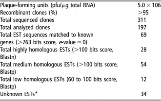
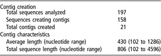
The characteristics of the constructed cDNA library are shown in Table 1. Library size was 5.0 × 10⁶ pfu/μg total RNA with the percentage of recombinant clones more than 95%. The total of the clones sequenced was 311, of which 197 clones had longer than 250 nucleotides (nt) with Phred quality value >20. The average insert size of all analyzed ESTs after removal of vector-based sequences was larger than 745 nucleotides. Fifty-six percent (110 of 197 clones) showed 5′-end DNA sequence, 44% (87 of 197 clones) have 3′-end sequence. Assembly of the 197 sequences generated 39 single-copy clones and 158 overlapping clones, which generate 21 contigs (Table 2) with an average length of 430 nt (min. 102, max. 1286). Table 2 also includes the number of sequences creating a contig and the corresponding number of contigs generated from the EST sequences. A total of 160 unique sequences have been identified.
Summary of cDNA library and BLAST scores

*Includes ESTs with low homology (Blast score <50) and sequences with no significant matches to DNA databases.
Summary of contig creation and characteristics

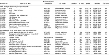
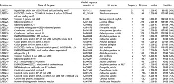
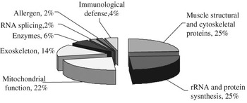
The EST sequences were searched with the Blastn and Blastp programs in the NCBI database. According to the degrees of homologies to the known genes in the Blast report, we summarized the EST sequences to three groups of classifications (Tables 3 and 4). The EST sequences from Tables 3 and 4 were submitted to the NCBI dbEST, and accession numbers were listed in the tables. The first group is the sequences with 100% identity to the known genes (e-value = 0), which include 12 unique ESTs. Muscle structural protein – actin 2 (20 copies) and 12S ribosomal RNA (12 copies) – are the most abundant transcripts. Eleven unique ESTs match the EST sequences from shrimp species and one matches the lobster’s sequence. The second high homologous (>100 bits from Blastn) group distinguished 12 unique EST sequences. Muscle structural proteins of troponin I (7 copies) and sarcoplasmic calcium-binding protein (6 copies) are most abundant transcripts. Seven unique ESTs hit genes from shrimp, crayfish, lobster and crab. Four unique ESTs hit mRNA sequences from insects of honey bee, fruit fly and mosquito. One unique EST hits genes from vertebrate fish. The third medium to low homologous (64.7 to 176 bit score from Blastp) group distinguished 25 unique ESTs. Muscle structural proteins of myosin light chain (21 copies) and myosin (20 copies) are the most abundant transcripts. Eight unique ESTs hit genes from the insects of silk worm, tribolium, lonomia, fruit fly, mosquito, honey bee and cockroach. Six unique ESTs hit genes from crab, shrimp and crayfish. One unique EST hits genes from vertebrate fish. Five unique ESTs hit genes from cetaceans. Five unique ESTs hit insect genes. Two ESTs have DNA satellite marker-like sequences with short TG, GA or CAA repeats. Based on the partial EST sequence data, the most abundant EST sequences excluding 37 unknown ESTs are from muscle structural and cytoskeletal protein (25%) and rRNA and protein synthesis (25%), mitochondrial function (22%), exoskeleton (14%) and others (14%). A summary of the classification of the reported ESTs by their biological function is shown in Figure 1.
List of shrimp ESTs to the known genes in Blastn

Shrimp ESTs with medium to low homologies to the known proteins in Blastp

Classification of the reported ESTs by their biological function.

Currently, only a few thousand of ESTs have been identified from invertebrate species – L. vannamei. An EST project offers a high degree of chance to reveal unique genes in shrimp species. Dhar and Alcivar-Warren (1997) had previously reported 10 ESTs from L. vannamei abdominal muscle. Primary hit species of each query of 49 single sequences from blast search in this report are organized as 18 from penaeid and vannamei species, 16 from insects, 13 from crustaceans excluding shrimp and two from fish. It makes sense that the highest hit species come from the shrimp species. More sequences hit insects than vertebrates since the biological classification of shrimp is much closer to insects than to vertebrates. Insects are in the class Insecta within Phylum Arthropoda. Also in the phylum are crustaceans. Shrimp, crabs and lobsters are in the class Malacostraca of crustaceans. In addition, the results also suggest that shrimp muscle tissue apparently expresses a considerable diversity of transcripts. The homologous transcripts from the cDNA library primarily include muscle structural proteins, rRNA and protein synthesis and mitochondrial function. As expected, the most abundant transcripts expressed in shrimp abdominal muscle are related to muscle structural proteins, the primary component of the muscle. The transcripts related to protein synthesis are found to be the second abundant group. We have recently reported that the mRNAs encoding for ubiquitin and heat shock protein 70 in the abdominal muscle did not increase significantly in premolt stages, which is typically associated with claw muscle degradation (Cesar and Yang, Reference Cesar and Yang2007). Results from this study by the randomly selected clone sequencing did not indicate transcripts directly related to protein degradation. The low frequency of mRNA transcripts of protein degradation may indicate that the protein synthesis is primary biological activity in the shrimp abdominal muscle tissue in the 1-month old shrimp.
In summary, analysis of the sequences through the Blast search indicates that the established cDNA library represents the abundance of the transcripts in the shrimp abdominal muscle tissue. The muscle ESTs in the shrimp species are highly rich in the transcripts of muscle structure and protein synthesis and closely related to insects over vertebrates. The results also suggest that shrimp muscle EST sequences are overlapping with other species as well as unique from insect and vertebrate species. Shrimp muscle EST sequences are complementary to many other species in the Phylum Arthropoda. Further research to continue DNA sequencing of the positive colonies from the cDNA libraries is needed. A large-scale systematic approach of shrimp muscle EST and functional genomics will provide valuable resources for the genetic improvement of shrimp growth.
Acknowledgments
We acknowledge Dr Shaobin Hou from the Biotechnology Core Facilities of the University of Hawaii at Manoa for his technical support on the DNA sequencings and assistance with the sequence analysis presented in this work. Our appreciation is also extended to USDA-CSREES-TSTAR program and CAPES (Coordenacao de Aperfeiçoamento de Pessoal de Nível Superior, Brazil) for their financial supports.