Introduction
A pseudoaneurysm is a tear through all the layers of an artery, through which blood can flow and enter a collection encapsulated by surrounding tissues.Reference Garino and Ryan1 Carotid artery pseudoaneurysm is reported as a rare complication following neck dissection. Haemorrhage of a carotid artery, known as ‘carotid blow-out’, is associated with a high mortality rate.Reference Schwartz, Fisher, Sako, Castaneda, Grage and Nicoloff2
Following pharyngolaryngectomy, reconstruction of a neo-pharynx using a free jejunal graft depends on an intact blood supply from a branch of the external carotid artery. Endovascular repair of carotid artery pseudoaneurysms has become increasingly popular, as such repair is minimally invasive and is positioned distal to the site of the causative infection or previous surgery or radiotherapy.Reference Baril, Ellozy, Carroccio, Patel, Lookstein and Marin3 Endovascular repair, utilising coils and stents, has also been used to treat carotid artery dissection.Reference Biggs, Chiou, Hagino and Klucznik4
Mycotic pseudoaneurysm refers to a pseudoaneurysm that is caused by any micro-organism, and is not specific to fungi as the name implies. Osler's description is in fact a misnomer, as the majority are not caused by fungi.Reference Osler5
This paper reports a case of a mycotic external carotid artery pseudoaneurysm that was successfully treated with endovascular coiling, while maintaining the patency of the superior thyroid artery supplying the patient's jejunal graft anastomosis.
Case report
A 66-year-old man, who three years previously had received radiotherapy for a tumour-node-metastasis (TNM) stage T2 N0 M0 soft palate squamous cell carcinoma, was diagnosed with a 5.8 cm, concentric, bulky, neoplastic lesion of the post-cricoid region and upper oesophagus, involving the left hemithyroid, with an 11 mm neck node at left level II. This was staged as a cT4a N0 M0 (pT4a N1 M0) squamous cell carcinoma of the hypopharynx and upper oesophagus. Neoadjuvant chemotherapy was given to reduce the size of the tumour.
The patient then underwent a pharyngolaryngectomy, left hemithyroidectomy, left selective neck dissection of levels I to IV, and free jejunal graft reconstruction. The jejunal graft was anastomosed end-to-end to the left superior thyroid artery, and end-to-side to the internal jugular vein. The patient had a gastrostomy feeding tube inserted during the same procedure.
Over the ensuing weeks, the patient had a stormy post-operative recovery, suffering fits and also stoma and neck wound dehiscence, which eventually healed. No cause was found for the patient's fits.
Two months later, the patient suffered minor bleeding episodes from his oropharynx. Soon afterwards, a small area of his left neck wound began to dehisce and discharge. Clinical examination did not reveal the source of the bleeding. The patient's haemoglobin levels remained stable, and swabs of the neck wound discharge grew Candida albicans, which was treated with appropriate antifungals.
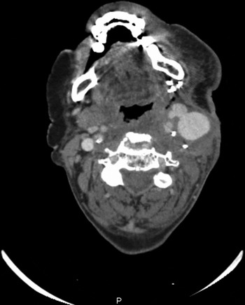
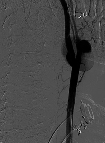
A computed tomography (CT) scan of the neck demonstrated a 3.1 × 2.4 cm, bilobed pseudoaneurysm of the left external carotid artery (Figure 1). The origin of the pseudoaneurysm was distal to the superior thyroid artery, to which the graft was anastomosed. Under general anaesthesia, the CT findings were considerably clarified by digital subtraction angiography (Figure 2).
Axial computed tomography neck scan showing the bilobed pseudoaneurysm of the left external carotid artery.

Digital subtraction angiography image showing the pseudoaneurysm just distal to the superior thyroid artery branch.

Treatment options were discussed, and a decision was made to treat the pseudoaneurysm with endovascular coiling.
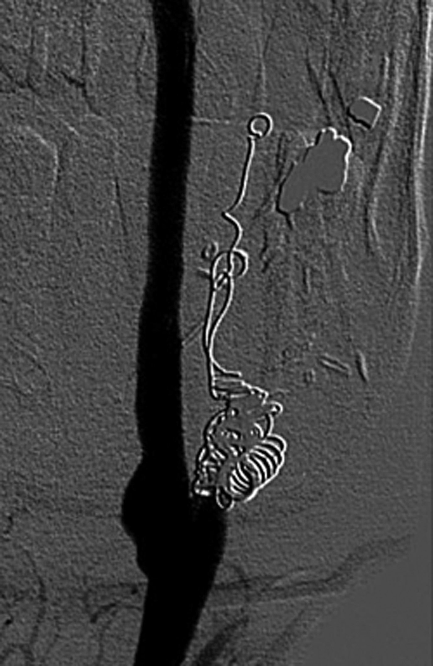
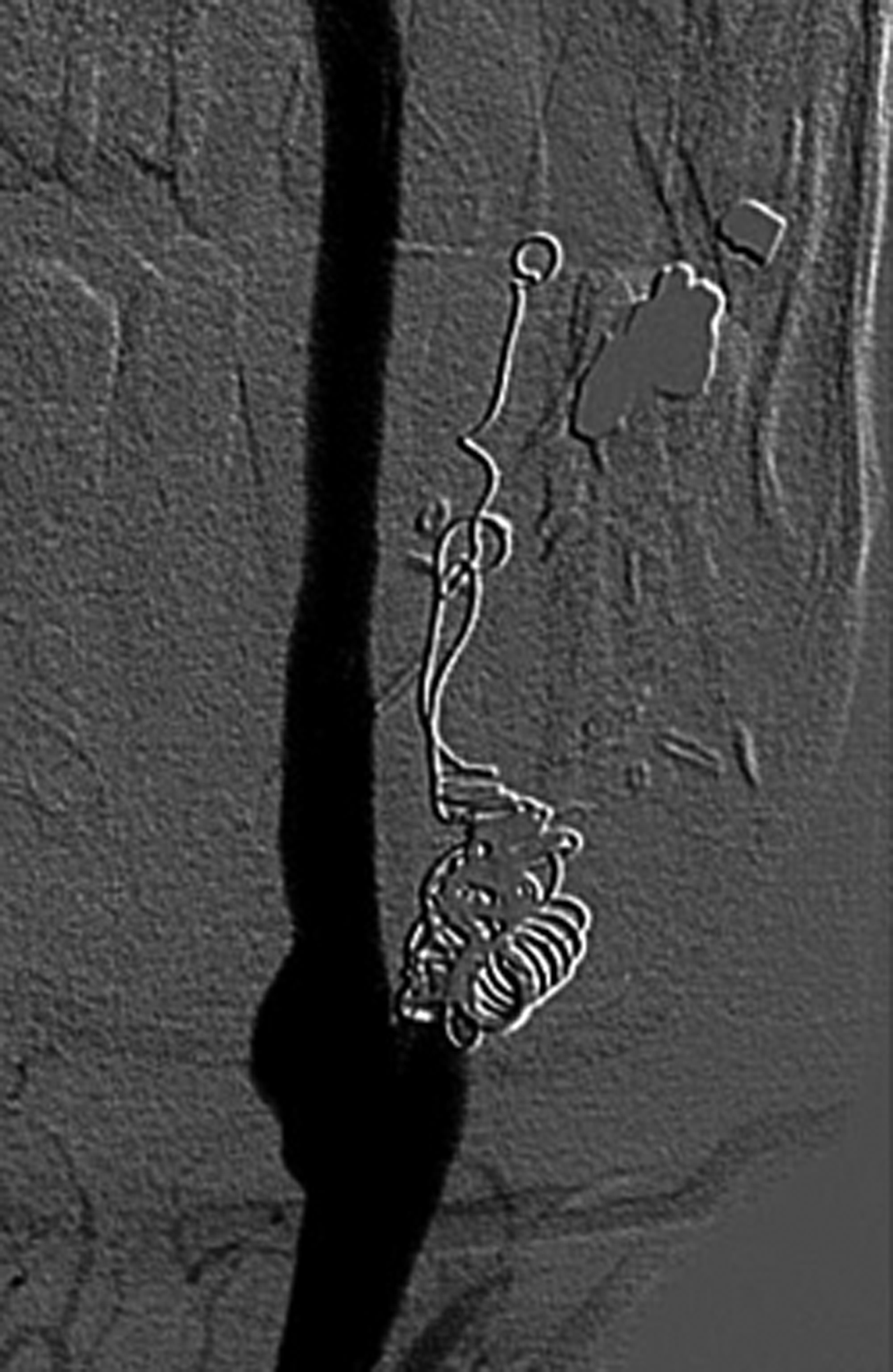
Subsequently, via a right common femoral artery approach, six fibred platinum coils (Boston Scientific, Boston, Massachusetts, USA) were inserted through a micro-catheter into the patient's left external carotid artery, from the branches distal to the neck of the pseudoaneurysm to the proximal rim of the neck of the pseudoaneurysm. Digital subtraction angiography images showed complete occlusion proximally, and ultrasonography showed stasis within the pseudoaneurysm. The left superior thyroid artery and its anastomosis to the jejunal graft were shown to remain patent (Figure 3).
Digital subtraction angiography image showing complete occlusion of the external carotid artery distal to the superior thyroid artery.
The patient's condition improved. Apart from a small dehiscence of his neck wound, he was fit for discharge two weeks after endovascular coiling of the pseudoaneurysm.
Discussion
Carotid pseudoaneurysms and blow-outs are life-threatening complications of major head and neck surgery, and must be dealt with in order to prevent significant morbidity and probable mortality.
Our presented patient had previously undergone radiotherapy and neoadjuvant chemotherapy. He developed an external carotid pseudoaneurysm, nearly two months after pharyngolaryngectomy, left selective neck dissection and free jejunal graft reconstruction for a T4 N1 M0 squamous cell carcinoma of the hypopharynx and upper oesophagus. Neck wound dehiscence with discharge and minor episodes of oral bleeding preceded diagnosis of the pseudoaneurysm, which was confirmed by CT scanning of the neck and digital subtraction angiography.
In this case, we decided to use endovascular coiling for two reasons. Firstly, the pseudoaneurysm was thought to be mycotic, due to infection in the overlying wound. It was thought that inserting a Dacron® covered stent was more likely to promote adhesion and biofilm formation, compared with endovascular coiling. In this patient, infection of an endoprosthesis could have caused significant complications. We noted that use of a covered stent to treat mycotic pseudoaneurysm had not previously been reported.
Our second reason for endovascular coiling related to the anatomy of the external carotid artery. In our patient, the lingual, facial, occipital and posterior auricular arteries branched at almost the same point. Therefore, it would have been difficult to use a stent to repair the pseudoaneurysm while still maintaining patency of the graft arterial supply.
The fibred embolisation coils used in this patient were attached to the pushing wire by an overlock which released when the wire was out of the catheter, but which, until that point, could be withdrawn if coil position was sub-optimal. Thus, it was easier to achieve a more accurate final placement. This was essential, as the distance from the superior thyroid artery and the internal carotid artery to the neck of the pseudoaneurysm was less than 8 mm, and at least a few millimetres of that distance needed to be occluded to prevent recannalisation.
• Carotid pseudoaneurysms and ‘blow-outs’ are life-threatening complications of major head and neck surgery
• In the presented patient, endovascular coiling of the external carotid artery was the only definitive treatment for a life-threatening mycotic pseudoaneurysm
• Endovascular repair is becoming more popular as its reported success rates rise
Our patient's pseudoaneurysm was bilobed; this was thought to be due to its herniation through the carotid sheath. Indeed, angiography showed low flow in the larger part of the pseudoaneurysm which was situated more laterally from the artery.
This case is similar to that reported by Goddard et al., who described a pseudoaneurysm of the left external carotid following pharyngolaryngectomy and jejunal grafting. In this latter case, the graft was anastomosed to the lingual-facial artery. Unfortunately, endovascular coiling was unsuccessful in this case, and the patient was returned to the operating theatre to undergo aneurysectomy and carotid sacrifice, which was fatal.Reference Goddard, Lenthall and Bradley6
Mycotic pseudoaneurysms are commonly associated with Staphylococcus aureus and salmonella species; other microbial species have also been reported. However, to our best knowledge, the involvement of candida species has not been reported. In our case, candida was grown from a swab from the neck wound; there was no positive candidal blood culture.
Reports have shown that mycotic pseudoaneurysms develop from soft tissue infections surrounding the artery, as well as from metastatic spread of infection and primary microbial arteritis.Reference Worley, Hern, O'Sullivan, Tassone and Hinton7 Previous irradiation may have been a contributing factor in our patient, as previous reports have linked radiation to pseudoaneurysm formation.Reference Girishkumar, Sivakumar, Andaz, Santosh, Solomon and Brown8
Conclusion
Following a pharyngolaryngectomy with jejunal free graft reconstruction, the presented patient developed a life-threatening mycotic pseudoaneurysm, for which endovascular coiling of the external carotid artery was considered to be the only definitive treatment. Without this intervention, the patient would probably have suffered a fatal carotid blow-out. Endovascular repair is becoming more popular as its reported success rates rise. Further case series, with longer follow-up periods, are needed to establish the usefulness of this treatment modality.