Introduction
The possibility of ruminant embryos surviving freezing-thawing and/or transfer procedures increases with more advanced developmental stages (Cocero et al., Reference Cocero, López Sebastián, Barragán and Picazo1996; Massip, Reference Massip2001). It is possible to hypothesize, therefore, that final outputs from in vitro reproductive techniques would be improved by culturing zygotes to the blastocyst stage prior to cryopreservation or transfer. However, developmental rates to blastocyst stage are still limited. Some oocytes fail to undergo in vitro nuclear maturation, fertilization and/or first cleavage, but the main sticking point has been identified as failure to reach the blastocyst stage (Greve et al., Reference Greve, Madison, Avery, Callesen and Hyttel1993; Bavister, Reference Bavister1995; Mermillod et al., Reference Mermillod, Oussaid and Cognie1999; Lonergan et al., Reference Lonergan, Rizos, Ward and Boland2001). The cause seems to be related to in vitro culture. Embryos have to overcome the so-called developmental block described at the 8- to 16-cell stage in ruminants (Wright & Bondioli, Reference Wright and Bondioli1981), when the control of embryo development switches from the maternal to the embryonic genome (Bavister, Reference Bavister1988). Currently, it is assumed that most of the early developmental failures in in vitro culture arise before or during the time of the developmental block (Eyestone & First, Reference Eyestone and First1991). Nevertheless, knowledge of the mechanisms involved in early developmental processes and the in vitro developmental block is still scarce (Rief et al., Reference Rief, Sinowatz, Stojkovic, Einspanier, Wolf and Prelle2002). The first objective of the current study was therefore to elucidate the influence of different early developmental stages at the onset of culture on the final number and quality of the blastocysts subsequently produced. Influences of possible defective in vitro maturation and/or fertilization were avoided by using embryos fertilized in vivo and recovered from the maternal oviduct (Rizos et al., Reference Rizos, Ward, Duffy, Boland and Lonergan2002).
Previous authors have attributed failures in culture to the in vitro culture system employed (Gardner et al., Reference Gardner, Vella, Lane, Wagley, Schelenker and Schoolcraft1998; Thompson, Reference Thompson2000). Culture of embryos either with somatic cells (Gandolfi & Moor, Reference Gandolfi and Moor1987; Rexroad & Powell, Reference Rexroad and Powell1988) or in semi-defined medium (Walker et al., Reference Walker, Lampe and Seamark1989; Pinyopummintr & Bavister, Reference Pinyopummintr and Bavister1991) is suitable to overcome developmental arrest and enhance blastocyst production; but co-culture with somatic cells seems to be more efficient in terms of embryo quality (Rizos et al., Reference Rizos, Ward, Boland and Lonergan2001) and has also proved to be a suitable model for studying embryo–maternal interactions (Rief et al., Reference Rief, Sinowatz, Stojkovic, Einspanier, Wolf and Prelle2002). The major inconvenience of this system is the high variability between replicates due to the unfeasibility of establishing cellular lines; cells undergo alterations in morphology and growth after several passages (Ouhibi et al., Reference Ouhibi, Menezo, Benet and Nicollet1989). In the current study, a second objective was to minimize such variability by defining a standardized and repeatable system of co-culture of embryos from oviduct recoveries with oviduct epithelial cells. Standardization of the culture system was carried out by establishing the influence of the monolayer age and the effect of frozen-thawed oviduct cells on developmental yield when compared with results obtained from later-stage embryos cultured in semi-defined medium.
Materials and methods
Experimental design
A total of 342 in vivo derived sheep embryos were used to assess possible influence of embryo developmental stage and in vitro culture conditions on the yield and quality of blastocysts obtained. Early cleavage stage embryos classified as excellent or good after recovery, on the basis of their morphology and synchrony between their stage and retrieval time, were divided into three experimental groups according to developmental stage: 2- to 4-cell (group A; n = 93), 5- to 8-cell (group B; n = 92) and 9- to 12-cell (group C; n = 85). In order to evaluate the effect of the age and preservation of oviduct cell monolayers, embryos were randomly allocated into one of four groups: three groups of culture with monolayers derived from fresh cells (2-, 4- and 5-day-old monolayers) and a fourth group with a 2-day-old monolayer derived from frozen-thawed oviduct cells. Blastocyst quality was also assessed by the ability to achieve the hatched blastocyst stage following freezing-thawing (Rizos et al., Reference Rizos, Ward, Boland and Lonergan2001; Dobrinsky, Reference Dobrinsky2002). Blastocysts derived from the different groups of culture of early stage embryos (group A, n = 41; group B, n = 38; group C, n = 39) were frozen. In agreement with previous studies (Leibo & Loskutoff Reference Leibo and Loskutoff1993; Del Campo et al., Reference Del Campo, Donoso, Palasz, Garcia and Mapletoft1993), only blastocysts that hatched out of the zona pellucida within a maximum of 3 days after thawing were classified as viable. Furthermore, the kinetics of blastocyst appearance (Van Soom et al., Reference Van Soom, Boerjan, Bols, Vanroose, Lein, Coryn and De Kruif1997) in each monolayer were used to assess the ability of the monolayer to support developmental competence of embryos. Two controls were used to determine any influence of embryo developmental stage: first, a group of more advanced developmental stages (morulae and blastocysts) obtained in vivo were cultured in semi-defined medium to the hatched stage (n = 29) to assess blastocyst rate, and second, a group of blastocysts produced in vivo (n= 43) were cryopreserved to assess blastocyst quality.
Superovulation and embryo recovery
The embryos were recovered from 4- to 7-year-old Manchega ewes, maintained outdoors with access to indoor facilities, at the experimental farm of the INIA Animal Reproduction Department in Madrid, Spain, at latitude of 40° N. The animals were superovulated with the protocols commonly used in our laboratory (Gonzalez-Bulnes et al., Reference Gonzalez-Bulnes, Garcia-Garcia, Santiago-Moreno, Lopez-Sebastian and Cocero2002). In brief, oestrus was synchronized by the insertion of an intravaginal progestagen-impregnated sponge (40 mg fluorogestone acetate, FGA, Chronogest, Intervet International, Boxmeer, The Netherlands) on day 0, exchanged for a new one that was maintained from day 7 to day 14. Ewes were also injected with a single dose of cloprostenol (Estrumate, 125 μg, i.m., Mallinckrodt Vet, Friesoythe, Germany) at the time of the first FSH treatment on day 12. The superovulatory treatment consisted of eight decreasing doses (1.5 ml × 3, 1.25 ml × 2, 1 ml × 3) of FSH, i.m. (OvAGEN, ICP, Auckland, New Zealand) administered twice daily starting 60 h before sponge removal and finishing 24 h after progestagen withdrawal. Progestagen removal was coincident with the sixth FSH treatment and oestrus detection was performed with adult Manchega rams after 24 h. Mating was repeated at 36 and 48 h after sponge withdrawal.
The embryos were obtained by surgical access to the genital tract, through prepubian laparotomy under general anaesthesia with xylazine (Rompun, Bayer, Leverkusen, Germany, 6 mg i.m.) and ketamine (Imalgène 1000, Merial, Lyon, France, 130 mg i.m.). The ovarian response in terms of the number of corpora lutea was determined by laparoscopy just before the laparotomy, thus avoiding surgery in non-responding females. Oviduct retrievals for early-staged embryos were performed at different intervals after sponge removal to obtain the different developmental stages. Embryos were therefore collected on day 4 to obtain 2- to 4-cell embryos, on day 4.5 for 5- to 8-cell embryos, and on day 5 for 9- to 12-cell embryos. At all time points both oviducts were flushed with PBS (Dulbecco's phosphate-buffered saline) supplemented with 0.2% BSA (bovine serum albumin Fraction V, Sigma Chemical, St Louis, MO) and 10 mg/ml of gentamicin (Sigma Chemical) from a catheter fed through a blunt needle inserted at the uterotubal junction. The catheter was fixed with silk to avoid backflow of the flushing medium, towards another catheter without a needle introduced into the fimbria and also fixed with silk. Uterine recoveries for morulae and blastocysts were performed on day 6 and day 7 after sponge withdrawal. Embryos were obtained by flushing both uterine horns with PBS-BSA from a blunt needle inserted at the major curvature of the horn, kept fixed with forceps to avoid backflow of flushing medium, towards another catheter provided with a blunt needle and fixed with silk at the utero-tubal junction.
Preparation of monolayers derived from fresh oviductal cells
Ewe oviducts were collected in a local abattoir and transported at 37 °C in Dulbecco's PBS supplemented with 10 mg/ml of gentamicin (Sigma Chemical). In the laboratory, oviducts were washed twice with the same solution. They were trimmed out from adjacent connective tissue and longitudinally dissected; then two small portions of oviduct (1–1.5 cm) were selected (one close to the utero-tubal junction and one close to the fimbria), slit lengthways to open the ducts and gently scraped at the luminal surface with a scalpel blade to collect epithelial cells. The cells were suspended in complete medium that consisted of the culture medium TCM 199 supplemented with 10% fetal calf serum (FCS), 1% l-glutamine and 1% antibiotic antimycotic solution (all from Sigma Chemical). The cell suspension was aspirated and expelled several times through a needle to break up the sheets of cells into smaller clumps. After a centrifugation step at 1000 rpm the concentrated pellet was re-suspended in complete medium at 37 °C. The cells were stained with 4% trypan blue to assess their viability and the concentration of viable cells was counted with a haemocytometer. The concentration of the viable cells was adjusted to 5 × 10⁵ ml−1 by dilution in culture media, prior to seeding into a four-well dish (Nunclon, Nunc International, Roskilde, Denmark) in a volume of 500 μL per well. Cells were cultured at 38.5 °C in a humidified atmosphere of 5% CO2.
Freezing-thawing procedure of oviduct cells and monolayer preparation
Frozen cells were processed in a similar way to fresh ones until the centrifugation step. Thereafter, the final pellet was re-suspended in complete medium with 10% dimethylsulfoxide (DMSO). The concentration of cells was adjusted to 1 × 10⁶ cells ml−1 and the pellets placed in cryotubes (Nunc International, Roskilde, Denmark). The cryotubes were then plunged into liquid nitrogen and stored at −196 °C. Thawing was performed in a water bath at 37 °C for 3 min and cells centrifuged for 5 min at 1000 rpm to remove cryoprotectant. Viability of frozen-thawed cells was assessed as described previously. The concentration of viable cells was adjusted to 5 × 10⁵ ml−1 by dilution in culture medium, prior to seeding into a four-well dish (Nunclon, Nunc International, Roskilde, Denmark) in a volume of 500 μl per well 48 h prior to placement of the embryos. Cells were cultured at 38.5 °C in a humidified atmosphere of 5% CO2.
Embryo culture
Culture of early-staged embryos to the blastocyst stage was carried out on ovine oviduct cell monolayers derived from fresh cells prepared 48 h, 4 days or 5 days before the start of culture, or frozen-thawed cells prepared 48 h prior the start of culture, as described above. Culture of morulae and blastocysts to the hatched blastocyst stage was performed in TCM 199 supplemented with 10% FCS, 1% l-glutamine and antibiotic-antimycotic solution (all from Sigma Chemical). Culture plates were maintained at 38.5 °C in a humidified environment of 5% CO2. Embryos were examined with an inverted microscope (Nikon Eclipse TE 300, Tokyo, Japan).
Embryo freezing and thawing
Freezing of blastocysts were carried out with a two-step conventional method with ethylene glycol (ETG) as the cryoprotectant. All cryoprotectant solutions were prepared in PBSS (PBS plus 20% FCS, Sigma Chemical). Embryos were first equilibrated in 0.75 M ETG at 32 °C for 10 min and then placed for a further 10 min in 1.5 M ETG under the same conditions. Embryos were loaded in groups of two to four into 0.25 ml straws (IMV, L'Aigle, France) and placed in a middle column of cryoprotectant-PBSS separated by air bubbles from similar columns of cryoprotectant-PBSS. Afterwards, the straws were placed in a controlled-rate freezer (Lauda E300, Konigshofen, Germany). The slow freezing programme carried out was similar to that described by Willadsen et al. (Reference Willadsen, Polge, Rowson and Moor1976): 1 °C min−1 to −7 °C, seeding, 0.3 °C min−1 to −30 °C, and 0.1 °C min−1 to −35 °C. The straws were then plunged into liquid nitrogen and stored for 1 month to 1 year at −196°C.
Thawing of straws was performed with the protocols commonly used in our laboratory (Cocero et al., Reference Cocero, López Sebastián, Barragán and Picazo1996). In brief, straws were immersed in water at 32 °C for 30 s. Embryos were then transferred to a 1 M sucrose solution in PBSS for 10 min and washed three times in PBSS for 5 min.
Statistical analysis
Differences in developmental rates between groups were analysed using the chi-squared test. Yates' correction was applied when necessary. A log-linear model was used to determine possible interactions between variables. Differences in distribution functions of timing of blastocyst appearance in each culture group were studied using the two-sample Kolmogorov–Smirnov test. Statistical significance was accepted at p < 0.05.
Results
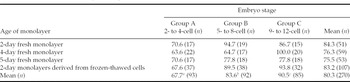
The blastocyst rate was significantly influenced by the embryo developmental stage at the beginning of culture (p < 0.001), being higher with the more advanced of the early developmental stages regardless of the monolayer used for culture (Table 1). Fewer embryos from group A developed to the blastocyst stage (67.7%) compared with embryos from group B (83.6%; p < 0.001) and group C (90.5%; p < 0.0001). Developmental competence also tended to be lower in group B than group C (p = 0.07). The probability of reaching the blastocyst stage was 2.4-fold higher for 2- to 4-cell embryos than for 5- to 8-cell embryos, and 4.5-fold lower than for 9- to 12-cell embryos. These differences decreased to 1.86-fold for the 5- to 8-cell embryos compared with the 9- to 12-cell embryos. Also, a critical developmental failure was found in culture of embryos between the 2- to 4-cell stage and late developmental stages of morulae and blastocysts (67.7% vs. 89.6%; p < 0.05). The results of the present study show that the ability of blastocysts produced by in vitro culture of 2- to 4-cell embryos to withstand freezing-thawing is lower than that of blastocysts obtained in vivo and frozen without culture (56.0 vs. 83.7%, p < 0.005). However, differences in viability after thawing between blastocysts obtained in vivo and those derived from groups B and C (71.0% and 71.7%, respectively) did not reach statistical significance.
Developmental rate (%) of 2- to 4-cell, 5- to 8-cell and 9- to 12-cell embryos cultured in 2-, 4- and 5-day monolayers derived from fresh cells and 2-day monolayers derived from frozen-thawed cells.

a,b Columns with different superscripts differ at p < 0.001.
a,c Columns with different superscripts differ p < 0.0001.
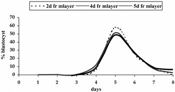
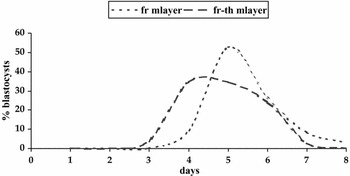
The data presented in Table 1 indicate that the rate of development to blastocyst is not affected by the age of the monolayer. Although 2-day monolayers yielded a higher rate of blastocyst than 4- and 5-day monolayers (84.3%, 76.3% and 75.5%, respectively), differences did not reach statistical significance. The kinetics of blastocyst appearance for early developmental stage embryos was similar in the different groups of embryos irrespective of the monolayer used for their culture (Fig. 1). Interestingly, there was detected an effect related to the management of the monolayer cells. The percentage of embryos developing into blastocysts was similar between monolayers derived from fresh or frozen-thawed cells (83.2% vs. 78.5%; p > 0.05), but the day of blastocyst appearance was different between the two types of derived monolayers (p < 0.0001; Fig. 2), mainly influenced by the group of 9- to 12-cell embryos (p < 0.0001).
Timing of blastocyst formation (days) of early-stage embryos co-cultured in 2-, 4- and 5-day fresh monolayers (2d fr mlayer, 4d fr mlayer, 5d fr mlayer, respectively).

Time to achieve blastocyst stage (days) of early embryos co-cultured in monolayers derived either from fresh-cells (fr mlayer) or frozen-thawed oviduct cells (fr-th mlayer).

Discussion
The results of the present study indicate that the developmental stage of early-stage embryos at the beginning of culture influences the blastocyst yield and quality. Embryo viability increased progressively from 2- to 4-cell embryos to 9- to 12-cell embryos. These data are in agreement with those reported for bovine (Camous et al., Reference Camous, Heyman, Méziou and Ménezo1984) and equine embryos (Ball et al., Reference Ball, Brinsko, Thomas, Miller and Ellington1993) in co-culture with somatic cells and for ovine embryos in semi-defined medium (Thompson et al., Reference Thompson, Parton, Cruickshank, Smith and Wales1989). Conversely, Eyestone & First (Reference Eyestone and First1991) showed a lower significant developmental yield for 3- to 4-cell than for 1- to 2-cell and 5- to 16-cell bovine cultured embryos.
Previous authors have identified these differences in developmental competence with the ‘developmental block’, an expression of embryo exposure to in vitro conditions during the late 4-cell or early 8-cell stage, which is considered as a very sensitive period of the embryo's existence (Eyestone & First, Reference Eyestone and First1991). In the present study, the critical period of this arrest was mainly overcome in vitro by 2- to 4-cell embryos, which were able to successfully resume growth to the morula stage (23/30, 76.6%). However, these earlier staged embryos showed a low blastocyst rate and a lower cryotolerance. Current data indicate temporal sensitivities of sheep embryos to the culture environment that are similar to those of cattle (Lonergan et al., Reference Lonergan, Rizos, Kanka, Nemcova, Mbaye, Kingston, Wade, Duffy and Boland2003). It seems reasonable to assume that the culture environment can be affecting mechanisms required to activate or complete cell compaction and blastocoele formation (i.e. cellular polarization and ion transport), as described by previous authors (Wrenzycki et al., Reference Wrenzycki, Herrmann, Carnwath and Niemann1998; Rief et al., Reference Rief, Sinowatz, Stojkovic, Einspanier, Wolf and Prelle2002). However, although further studies would be necessary, the addition of different substances to comply with the requirements of the earliest stages of development may enhance embryo developmental competence.
On the other hand, the present results did not show any influence of monolayer age or preservation on the developmental ability of early-stage sheep embryos. Embryo viability was similar between monolayers of different ages, in a similar way to bovine embryos cultured in oviduct cells either in suspension or on a near-confluent monolayer (McCaffrey et al., Reference McCaffrey, McEvoy, Diskin, Gwazdauskas, Kane and Sreenan1991). In our study, 2-day monolayers were chosen to avoid possible degenerative processes and deficient secretion of embryotrophic substances observed in more aged monolayers (Thibodeaux et al., Reference Thibodeaux, Menezo, Roussel, Hansel, Goodeaux, Thompson and Godke1992; Nancarrow & Hill, Reference Nancarrow and Hill1995; Wiemer et al., Reference Wiemer, Cohen, Tucker and Godke1998). Ability of monolayers to support embryonic development was not affected by freezing-thawing of oviduct cells. Moreover, the kinetics of blastocysts appearance were even better in monolayers derived from frozen cells than fresh ones. These data corroborate previous findings reporting successful results either with frozen-thawed monolayers in in vivo produced embryos (Rexroad & Powell, Reference Rexroad and Powell1988; Ellington et al., Reference Ellington, Carney, Farrel, Simkin and Foote1990), with frozen-thawed cells in in vitro produced embryos (Katska et al., Reference Katska, Rynska and Smorag1995) or with frozen conditioned medium in in vivo derived embryos (Van Langendonckt et al., Reference Van Langendonckt, Vansteenbrugge, Donnay, Van Soom, Berg, Semple, Grisart, Mermillod, Brem, Massip and Dessy1996). Freezing-thawing procedures seem to be an adequate alternative in somatic cell co-culture since they provide homogeneity to the culture system by selection of cells, and repeatability by using the same stock of cells in different replicates.
In conclusion, in vitro embryo viability and quality is strongly determined by developmental stage at the beginning of culture. Survival rate is higher with the more advanced early-stage embryos regardless of culture conditions. Improvement of blastocyst yield and embryo quality from early developmental stages would require, as a next step, further studies focused on the identification of the intrinsic features and specific requirements of these developmental stages. From the results of this study we can also conclude that the elaboration of monolayers from a pool of frozen cells is a good strategy for the implementation of a standardized system to be applied in practice.
Acknowledgements
The authors wish to thank Dr R. Calvo for her kind help with the statistical analysis of data. The valuable critical revision of this manuscript by Dr C.J.H. Souza is gratefully acknowledged. This work was supported by funds from the project INIA SC00-051-C3.1. R.M.G.G. and A.V.L. were supported by a pre-doctoral INIA research grant and A.G.B. by the Programme Ramon y Cajal.