Introduction
The distinct reproductive strategies of fish results, among other features, in different sizes of eggs and/or oocytes between species, thereby leading to differential availability of energetic reserves for oogenesis and influencing the duration of embryonic development (Depêche & Billard, Reference Depêche and Billard1994).
Differences in the reproductive behaviour and oocyte diameter might be inter-specific or intra-specific, latitudinal, related to spawning period or else to individuals within the same reproductive period (Vazzoler, Reference Vazzoler1996).
The embryonic process starts with fertilization of the oocyte by the spermatozoon in the micropyle and involves the reorganization of egg components. In order to be understood, topological studies of fertilized eggs should be combined (Depêche & Billard, Reference Depêche and Billard1994).
The successful rearing of a fish species depends on knowledge about its early biology, including fertilization and embryonic development traits, which directly affect both the fertilization and the hatching rates. The description of embryonic stages in teleosteans, besides providing support to basic biology features of a given species, can be applied to fish culture and fisheries (Matkovic et al., Reference Matkovic, Cussac, Cukier, Guerrero and Maggese1985), by gathering useful information for fish mass production in laboratory or for systematics and environmental inventory (Reynalte-Tataje et al., Reference Reynalte-Tataje, Zaniboni-Filho and Esquivel2004; Ninhaus-Silveira et al., Reference Ninhaus-Silveira, Foresti and Azevedo2006).
The species Brycon amazonicus is native from Brazilian Amazon (Lima, Reference Lima, Reis, Kullander and Ferraris2003) and had been cited as Brycon cephalus until recently, because of divergences in the taxonomic classification (Gomes & Urbinati, Reference Gomes, Urbinati, Baldisserotto and Gomes2005). The broodstock of B. amazonicus in this study are from Guaporé River, Mato Grosso, where the species is locally known as ‘jatuarana’. It is a large and humpbacked Brycon fish (up to 1 m in length and 8 kg in weight) with a goldish colouration on sides and dark on the back, found mainly in lakes downstream waterfalls (Feitosa, Reference Feitosa2004).
Several research groups have studied the viability of fishes of the genus Brycon in both fish culture and repopulation of regions where they became threatened by margin deforestation, construction of dams and river pollution (Oliveira et al., Reference Oliveira, Conte, Cyrino, Cyrino, Urbinati, Fracalossi and Castagnolli2004).
Taking into account the economic and ecological relevance of Brycon amazonicus, the goals of the present work were to describe the diameter of eggs and oocytes of this species, as well as the chronology of its embryonic development under controlled conditions in order to provide information about early biological features of this species that might be helpful for management and production programmes.
Material and methods
The samples were collected by Buriti fish farm, in the municipality of Nova Mutum, Mato Grosso, in December 2004.
During the studied period, the following physical–chemical water parameters were evaluated: temperature (mercury-in-glass thermometer), pH (digital potentiometer), dissolved oxygen (digital oxygen meter), alkalinity and ammonium, both determined according to Goltermann et al. (Reference Goltermann, Clymo and Ohnstad1978). The temperature was monitored daily at 6:00 a.m., 1:00 p.m. and 6:00 p.m., while the other variables were analyzed every 3 days early in the morning. To determine ammonium values, samples of water were frozen after collection and transported to the central laboratory of Centro de Aquicultura at UNESP for further analyses. The other analyses were carried out in the fish farm facility.
Induced spawning
Adult reproductively mature fish were induced to spawn, being the selected females and males placed separately in brick-made 2.0 m2 tanks, with continuous water flow and covered with nets. The spawning was induced using commercial common carp hypophysis extract (dried in acetone). The females received the first hormonal dose at the time they arrived at the laboratory (0.5 mg/kg) and the second one after a 10-h interval (5.0 mg/kg), at the same time as the single application to males (1.0 mg/kg).
When courtship behaviour was noticed, the fish were anesthetized by immersion in benzocaine solution (2 g dissolved in 150 ml of 96% alcohol diluted in 20 l of water), being then submitted to abdominal massage for gamete extrusion. Dry fertilization (Woynarovich & Hórvath, Reference Woynarovich and Horváth1983) was carried out by adding semen to oocytes followed by gentle mixing. Afterwards water was added for gamete activation and egg hydration, the eggs were rinsed and placed into 200-l incubators.
Sampling
A randomized experimental design was used. Biometric parameters, such as total length and weight, and reproductive data, such as volume of released oocytes, number of oocytes per ml and hatching rate, were obtained from 10 females and their respective offspring.
A sample comprised of 1 ml of oocytes was taken at the moment of extrusion from the 10 females induced to spawn, then fixed in 10% buffered formaldehyde and transferred to 70% alcohol after 24 h.
Samples that contained 10 oocytes and/or eggs were obtained at the following periods: extrusion; fertilization (when gametes were activated in water); 10 s, 20, 30, 60 and 90 s after fertilization (PF); each minute up to 10 min PF, each 5 min up to 30 min PF, 45 min PF and each hour up to hatching. Samples from five females were fixed in Karnovsky's solution (2.5% glutaraldehyde and 2.5% paraformaldehyde) for 24 h, then washed, transferred into cacodylate buffer, and stored at 4°C; the samples from the other five females were fixed in 10% buffered formaldehyde and transferred into 70% alcohol after 24 h.
Sample processing
The material was transported to the Faculdade de Ciências Agrárias e Veterinárias at Universidade Estadual Paulista (FCAV/UNESP), Jaboticabal Campus, São Paulo, and partly processed in the histology laboratory in the Animal Morphology and Physiology Department and partly in the electron microscopy laboratory.
To estimate the total number of extruded eggs, the number of cells in 1 ml, obtained and fixed when striping, was multiplied by the total volume of oocyte mass released by the 10 females induced to spawn. To calculate the frequency distribution of oocytes in diameter groups, 30 gametes from each female, collected at the moment of extrusion and fixed, were measured (in mm) using a light stereomicroscope equipped with a micrometer (Leica MZ 8).
The size of eggs during incubation was analyzed using the offspring of six females, and defined by two measurements taken between fertilization and hatching, as follows: total diameter of eggs with perivitelline space (distance between yolk mass and chorion; CEP) and yolk diameter, disregarding the perivitelline space (SEP), both in millimetres.
To analyze the fertilization events based on scanning electron microscopy (SEM), four offspring were fixed in Karnovsky's solution, transferred to sodium cacodylate buffer, post-fixed in 2% osmium tetroxide for 2 h, washed again in buffer and dehydrated in a graded series of ethanol at 30, 50, 70, 80, 90, 95 and 100%. Afterwards, the samples were dried to the critical point in a liquid CO2 drier (BAL-TEC), mounted on a copper grid, metalized with gold, and electron micrographed under a scanning electron microscope (JEOL-JSM 5410).
The alterations observed from oocyte extrusion up to larvae hatching were recorded by a light camera attached to the stereomicroscope (Leica MZ 95).
Results
The mean values of physical–chemical water parameters during the sampling period are shown in Table 1.
Mean values of physical–chemical water parameters during the sampling period, followed by their respective standard deviation (S), and maximum and minimum values

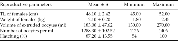
The biometric and reproductive data obtained during the induced spawning of B. amazonicus are presented in Table 2. At the moment of extrusion, the eggs were bluish green, spherical, translucent, semi-dense and presented a mean diameter among repetitions (n = 30) of 1.21 ± 0.06 mm.
Reproductive parameters of Brycon amazonicus used in the induced spawning, followed by their respective standard deviation (S), minimum and maximum values
TL = total length, n = 10.
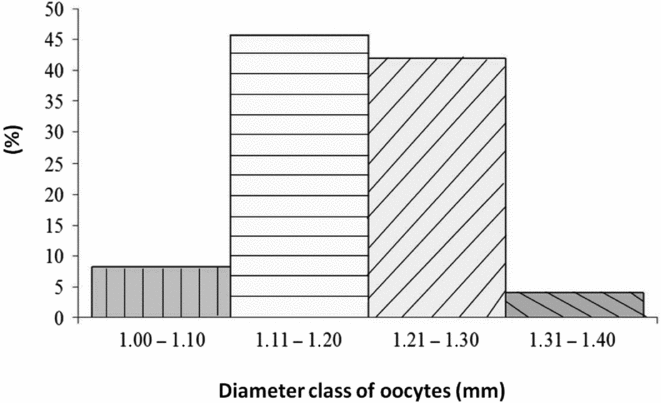
The frequency distribution of oocytes released by B. amazonicus females, in length classes (Fig. 1), showed that nearly half of them (45.70%) had a diameter between 1.11–1.20 mm, with the great majority of female gametes (87.7%) being between 1.11–1.30 mm at the moment of extrusion.
Frequency of oocytes per diameter class immediately after extrusion in Brycon amazonicus.
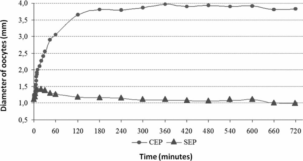
From fertilization onwards, the total diameter of eggs of B. amazonicus increased quickly up to 60 min PF, changing from 1.22 ± 0.04 mm to 3.06 ± 0.46 mm within this period. This enlargement was progressive, but less accentuated, between 60 and 120 min PF, and reached 3.65 ± 0.42 mm, and continued slowly up to 180 min PF when it became stable (Fig. 2).
Mean diameter of eggs of Brycon amazonicus during incubation. (•) CEP: with perivitelline space; (▴) SEP: without perivitelline space.
As the yolk (SEP) and total (CEP) diameter of eggs at 180 min PF remained the same as that observed at fertilization (1.16 ± 0.13 mm and 3.81 ± 0.40 mm, respectively), the increase in the total diameter indicated a period of progressive hydration of eggs, leading to a larger perivitelline space after 3 h of incubation (Fig. 2).
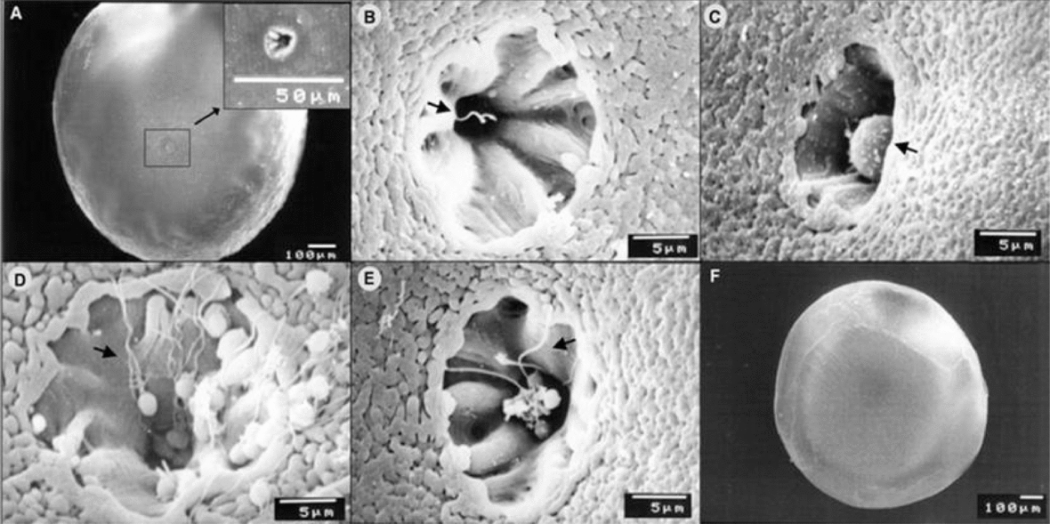
The description of the events related to fertilization of B. amazonicus under scanning electron microscope revealed a single micropyle in each oocyte (Fig. 3A), characterized by a depression of chorion in the animal pole of female gametes, and resembled a funnel-like canal, with longitudinal fringes along the walls. At 10 s PF the penetration by male gametes had already taken place in several oocytes, as visualized in Fig. 3B, in which the spermatozoon tail can be seen at micropyle vestibule.
Electron micrograph of fertilization in Brycon amazonicus. (A) Morphology of micropyle (arrow). (B) Spermatozoon tail (arrow) penetrating the oocyte 10 s post fertilization (PF). (C) Formation of fertilization cone (arrow) at 10 s PF. (D) Spermatozoa (arrow) migrating towards the micropyle at 30 s PF. (E) Several spermatozoa (arrow) in the micropyle vestibule at 90 s PF. (F) Fertilized egg.
Also at 10 s PF, some eggs had already been fertilized and protected from polyspermy by the formation of a fertilization cone through the micropyle canal (Fig. 3C). At 30 s PF, many spermatozoa could be seen in the micropyle walls of some oocytes or moving towards this region (Fig. 3D), although the masculine gamete had already penetrated the cell and the fertilization cone was present earlier in most of sampled gametes.
Even though most analyzed eggs had been fertilized between 10 and 30 s PF, the fertilization events in B. amazonicus occurred asynchronically; at 90 s PF (Fig. 3E) some oocytes with spermatozoa in the micropyle vestibule were detected, but the tails of these masculine gametes were extended and detached from the oocyte surface, differing from the pattern observed in the first 30 s PF, and their viability was unknown. In Fig. 3F, a fertilized egg can be observed (10 min PF) displaying a flattened shape because of the cell movement towards the animal pole, giving rise to the blastodisc.
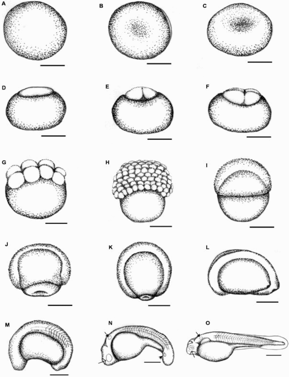
In Fig. 4, a few morphological alterations are observed in the female gametes between extrusion (Fig. 4A) and fertilization (Fig. 4B). A remarkable flattening of eggs was observed 10 min PF (Fig. 4C). The differentiation of blastodisc that typifies the animal pole, arranged over a large amount of yolk from the vegetative pole was confirmed 20 min PF (Fig. 4D).
Embryonic development of Brycon amazonicus. (A) Oocyte (extrusion). (B) Fertilized egg. (C) 10 min post fertilization (PF). (D) 20 min PF. (E) 30 min PF. (F) 45 min PF. (G) 1 h PF. (H) 2 h PF. (I) 3 h PF. (J) 5 h PF. (K) 6 h PF. (L) 7 h PF. (M) 8 h PF. (N) 10 h PF. (O) 13 h PF (hatching). Otic vesicle ( ), optic vesicle (→) and Küpffer's vesicle (
 ). Scale bar = 0.5 mm.
At 30 min PF (Fig. 4E), the first division of blastodisc had already occurred, showing two blastomeres of similar sizes, followed by the formation of four and eight blastomeres at 45 min (Fig. 4F) and at 1 h PF (Fig. 4G), respectively. Successive cell divisions resulted in overlapping layers of blastomeres arranged in a ‘half-berry’ shape, characterizing the morula stage 2 h PF (Fig. 4H). At 3 h PF (Fig. 4I), a cell proliferation was detected with divergent movements between animal and vegetative poles and indicated the beginning of the epiboly and gastrula stages, covering about 30% of the yolk at this moment and reaching nearly 70% 2 h later (Fig. 4J).
The formation of the germ ring and closure of the blastopore were almost complete 6 h PF (Fig. 4K) and the development of embryonic axis and the neural tube was reported 7 h PF (Fig. 4L), being the cephalic and caudal regions defined 1 h later (Fig. 4M), when 8 –12 somite pairs were detected. The otic and optic vesicles, as well as the Küpffer's vesicle, could be identified 10 h PF, simultaneously to the detachment of tail from the yolk sac (Fig. 4N). The chorion rupture took place at 13 h of embryonic development, when non-pigmented larvae were released with undifferentiated organic systems (Fig. 4O).
Discussion
No remarkable variation in the physical–chemical parameters of water was observed during the studied period, and the mean values were close to optimal for development of tropical fish (Sipaúba-Tavares, Reference Sipaúba-Tavares1995).
The number of released oocytes of females of B. amazonicus after induced spawning was comparable with that reported in previous studies for B. cephalus (Gomes, Reference Gomes1998), in which the oocyte mass corresponds to nearly 10% of body weight. The quantity of oocytes per gram is inversely correlated to their size (Vazzoler, Reference Vazzoler1996).
The overall aspect of B. amazonicus oocytes at the moment of extrusion was similar to that observed in fish of this genus. Spherical, translucent, semi-dense and greenish eggs were also detected in B. cephalus (Lopes et al., Reference Lopes, Senhorini and Soares1995; Romagosa et al., Reference Romagosa, Narahara and Fenerich-Verani2001; Vasques, Reference Vasques2003), B. insignis (Andrade-Talmelli et al., Reference Andrade-Talmelli, Kavamoto, Romagosa and Fenerich-Verani2001b) and B. orbignyanus (Ganeco, Reference Ganeco2003; Reynalte-Tataje et al., Reference Reynalte-Tataje, Zaniboni-Filho and Esquivel2004).
The small diameter of oocytes at extrusion is another common feature as, in this study, the mean diameter was equal to 1.21 ± 0.06 mm, with nearly 90% of gametes ranging from 1.11–1.20 mm, close to the values reported for the same species by Pardo-Carrasco et al. (Reference Pardo-Carrasco, Arias-Castellanos, Suárez-Mahecha, Cruz-Casallas, Vásques-Torres, Atencio-Garcia and Zaniboni-Filho2006a), in which a diameter varying from 1.00–1.45 mm was detected, with modal values between 1.09–1.18 mm, in females induced with carp pituitary extract. These diameter values were close to those observed for B. cephalus, with a mean diameter of 1.010 mm (Romagosa et al., Reference Romagosa, Narahara and Fenerich-Verani2001) and 0.91 ± 0.015 mm (Vasques, Reference Vasques2003); B. insignis in which the most frequent diameter was equal to 1.45 mm (Andrade-Talmelli et al., Reference Andrade-Talmelli, Kavamoto, Narahara and Fenerich-Verani2002); and B. orbignyanus, with a mean value of 1.59 ± 0.15 mm (Landinez et al., Reference Landinez, Senhorini, Sanabria, Baldan and Urbinati2004) with slight variation among species.
The oocyte size is related to the reproductive behavior, and both fecundity and diameter of mature oocytes are labile strategies, characterized by latitudinal, season, inter-specific and intra-specific variation, including differences among individuals of the same size within the spawning period. Migratory species with full spawning, external fertilization and no parental care, as the studied species, produce a larger number of small eggs (Vazzoler, Reference Vazzoler1996).
The fast hydration and development of a relatively large perivitelline space in the eggs of B. amazonicus after fertilization is another trait associated with the reproductive behaviour of Brycon fish, characterized by external fertilization and development without parental care, being the offspring left in the water column. The increased perivitelline space of eggs directly after fertilization indicates that the embryos are likely to be protected from environmental injuries (Ribeiro et al., Reference Ribeiro, Santos and Bolzan1995). The perivitelline space is formed even in non-fertilized eggs but activated by the contact between female gametes and water, which triggers out the cortical reaction that separates the chorion from the yolk once cortical alveoli are ruptured (Kunz, Reference Kunz2004).
Eggs with large perivitelline spaces after hydration were also reported in B. cephalus (Romagosa et al., Reference Romagosa, Narahara and Fenerich-Verani2001), B. orbignyanus (Reynalte-Tataje et al., Reference Reynalte-Tataje, Zaniboni-Filho and Esquivel2004), and B. insignis (Andrade-Talmelli et al., Reference Andrade-Talmelli, Kavamoto, Narahara and Fenerich-Verani2002), as well as in other species from distinct genera of Characiformes with similar reproductive strategies, such as Prochilodus lineatus (Castellani et al., Reference Castellani, Tse, Leme dos Santos, Faria and Santos1994) and Leporinus piau (Borçato et al., Reference Borçato, Bazzoli and Sato2004). Those species that undergo reproductive migration in a specific season and produce free eggs of fast hatching without parental care should be carefully monitored once any modification of hydrographic systems might affect their biological diversity (Andrade-Talmelli et al., Reference Andrade-Talmelli, Kavamoto, Narahara and Fenerich-Verani2002).
In teleosteans, the eggs are characterized as telolecithal because of the presence of animal and vegetative poles, being also referred to as polylecithal, once large amounts of yolk are detected, undergoing partial or meroblastic segmentation, restricted to the animal poles (Ribeiro et al., Reference Ribeiro, Santos and Bolzan1995). These features were observed in eggs of B. amazonicus in this study.
The asynchrony in the fertilization events observed in B. amazonicus has also reported in B. orbignyanus (Ganeco & Nakaghi, Reference Ganeco and Nakaghi2003) and B. orthotaenia (Sampaio, Reference Sampaio2006). This behaviour along with micropyle morphology, the presence of a funnel-like micropyle vestibule and several straight-arranged grooves, are common reproductive features for these Brycon species. Funnel-like micropyles are observed in most teleosteans and they allow the passage of a single spermatozoon through the internal opening (Rizzo & Bazzoli, Reference Rizzo and Bazzoli1993). The micropyle microstructure is noteworthy as useful for both identification and phylogenetic studies of fishes, once differences can be found among species in the same genus or family, putatively acting as mechanisms to prevent interspecific hybridization (Chen et al., Reference Chen, Shao and Yang1999).
The non-adhesive smooth chorion with regular pores reported in B. orbignyanus and B. amazonicus in this work is associated with the reproductive strategy of these species, i.e. migratory fish with free eggs (Rizzo et al., Reference Rizzo, Sato, Barreto and Godinho2002). The micropyle morphology varies in freshwater teleosteans and these differences are related to variation in the morphology of spermatozoa, although a similar pattern can be observed within certain systematic groups (Ricardo et al., Reference Ricardo, Aguiar, Rizzo and Bazzoli1996).
As observed in B. amazonicus, the first event after gamete activation in Prochilodus lineatus was the entrance of a spermatozoon through micropyle, followed by the formation of the fertilization cone (Brasil et al., Reference Brasil, Nakaghi, Santos, Grassiotto and Foresti2002). However, these events occurred asynchronically in the egg mass, i.e. different events were observed within the same sample as has been reported in B. amazonicus, in which spermatozoa were detected close to the micropyle vestibule in some oocytes at 30 s PF, while most eggs had already been fertilized and presented the fertilization cone.
The spermatozoa observed in the micropyle vestibule at 90 s after fertilization, displaying extended tail and detached from oocyte surface (a different disposition in relation to earlier moments) were probably unviable. Motility studies in spermatozoa of B. amazonicus revealed a short activity period of these cells, ranging from 26 to 50 s (Pardo-Carrasco et al., Reference Pardo-Carrasco, Zaniboni-Filho, Arias-Castellanos, Suárez-Mahecha, Atencio-Garcia and Cruz-Casallas2006b). Other studies on male gametes in fish of the genus Brycon have also reported a short activity period, being nearly 40 s in B. insignis (Andrade-Talmelli et al., Reference Andrade-Talmelli, Kavamoto and Fenerich-Verani2001a) and 41 ± 7 s in B. siebenthalae (Cruz-Casallas et al., Reference Cruz-Casallas, Lombo-Rodríguez and Velasco-Santamaria2005). Analysis of seminal traits in Brycon cephalus classified the spermatozoa of this species as aquasperm, characterized by a small round head without acrosomal vesicle (Ninhaus-Silveira et al., Reference Ninhaus-Silveira, Foresti and Azevedo2006).
The formation of the fertilization cone right after the penetration of spermatozoon in the micropyle canal, as observed in B. amazonicus, occurs by cytoplasm movements that lead to cytoplasm projection into the micropyle canal, giving rise to a bubble-like structure (Kudo, Reference Kudo1980). When the cytoplasm content is projected into the micropyle canal, the spermatozoon nucleus stays in the base of expanding cone, being inserted in the oocyte cytoplasm (Kudo, Reference Kudo1980). The formation of this cone reinforces the evolution of strategies to avoid the access of extra spermatozoa that might potentially cross the micropyle canal even after the entrance of the fertilizing spermatozoon (Brasil et al., Reference Brasil, Nakaghi, Santos, Grassiotto and Foresti2002).
The time of embryonic development in B. amazonicus described in this work was similar to that observed for the same species in a previous study (Mira-López et al., Reference Mira-López, Medina-Robles, Velasco-Santamaría and Cruz-Casallas2007), which reported an embryogenesis period of 13.5 h at 28.2 ± 1.5°C. The sequence of embryogenesis events in the species analyzed in the present work is also similar to that observed in other species of this genus, such as B. insignis (Andrade-Talmelli et al., Reference Andrade-Talmelli, Kavamoto, Romagosa and Fenerich-Verani2001b), B. cephalus (Lopes et al., Reference Lopes, Senhorini and Soares1995; Romagosa et al., Reference Romagosa, Narahara and Fenerich-Verani2001; Vasques, Reference Vasques2003), B. orbignyanus (Ganeco, Reference Ganeco2003; Landinez et al., Reference Landinez, Senhorini, Sanabria, Baldan and Urbinati2004; Reynalte-Tataje et al., Reference Reynalte-Tataje, Zaniboni-Filho and Esquivel2004) Brycon orthotaenia (Sampaio, Reference Sampaio2006) and Brycon gouldingi (Faustino et al., Reference Faustino, Nakaghi and Neumann2010), as well as in other Characiformes, like Piaractus mesopotamicus, Colossoma macropomum (Ribeiro et al., Reference Ribeiro, Santos and Bolzan1995), Prochilodus lineatus (Castellani et al., Reference Castellani, Tse, Leme dos Santos, Faria and Santos1994; Ninhaus-Silveira et al., Reference Ninhaus-Silveira, Foresti and Azevedo2006), Leporinus piau (Borçato et al., Reference Borçato, Bazzoli and Sato2004), Hoplias unitaeniatus, H. malabaricus and H. lacerdae (Gomes et al., Reference Gomes, Scarpelli, Arantes, Sato, Bazzoli and Rizzo2007). On the other hand, differences in the chronology of events were observed, even within Brycon species, what can be related to inter-specific variation or else to different water temperature during incubation.
In B. amazonicus, the beginning of somitogenesis took place after epiboly and closure of the blastopore, as has been reported in other Characiformes (Castellani et al., Reference Castellani, Tse, Leme dos Santos, Faria and Santos1994; Lopes et al., Reference Lopes, Senhorini and Soares1995; Ribeiro et al., Reference Ribeiro, Santos and Bolzan1995; Andrade-Talmelli et al., Reference Andrade-Talmelli, Kavamoto, Romagosa and Fenerich-Verani2001b; Ganeco, Reference Ganeco2003; Borçato et al., Reference Borçato, Bazzoli and Sato2004; Sampaio, Reference Sampaio2006; Ninhaus-Silveira et al., Reference Ninhaus-Silveira, Foresti and Azevedo2006; Gomes et al., Reference Gomes, Scarpelli, Arantes, Sato, Bazzoli and Rizzo2007; Faustino et al., Reference Faustino, Nakaghi and Neumann2010). In fact, epiboly movements in small eggs finish before the formation of somites (Ribeiro et al., Reference Ribeiro, Santos and Bolzan1995). In Oreochromis niloticus, the two first somite pairs appear before the end of gastrulation, and the tail portion of the embryo did not reach the vegetative pole, which according to the authors would be related to the large size of eggs in this species, typical of fish with parental care as cichlids (Perciformes; Morrison et al., Reference Morrison, Miyake and Wright2001).
Conclusion
The results of this study showed that B. amazonicus presents a reproductive strategy that is common to most rheophilic species that perform spawning migration. They release a large number of small eggs in the water column and are susceptible to predators and adverse environmental conditions that fast develop a large perivitelline space that putatively acts as a mechanical protective mechanism for embryos that undergo a short embryonic period and thereby enhances their survival chances. The larvae hatch with undifferentiated organic systems, lack fins and present rudimentary eyes and large yolk sacs; they are unable to face environmental risks such as escape from predators and searching for food.
Acknowledgements
The authors are grateful to Buriti fish farm for providing the biological material, Ms Cláudia Aparecida Rodrigues, the technician in the Electron Microscopy Laboratory (at FCAV/UNESP) for assistance in sample processing, CNPq (473712/2007–5) and Fapesp (2006/51326–0) for the financial support.
Declaration of Interest
The authors declare that they have no competing interests.