Introduction
Several snails within the family Lymnaeidae can act as intermediate hosts in the life cycle of Fascioloides magna. In North America, Dunkel et al. (Reference Dunkel, Rognlie, Johnson and Knapp1996) listed five snail species as natural intermediate hosts of F. magna and five other species as potential hosts because of their successful infections under laboratory conditions. In Europe, Galba truncatula is the most common intermediate host of F. magna (Erhardová-Kotrlá, Reference Erhardová-Kotrlá1971; Vignoles et al., Reference Vignoles, Novobilský, Rondelaud, Bellet, Treuil, Koudela and Dreyfuss2006). However, six other species – Lymnaea palustris (Chroustová, Reference Chroustová1979), Lymnaea (Omphiscola) glabra (Rondelaud et al., 2006), Lymnaea fuscus (Novobilský et al., Reference Novobilský, Kašný, Pankrác, Rondelaud, Engström and Höglund2012), Radix peregra (Faltýnková et al., Reference Faltýnková, Horáčková, Hirtová, Novobilský, Modrý and Scholz2006), Radix labiata and Radix lagotis (Huňová et al., Reference Huňová, Kašný, Hampl, Leontovyč, Kuběna, Mikeš and Horák2012) – can also sustain larval development of this parasite under laboratory conditions. In all of these lymnaeids, larval digenean development is similar, although some differences in intensity of F. magna infections have been noted. In France, Vignoles et al. (Reference Vignoles, Novobilský, Rondelaud, Bellet, Treuil, Koudela and Dreyfuss2006) reported an increase in the redial and cercarial production in laboratory-infected G. truncatula which was correlated with snail shell growth during the experiment. According to these authors, a mean of 44 free rediae and 210 cercariae were found in large snails (10–11 mm in height) subjected to single-miracidium infections and raised at 20°C according to Abrous et al. (Reference Abrous, Roumieux, Dreyfuss, Rondelaud and Mage1998). In contrast, the respective mean numbers were only 40 rediae and 164 cercariae in somewhat smaller snails (9–10 mm high). This variation in parasite burdens was particularly marked for second-generation rediae (Vignoles et al., Reference Vignoles, Novobilský, Rondelaud, Bellet, Treuil, Koudela and Dreyfuss2006), with a mean of ≈ 4 second-generation rediae produced by each redia of the first generation in the two examples given above.
Wider intervals in the numbers of first-generation rediae per snail (1–6) and second-generation rediae per fully grown redia of the first generation (3–6) were reported by Erhardová (Reference Erhardová1968) and Erhardová-Kotrlá (Reference Erhardová-Kotrlá1971). However, it is unclear if this variability in the shell growth of G. truncatula populations occurs in response to F. magna irrespective of environmental conditions (Rondelaud et al., Reference Rondelaud, Vignoles and Dreyfuss2009, Reference Rondelaud, Hourdin, Vignoles, Dreyfuss and Cabaret2011). Furthermore, the precise numerical distribution of first- and second-generation rediae in snails subjected to single-miracidium infections is unknown. To answer these questions, snails originating from four different field populations of G. truncatula differing in their shell height at the adult stage (from 6.5 to 12 mm) were subjected to single-miracidium infections with F. magna in the laboratory. Resulting rediae and cercariae were counted after dissection at day 75 post-exposure (pe) at 20°C. Non-exposed snails were used as controls for determination of survival rate variation and shell growth in the absence of the parasite.
Materials and methods
Collection and examination of snails
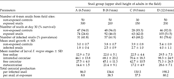
Four French populations of G. truncatula differing in shell height at the adult stage (table 1) were investigated. Population A originated from acid soils on a river bank (La Vienne) and the upper shell height of the adult snails was < 7 mm (Dreyfuss et al., 1997). Populations B and C were also from acid soil but were collected from road ditches. The maximum heights of these adults were: 8 and 9 mm, respectively. Finally, population D colonized a road ditch on predominantly clay soil and had adults with an upper shell height of 12 mm. The longest distance (132 km) between snail habitats was noted for populations C and D. Snails of height 4 mm belonging to the spring (populations B, C and D) or summer (A) generation were collected from each site: a total of 350, 250, 180 and 250 snails from populations A, B, C and D, respectively. These snails were then progressively acclimatized to laboratory conditions for 48 h at a constant temperature of 20°C before miracidial exposure as described below. Furthermore, 30 additional adult snails were collected from each population and dissected under a stereomicroscope to verify the absence of trematode larval forms.
Main characteristics of the four populations of G. truncatula used for experimental infections with F. magna. The size of field snails for each of groups A, B, C and D relates to the maximum height (mm) of adults and the snails used in all experimental infections were 4 mm high.

Collection of F. magna eggs and hatching of miracidia
Eggs of F. magna were collected from adult flukes recovered from the livers of naturally infected red deer (Cervus elaphus) hunted in the district of Krivoklátsko (Central Bohemia, Czech Republic). Prior to hatching, these eggs were washed several times with spring water and were incubated for 20 days at 20°C in the dark (Ollerenshaw, Reference Ollerenshaw1971).
Experimental infections of snails
Thirty (population C) or 50 (populations A, B and D; table 1) snails from each population were used as non-infected controls, while the remaining snails (table 1) were subjected to infection with F. magna by exposure to a single miracidium per snail for 4 h at 20°C in 3.5 ml of spring water. Snails were then maintained for 30 days in groups of 10 individuals in 14-cm Petri dishes filled with 60 ml of spring water, according to the method of Rondelaud et al. (Reference Rondelaud, Fousi, Vignoles, Moncef and Dreyfuss2007). Snails were fed on dried lettuce leaves and dead grass (Molinia caerulea), while several stems of fresh spring moss (Fontinalis sp.) ensured oxygenation of the water. The spring water contained 35 mg/l of dissolved calcium. The Petri dishes were placed at a constant temperature of 20°C ( ± 1°C) and a natural photoperiod of 10 h light.
At day 30 pe, all surviving snails were isolated one by one in 35-mm Petri dishes with spring water and pieces of dead grass, lettuce and spring moss at 20°C. A daily inspection was made to change spring water and food if necessary, and to count any cercariae that were released in the dishes. From day 49 pe, the exposed snails were subjected to a thermal shock every 3 days by placing their Petri dishes at 10–13°C for 3 h (outdoors) to stimulate natural cercarial shedding (Sanabria et al., Reference Sanabria, Mouzet, Pankrác, Djuikwo Teukeng, Courtioux, Novobilský, Höglund, Kašný, Vignoles, Dreyfuss, Rondelaud and Romero2013).
At day 75 pe, the shell height of each surviving G. truncatula was measured to the nearest 0.1 mm using callipers. At the same time, all surviving snails were dissected and free rediae were counted under a stereomicroscope. The identification of redial generations was made by light microscopic observations using a concave slide and was based on the gross morphology of the pharynx, and the presence or the absence of a muscular ring, according to criteria reported by Vignoles et al. (Reference Vignoles, Novobilský, Rondelaud, Bellet, Treuil, Koudela and Dreyfuss2006). Intraredial cercariae, free cercariae within the snail's body tissues, and cercariae that had been shed from the snails before day 75 pe (which immediately encysted into metacercariae) were also taken into account.
Data analysis
The two variables calculated were snail survival at day 30 pe and prevalence of F. magna infection in relation to the number of surviving snails at day 30 pe. For each factor studied, the difference between the values recorded for the four snail populations was analysed using a χ2 test. Another variable was the growth rate of non-exposed controls and infected snails from the date of exposure to the date of snail dissection at day 75 pe. The numbers of free rediae in relation to their first, second or third generations, as well as the quantities of intraredial, free or encysted cercariae were also counted. Individual values recorded for the growth rate of snails during the experiment, and the numbers of rediae and cercariae were averaged and their standard deviations were calculated for each snail population. A Spearman correlation test and two-way analysis of variance (ANOVA) were used to establish levels of statistical significance. All the analyses were carried out using the Statview 5.0 software (SAS Institute Inc., Cary, North Carolina, USA).
Results
General characteristics of F. magna infection in G. truncatula
Compared to non-exposed controls, survival of exposed snails at day 30 pe (table 2) was significantly lower in the four snail groups (A: χ2= 91.07, P< 0.001; B: χ2= 40.58, P< 0.001; C: χ2= 26.35, P< 0.001; D: χ2= 27.24, P < 0.001). In exposed snails, survival was also significantly lower (χ2= 43.70, P< 0.001) in group A but did not differ significantly in groups B, C and D. Prevalences of F. magna infection in groups B (61.9%) and C (68.2%) were significantly lower (χ2= 10.82, P< 0.05) than that observed in group D (78.6%) but significantly higher (χ2= 32.65, P< 0.001) than in A (41.8%). The mean shell growth of non-exposed controls at day 75 pe ranged between 3.0 and 5.5 mm in groups A and D, whereas that of the infected snails ranged from 1.4 mm in group A to 4.0 mm in group D. Both the origin of the snail population (F= 14.12, P< 0.01) and infection with F. magna (F= 7.23, P< 0.01) had a significant influence on snail shell growth in this experiment.
General characteristics of F. magna infection in four populations of G. truncatula. The size of field snails for each of groups A, B, C and D relates to the maximum height (mm) of adults, and the snails used in all experimental infections were 4 mm high.

The redial and cercarial burdens found in G. truncatula are shown in table 2. The number of free rediae within the G. truncatula body significantly increased (r= 0.91, P< 0.01) with the increasing shell growth of the snails during the experiment. Positive correlations were also noted for the numbers of intraredial cercariae (r= 0.84, P< 0.01), free cercariae (r= 0.87, P< 0.01) and metacercariae encysted after shedding or snail dissection (r= 0.74, P< 0.05). Total cercarial production of F. magna per infected snail, including intraredial and free cercariae, ranged between 86 and 198 in groups A and D, respectively. In contrast, when cercarial production was calculated from the total number of G. truncatula exposed to miracidia at the beginning of the experiment, the values were clearly lower: from a mean of 9 (group A) up to 80 cercariae (group D).
Number of F. magna rediae according to their generation
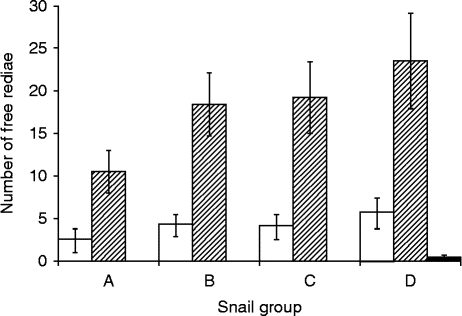
The numerical distribution of free rediae in the four groups of G. truncatula according to each redial generation is summarized in fig. 1. As for the total number of these rediae (table 2), the mean number of first-generation rediae significantly increased (F= 32.20, P< 0.001) from group A to group D. A similar finding was also noted for second-generation rediae (F= 6.30, P< 0.01). The number of third-generation rediae was low in group D, the group that had the highest shell sizes at day 75 pe.
Mean values and SD for free rediae of F. magna in four populations of G. truncatula (A, B, C and D) in relation to generation 1 (white bars), 2 (striped bars) or 3 (black bars). The upper shell heights of adults in the field were: A, 6.5 mm; B, 8.0 mm; C, 9.0 mm; D, 12.0 mm. For further details on snail populations, see table 1.
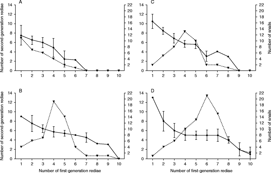
The distribution of first-generation rediae within each group of G. truncatula was wider (fig. 2). In group A, this number ranged from 1 to 6 rediae per snail and a single first-generation redia was found in 11 snails (out of 31). In groups B and C, the numerical range was from 1 to 9 first-generation rediae per snail and the number of snails peaked at 4 rediae per snail. In group D, this number ranged from 1 to 10, with a peak in the snail number at 6 rediae. If the production of second-generation rediae per fully grown redia of the first generation is considered (fig. 2), the number of second-generation rediae was 13 in group D when a single redia of the first generation per snail developed. This number correlatively decreased when the quantity of first-generation rediae increased within the snail. These trends were also recorded for the other snail groups (A, B and C).
Mean values and SD for second-generation rediae of F. magna per fully grown redia of the first generation (left y axis, ●) and numerical distribution of F. magna-infected snails (right y axis,▾) in relation to the number of first-generation rediae per snail in four groups of G. truncatula (A–D). The upper shell heights of adults in the field were: A, 6.5 mm; B, 8.0 mm; C, 9.0 mm; D, 12.0 mm. For further details on snail populations, see table 1.
In a total of 17 snails (out of 231 infected snails), small encapsulated pharynges of young rediae of the first and second generation were noted (data not shown). In these snails, we never found mummified cadavers of fully grown rediae or encapsulated residues of their pharynges.
Discussion
In this experimental study, we revealed that the prevalence of F. magna infection in G. truncatula was significantly influenced by the shell size of the intermediate snail host. In contrast, snail survival at day 30 pe was significantly lower in group A, whereas percentages noted in the other three groups did not significantly differ from each other. Two complementary hypotheses are proposed to explain these variations. The first relates to the individual susceptibility of each snail population to F. magna and their inherited capacity to sustain larval development of the parasite. Such variations in characteristics of snail infection relating to the origin of each population have already been reported for the closely related species Fasciola hepatica in G. truncatula (Vignoles et al., Reference Vignoles, Dreyfuss and Rondelaud2002). However, the necessity of sufficient snail growth during the experiment and, as a consequence, on that of a large body volume to obtain complete larval development of F. magna within the snail host must also be considered. This idea is supported by the presence of third-generation rediae exclusively in the largest snails (fig. 1).
Even though the four snail groups were raised in the laboratory using the same protocol, each population showed differential growth during the experiment (table 2). In the case of the non-exposed snails, it can be hypothesized that several successive generations (raised on a calcium-enriched soil) would be necessary to obtain a significant increase of the shell height for adult G. truncatula coming from acidic soils. Another explanation would be to relate this result to the genetic variability in growth potential of each snail population, although several studies (Jabbour-Zahab et al., Reference Jabbour-Zahab, Pointier, Jourdane, Jarne, Oviedo, Bargues, Mas-Coma, Angles, Perera, Balzan, Khallaayoune and Renaud1997; Meunier et al., Reference Meunier, Hurtrez-Boussès, Jabbour-Zahab, Durand, Rondelaud and Renaud2004) have reported that such genetic diversity between populations of G. truncatula is low, even in samples from different continents. Lower values of the shell height noted for infected snails in the present study correspond to the results obtained on G. truncatula infected by other digenean species (Rondelaud et al., Reference Rondelaud, Vignoles and Dreyfuss2009). This can be explained by intensive uptake of nutrients by the parasite and limited utilization of nutrients for snail somatic growth (Graczyk & Fried, Reference Graczyk, Fried and Dalton1999).
Although the effect of trematode infection on snail growth has been evidenced before, gigantism of infected snails is still a controversial phenomenon (Thompson, Reference Thompson, Fried and Graczyk1997). For instance, Wilson & Denison (Reference Wilson and Denison1980) reported an increased shell size and soft-tissue mass in F. hepatica-infected snails. In contrast, the present study indicates that shell height was decreased in infected G. truncatula compared to non-exposed controls. Similarly, inhibited growth of snails has also been described for L. fuscus experimentally infected with F. magna (Novobilský et al., Reference Novobilský, Kašný, Pankrác, Rondelaud, Engström and Höglund2012). Retarded growth caused by F. magna larvae may be explained by altered metabolism in infected snails.
In groups B, C and D of G. truncatula, the redial and cercarial production of F. magna ranged within the values previously reported both by Vignoles et al. (Reference Vignoles, Novobilský, Rondelaud, Bellet, Treuil, Koudela and Dreyfuss2006) and Sanabria et al. (Reference Sanabria, Mouzet, Pankrác, Djuikwo Teukeng, Courtioux, Novobilský, Höglund, Kašný, Vignoles, Dreyfuss, Rondelaud and Romero2013) from meadow populations of G. truncatula. According to these authors, the mean number of free rediae ranged between 37 and 44, while the total cercarial production of F. magna ranged from 164 to 231 cercariae per snail. The lower values in group A also corresponded to results recorded by the same team in the same snail population when infected with F. hepatica (Rondelaud et al., Reference Rondelaud, Dreyfuss, Vareille-Morel and Moukrim1997; Vignoles et al., Reference Vignoles, Rondelaud and Dreyfuss2011). All these results further support the idea that redial and cercarial production of F. magna is dependent on the shell growth of the snail host, as already reported for both Echinostoma revolutum (Zischke, 1967) and F. hepatica (Rondelaud & Barthe, Reference Rondelaud and Barthe1987).
Concerning redial generations of F. magna, the presence of scarce third-generation rediae in the highest size classes of group D confirmed the observations first reported by Vignoles et al. (Reference Vignoles, Novobilský, Rondelaud, Bellet, Treuil, Koudela and Dreyfuss2006). The values for first- and second-generation rediae are also in accordance with the report by Vignoles et al. (Reference Vignoles, Novobilský, Rondelaud, Bellet, Treuil, Koudela and Dreyfuss2006). However, the numerical distribution of each redial generation within these four snail groups requires further comment.
First, it was shown that a single sporocyst of F. magna can produce up to ten rediae of the first generation during the infection of G. truncatula, so that the distribution of these rediae within the snail's body showed great variability in the four snail groups studied. It is evident that the snail growth rate during the experiment plays a central role in redial development. However, additional factors are probably also important, because a total of 40 G. truncatula in the four groups (fig. 2) contained one or two first-generation rediae per snail. The most likely explanation of this finding is that some young rediae of this generation might migrate out from the digestive gland to the snail foot or tentacles after their exit from the sporocyst, which makes them unsuitable for further development. They may also disappear or degenerate, forming small mummified cadavers, as reported for rediae of F. hepatica by Barber (Reference Barber1962), Rondelaud & Barthe (Reference Rondelaud and Barthe1980) and Rondelaud et al. (Reference Rondelaud, Vignoles and Dreyfuss2009). This interpretation is supported by the finding of small, encapsulated residues of redial pharynges in several snails after their dissection at day 75 pe (data not shown).
Second, in snails harbouring only a single first-generation redia, the mean daughter redia production was higher than that noted for snails containing two or more fully developed first-generation rediae (fig. 2). This daughter redia production was higher than that reported by Erhardová-Kotrlá (Reference Erhardová-Kotrlá1971) in the same lymnaeid species when experimentally infected with F. magna. This suggests that production of daughter rediae of F. magna can follow adaptively the volume of the snail visceral cavity with respect to the number of growing mother rediae. An argument supporting this approach is the observed decrease in daughter redia production per fully grown redia of the first generation at the same time as the number of mother rediae increased within the snail (fig. 2).
In conclusion, we recorded inter- and intrapopulation variability in the numerical development of F. magna rediae, which was correlated with the snail growth capacity of each snail population investigated in this experiment. Absence of encapsulated pharynges of degenerated fully grown rediae indicates that redial generations of F. magna did not show any abnomal pattern of development in the studied G. truncatula populations, as has been observed in similar experiments with the related species F. hepatica (Rondelaud et al., Reference Rondelaud, Vignoles and Dreyfuss2009). Our findings clearly support the idea that development of F. magna redial generations is snail species dependent.
Acknowledgements
This study was supported by the Swedish Foundation for Agricultural Research (contract H1050003) and the Charles University in Prague (UNCE 204017, PRVOUK P41 and SVV 267210/2013). The authors thank Dr David A. Morrison for revising the English text.