I. INTRODUCTION
Composite films containing noble metal nanoparticles (NPs) have potential applications in nonlinear optical devices, all-optical switching devices, nanoelectronic devices, optical labeling, optical communication, and surface enhanced Raman scatteringReference Min and Veronis1–Reference Kim, Kim and Seung4 because of their chemical stability and their excellent absorption properties in the visible region or the near-infrared region,Reference Kreibig and Vollmer5,Reference Biswas, Eilers, Hidden, Aktas and Kiran6 which results from surface plasmon resonance (SPR) of metal NPs. In the past decades, composite films containing noble metal NPs have been studied widely, especially the single metal dispersed composite film. For single metal dispersed composite film, one SPR respond band is obtained, and the intensity and frequency of SPR band can be tuned by changing the metal NPs' shape, size, structure, content, and the surrounding environment.Reference Liu, Atwater, Wang and Huo7,Reference Kelly, Coronado, Zhao and Schatz8 However, the sole SPR band with narrow frequency variation limits their application in multiplex biosensor or other multichannel photodetectors.
To broaden the respond band or gain multi-band in the visible region, the composite film system has been expanded from a single metal NPs dispersed system to a bimetal NPs dispersed system. According to theoretical researches, the bimetal NPs dispersed system will exhibit adjustable SPR band or two SPR bands, which depends on the presence of the two metals in the composite, in their alloy, or in each metal state.Reference Belotelov, Carotenuto, Nicolais, Longo, Pepe, Perlo and Zvezdin9,Reference Suyal10 Now, much work has been done on the preparation of bimetal NPs dispersed system, including metal alloys,Reference Hubenthal, Zeigler, Hendrich, Alschinger and Trager11–Reference Alqudam, Annapoorni and Shivaparasd14 core–shell structures,Reference Hodak, Henglein, Giersig and Hartland15–Reference Yu, You, Polavarapu and Xu18 and mixed particles system.Reference Wang, Wang, Hang and Gong19 Especially, noble metal NPs, such as Au and Ag dispersed system, have attained a huge interest because of easy reductive preparation, water solubility, and high chemical stability.Reference Wang, Song, Wang, Zhang, Zhang and Sun20 Reddy et al.Reference Reddy, Varaprasad, Reddy, Raju and Reddy21 reported the fabrication of Ag–Au alloy nanocomposites obtained from the synthesis of acrylamide (AM) and 2-acrylamido-2-methyl-1-propanesulfonic acid (AMPS) based hydrogels for antimicrobial applications. Sun et al.Reference Sun, Wei and Li22 prepared Ag–Au alloy/SiO2 composite thin film by means of sol–gel method and deoxidized under ultraviolet radiation. Although the Au–Ag alloy dispersed composite exhibits unique electronic, optical, and catalytic properties compared with the single metal system,Reference Hirsch, Stafford, Bankson, Sershen, Rivera, Price, Hazle, Halas and West23 the results show one peak or two peaks, with one in the form of a shoulder peak in the extinction spectra. To the best of our knowledge, there are little reports where two clearly separated plasmon peaks are obtained by a chemical method. This is because it is difficult to prepare Au–Ag bimetal separately dispersed in composite films. The crystal structure of both Au and Ag are face-centered cubic with nearly identical bulk lattice constant, thus making them mutually miscible. When a physical method is employed, such factor is no longer applicable. Recently, Chakravadhanula et al.Reference Chakravadhanula, Elbahri, Schurmann, Takele, Greve, Zaporojtchenko and Faupel24 obtained equal intensity double plasmon resonance of bimetallic quasi-nanocomposites through a physical deposition method. However, this method needs high cost and vacuum conditions for preparation, as well as the intensity of double peaks was sensitive to the thickness of Ag and Au films, which was so difficult to obtain and control in the region of several nanometers to even a fraction of nanometers. In this article, we use a chemical approach to prepare Ag–Au bimetal dispersed SiO2 composite film and obtained equal intensity double SPR peaks. We also investigated the composite film by transmission electron microscopy (TEM), x-ray photoelectron spectroscopy (XPS), and ultraviolet-visible (UV-vis) absorption spectra and compared it with the calculated absorption spectra using the modified Mie theory, as well as the influence of Ag–Au content was studied.
II. EXPERIMENT
The Au–Ag NPs bimetal dispersed SiO2 composite films were obtained via a chemical solution approach combining with a sol–gel spin-coating process.Reference Lee, Kim and Choi25,Reference Jeon, Yi and Oh26
A. Preparation of Aux/(SiO2)0.5−x and Agy/(SiO2)0.5−y precursor solutions
Tetraethyl orthosilicate (Si(OC2H5)4, TEOS), silver nitrate (AgNO3), and chloroauric acid (HAuCl4·4H2O) were of analytical grade and were used as raw materials without further purification. Deionized water and absolute ethyl alcohol (C2H5OH) solution were used as solvents. TEOS, C2H5OH solution, and deionized water with a molar ratio of 1:15:5 were mixed and stirred for 1 h at room temperature. Several drops of nitric acid were added as hydrolysis catalyst before stirring for adjusting the pH value of solution between 2 and 3. Then the solution was divided into ten parts, and in each part, the required amount of AgNO3 or HAuCl4·4H2O was added into the solution with the molar ratios of HAuCl4·4H2O/(HAuCl4·4H2O + TEOS) = 4, 10, 16, 20 mol% or AgNO3/(AgNO3 + TEOS) = 36, 30, 24, 20 mol%. After further stirring for 1 h and aging for 2 days, Au–SiO2 and Ag–SiO2 transparent precursor solutions with required molar ratios were obtained, which were denoted as Aux/(SiO2)0.5−x and Agy/(SiO2)0.5−y with x = 2, 5, 8, 10; y = 20 − x mol%.
B. Fabrication of AuxAgy/(SiO2)0.8 composite films
AuxAgy/(SiO2)0.8 composite films were obtained by spin-coating Aux/(SiO2)0.5−x and Agy/(SiO2)0.5−y films one after another on a SiO2 glass substrate with a dimension of 20 × 20 mm, which was ultrasonically cleaned successively in an acetone, ethanol solution, deionized water, and dried in the oven in advance. Aux/(SiO2)0.5−x films with thickness of 50 nm were deposited from the precursor solutions at a spinning speed of 3000 rpm for 1 min, followed by pyrolyzing at 190 °C for 2 min. Agy/(SiO2)0.5−y films with thickness of 50 nm were coated on the surface of Aux/(SiO2)0.5−x films at the same spinning speed after those films were cooled to room temperature, and then the composite films were annealed at 500 °C for 30 min in air. In this process, along with the formation of metal particles from metal salts, the heat treatment promoted silica gel network formation and evaporation of the liquid phase in gel, and led to the homogeneous AuxAgy/(SiO2)0.8[Aux/(SiO2)0.5−x + Agy/(SiO2)0.5−y] film, where x + y = 0.2.Reference Lee, Kim and Choi25Figure 1 shows the schematic diagram of the formation of the bimetal dispersed AuxAgy/(SiO2)0.8 composite film.
Schematic diagram for formation of the bimetal dispersed AuAg/SiO2 composite film.

C. Characterization and calculation
TEM image was performed on JEM-100CXG electron microscope (JEOL, Tokyo, Japan). XPS data were obtained with a PHI-5300 ESCA electron spectrometer using Al monochromatic Kα radiation (hυ = 1486.6 eV) as the x-ray source. The binding energies were referenced to C 1s line at 285.0 eV from adventitious carbon. UV-vis absorption spectra were measured with a UNIC UV-2800 UV-visible spectrophotometer (UNICO Instruments Co., Ltd., China). The differential thermal analysis (DTA) was performed using a WCR-2C differential thermal analyzer in Ar atmosphere. The theoretical absorption spectra were calculated by Excel software.
III. RESULTS AND DISCUSSION
The process for obtaining silica-gel involves the hydrolysis of TEOS [Eq. (1)] and the condensation of hydrolysis products with the elimination of water or alcohol as indicated in reaction [Eq. (2) or (3)], depending on the H2O/TEOS ratio.Reference Stefanescu, Stoia and Stefanescu27 No matter if the hydrolysis process is carried out in a basic or acid condition, only providing high amount of acid and water as occurs in the present case, the fully hydrolyzed molecule obtained in the process is Si(OH)4.Reference Monsivais-Gámez, Ruiz and Martinez28 When the fully hydrolyzed silica sol is mixed with a high polarizable ion, such as Ag+ or Au+, the reaction [Eq. (4)] should be expected with the formation of nonbridging oxygen by a cation exchange process.Reference De, Licciulli, Massaro, Tapfer, Catalano, Battagin, Meneghini and Mazzoldi29–Reference Bansal31 Finally, the Ag/SiO2 or Au/SiO2 films were obtained by decomposition of Si(OH)4 and SiO(OH)3Ag or SiO(OH)3Au [Eq. (5)], eliminating several water molecules. The possible formation process is proposed as follows:
 $${\rm{Si}}{\left( {{\rm{O}}{{\rm{C}}_{\rm{2}}}{{\rm{H}}_{\rm{5}}}} \right)_4} + \left( {4 - n} \right){{\rm{H}}_{\rm{2}}}{\rm{O}} \to {\rm{Si}}{\left( {{\rm{O}}{{\rm{C}}_{\rm{2}}}{{\rm{H}}_{\rm{5}}}} \right)_n}{\left( {{\rm{OH}}} \right)_{4 - n}} + \left( {4 - n} \right){{\rm{C}}_2}{{\rm{H}}_5}{\rm{OH}}\quad \left( {0 \,\le \,n \,\le \,3} \right)\quad ,$$
 $${\rm{Si}}{\left( {{\rm{O}}{{\rm{C}}_{\rm{2}}}{{\rm{H}}_{\rm{5}}}} \right)_n}{\left( {{\rm{OH}}} \right)_{4 - n}} + {\left( {{\rm{HO}}} \right)_{4 - n}}{\left( {{{\rm{C}}_2}{{\rm{H}}_{\rm{5}}}{\rm{O}}} \right)_n}{\rm{Si}} \to {\left( {{\rm{HO}}} \right)_{3 - n}}{\left( {{{\rm{C}}_{\rm{2}}}{{\rm{H}}_{\rm{5}}}{\rm{O}}} \right)_n}\,{\rm{Si}}-{\rm{O}}-{\rm{Si}}{\left( {{\rm{O}}{{\rm{C}}_{\rm{2}}}{{\rm{H}}_{\rm{5}}}} \right)_n}{\left( {{\rm{OH}}} \right)_{3 - n}} + {{\rm{H}}_2}{\rm{O}}\quad {\rm{,}}$$
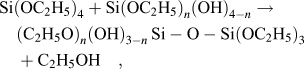 $${\rm{Si}}{\left( {{\rm{O}}{{\rm{C}}_{\rm{2}}}{{\rm{H}}_5}} \right)_4} + {\rm{Si}}{\left( {{\rm{O}}{{\rm{C}}_{\rm{2}}}{{\rm{H}}_5}} \right)_n}{\left( {{\rm{OH}}} \right)_{4 - n}} \to {\left( {{{\rm{C}}_2}{{\rm{H}}_5}{\rm{O}}} \right)_n}{\left( {{\rm{OH}}} \right)_{3 - n}}\,{\rm{Si}}-{\rm{O}}-{\rm{Si}}{\left( {{\rm{O}}{{\rm{C}}_{\rm{2}}}{{\rm{H}}_5}} \right)_3} + {{\rm{C}}_2}{{\rm{H}}_5}{\rm{OH}}\quad {\rm{,}}$$
 $${\rm{Si}}{\left( {{\rm{OH}}} \right)_4} + {\rm{A}}{{\rm{g}}^ + }\,\,{\rm{or}}\,\,{\rm{A}}{{\rm{u}}^ + } \to {\left( {{\rm{HO}}} \right)_3}{\rm{Si}}-{\rm{O}}-{\rm{Ag}}\,{\rm{or}}\,\,{\left( {{\rm{HO}}} \right)_3}{\rm{Si}}-{\rm{O}}-{\rm{Au}} + {{\rm{H}}^ + }\quad {\rm{,}}$$
 $${\rm{Si}}{\left( {{\rm{OH}}} \right)_4} \to {\rm{Si}}{{\rm{O}}_{\rm{2}}} + 2{{\rm{H}}_{\rm{2}}}{\rm{O}}$$
or
 $$2{\rm{SiO}}{\left( {{\rm{OH}}} \right)_3}{\rm{Ag}}\;\,{\rm{or}}\,\;{\rm{2SiO}}{\left( {{\rm{OH}}} \right)_3}{\rm{Au}} \to {\rm{2Si}}{{\rm{O}}_{\rm{2}}} + 3{{\rm{H}}_2}{\rm{O}} + {{\rm{O}}_{\rm{2}}} + 2{\rm{Ag}}\,\,{\rm{or}}\,\,{\rm{2Au}}\quad {\rm{.}}$$
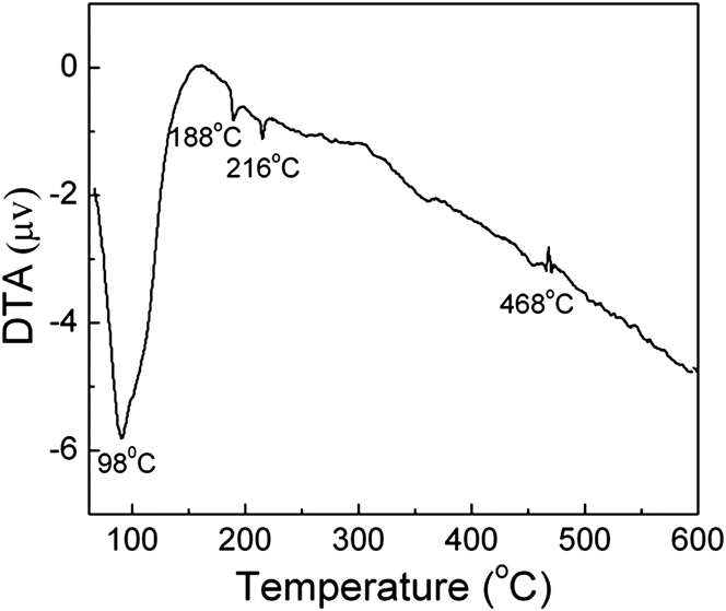
To determine the formation temperature of Au and Ag NPs in SiO2 matrix and to select proper annealing temperature, the DTA analysis was performed on Au0.1Ag0.1/(SiO2)0.8 gel powder. As can be seen from Fig. 2, there are three endothermic peaks at about 98, 188, 216 °C and one exothermic peak at 468 °C in the curve. The first broad endothermic peak at 98 °C is due to the volatilization of water and ethanol that come from the added solvent in the colloid preparation process or give off by hydrolysis and condensation reaction of Si(OC2H5)4Reference Stefanescu, Stoia and Stefanescu27 as indicated in the reactions of (1) and (2) or (3). Based on the mechanism analysis of AuAg/SiO2 film, it can be inferred that Si–O–Ag and Si–O–Au exist in the formation process of Au0.1Ag0.1/(SiO2)0.8 film. In addition, it has been reported earlier that the temperature formation of silver metal clusters by breaking Si–O–Ag is at about 200 °CReference De, Licciulli, Massaro, Tapfer, Catalano, Battagin, Meneghini and Mazzoldi29,Reference Khan, Kumar, Ahamed, Alrokayan, Alsalhi, Alhoshan and Aldwayyan32 ; our previous workReference Li, Zhang, Zhao and Zhao33 also confirmed the appearance of SPR peak resulting from Ag NPs when the Ag/SiO2 film annealed above 200 °C. Taking into account the fact that the ionic radius of Au+ is larger than the radius of Ag+, it can be deduced that the endothermic peaks at 188 and 216 °C are attributed to the formation of Au and Ag NPs by breaking Si–O–Au and Si–O–Ag. The exothermic peak at 468 °C may be attributed to the combustion of residual carbon and organics.Reference De, Licciulli, Massaro, Tapfer, Catalano, Battagin, Meneghini and Mazzoldi29,Reference Que, Zhou, Lam, Chan, Tan and Kam34 Evidently, the crystallization temperature of Au and Ag NPs is different. Therefore, we choose 190 °C as the pyrogenation temperature of Au/SiO2 film for the formation of Au NPs and 500 °C (>216 °C) as the annealing temperature of the composite film for the formation of Ag NPs.
DTA curve of the gel powders for Au0.1Ag0.1/(SiO2)0.8 sample. The sample is prepared by mixing Au0.1/(SiO2)0.4 and Ag0.1/(SiO2)0.4 gel powders before testing.
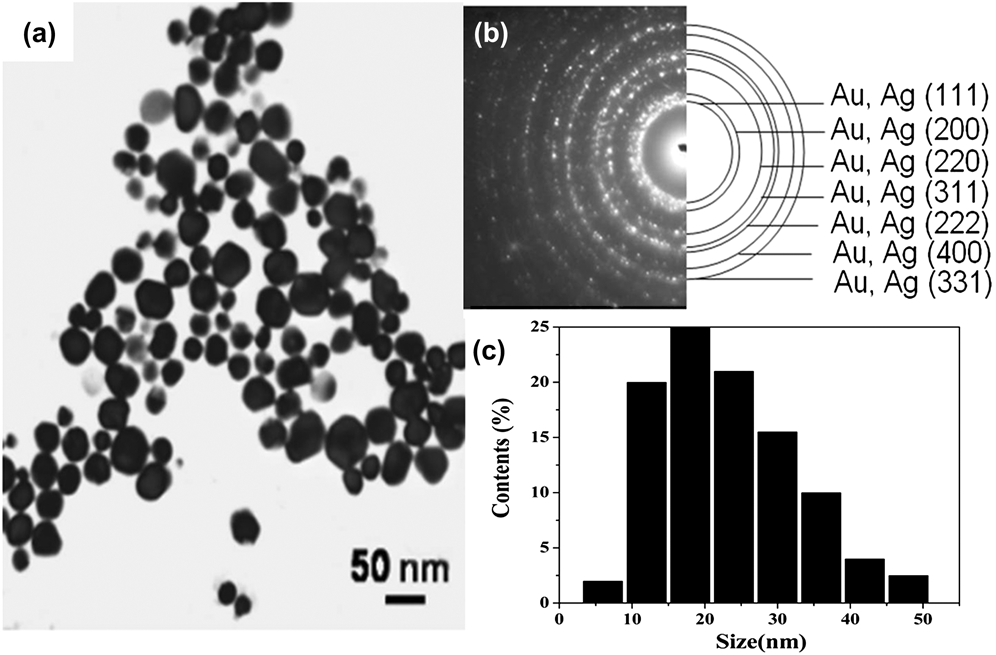
Figure 3 shows the TEM image of Au0.1Ag0.1/(SiO2)0.8 composite film. It can be seen that the considered Ag or Au NPs are almost in a spherical structure with large size distributions. The SAED pattern of the composite film in Fig. 3(b) shows the characteristic diffraction rings of metal Ag or Au, which from inner to outer correspond to the (111), (200), (220), (311), (222), (400), and (331) crystal planes of the cubic phases of Ag or Au. However, at this stage, it is hard to determine the existence form of the two metals in the composite film, whether it is in their alloy or in a separate NP mixed state, since Ag and Au have the same face-centered cubic structure with almost the same bulk lattice constant (Au: 4.0786 Å; Ag: 4.0862 Å).Reference Sun and Xia35Figure 3(c) shows the particle size distribution histogram corresponding to Fig. 3(a), which is mainly in the range of 10–30 nm, and the mean particle size is about 23 nm.
TEM micrograph and (a) the selected area electron diffraction pattern (b) of Au0.1Ag0.1/(SiO2)0.8 thin film, (c) the nanoparticle size distribution histogram of the sample corresponding to (a). The sample is prepared by spin-coating one Au0.1/(SiO2)0.4 film and one Ag0.1/(SiO2)0.4 film.
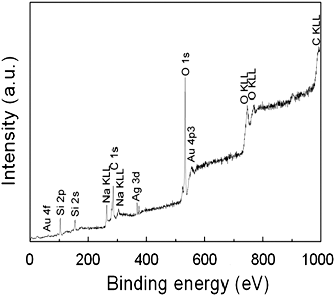
Figure 4 shows the XPS survey spectrum of Au0.1Ag0.1/(SiO2)0.8 composite film. The appearance of Au 4f, Au 4p, Ag 3d, Si 2p, Si 2s, O 1s, and O KLL photoelectron peaks confirm the existence of gold, silver, silicon, and oxygen species in the film. In addition, Na KLL, C KLL, and C 1s peaks are also observed in the spectrum, which originate from the diffusion of Na atoms that existed in the glass substrate into the sample surface during the annealing process and the air pollution or CO/CO2 adsorption on the sample surface, respectively. These are unavoidable for XPS analysis.
XPS survey spectrum of Au0.1Ag0.1/(SiO2)0.8 composite film. The sample is prepared by spin-coating four layers of Ag0.1/(SiO2)0.4 and four layers of Au0.1/(SiO2)0.4 film.
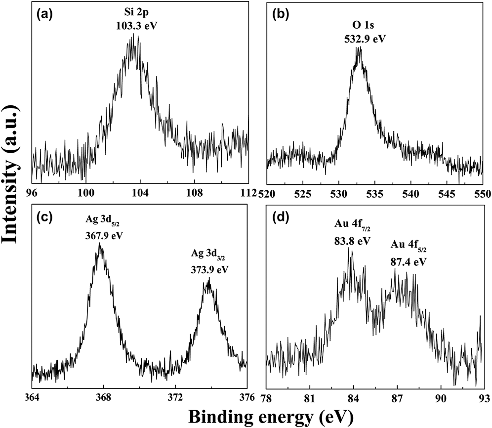
Figure 5 shows the high-resolution XPS spectra of (i) Si-2p, (ii) O-1s, (iii) Ag-3d, and (iv) Au-4f core level regions of Au0.1Ag0.1/(SiO2)0.8 composite film. In Figs. 5(a) and 5(b), the peaks centered at 103.3 and 532.9 eV belong to Si 2p and O 1s in SiO2.Reference Moulder, William and Peter36 The peaks located at 367.9 and 373.9 eV in Fig. 4(c) with a peak separation of 6.0 eV are attributed to Ag0 3d 3/2 and Ag0 3d 5/2, in spite of a little deviation from the values reported in the literature, i.e., the standard binding energies of Ag0 3d 3/2 and Ag0 3d 5/2 are 368.2 and 374.2 eV, respectively.Reference Moulder, William and Peter36 Similarly, two peaks at 83.8 and 87.4 eV in Fig. 5(d) can be referred to Au0 4f 7/2 and Au0 4f 5/2, respectively, in spite of a little deviation from the corresponding values of pure metallic Au (Au0 4f 7/2, 84.1 eV and Au0 4f 5/2, 87.7 eVReference Moulder, William and Peter36). The lower binding energies of Ag 3d and Au 4f may be attributed to a charge transfer between SiO2 and metal particles because the combination of Ag, Au, and SiO2 adjusts their Fermi level, similar to the case of the other metal-oxide composites.Reference Wang, Zhang, Yan and Deng37,Reference Sun, Qiao, Tan, Wang and Qiu38 However, the absence of the peaks located at 84.9 and 88.1 eVReference Sudipto and Goutam39 corresponding to the oxidized Au species implies that Au also remains in a metal state after coating the Ag0.1/(SiO2)0.4 layer. The above results indicate that Ag and Au NPs are formed in SiO2 film, and they exist in each metal state.
High-resolution XPS spectra of Au0.1Ag0.1/(SiO2)0.8 composite film: (a) Si 2p, (b) O 1s, (c) Ag 3d, and (d) Au 4f. The sample is prepared by spin-coating four layers of Ag0.1/(SiO2)0.4 and four layers of Au0.1/(SiO2)0.4 film.
The NPs formed in the composite film in its pure metal or alloy form can be exactly determined from its optical absorption spectra. In single metal dispersed system, the absorption spectra can be predicted by the well-known Mie scattering theory,Reference Mie40 which can be explained by Bohren and HuffmanReference Bohren and Huffman41 and have been calculated by various algorithms.Reference Du42–Reference Hosoya, Suga, Yanagawa and Kurokawa45 The absorbance (α) is proportional to the extinction cross section (σ), which can be obtained by the Mie scattering theory in the form asReference Hosoya, Suga, Yanagawa and Kurokawa45:
 $${\rm{\sigma }}\left( {\rm{\omega }} \right) = 9{{\rm{\omega }} \over c}{\rm{\varepsilon }}_{\rm{0}}^{3/2}{V_0}{{{\varepsilon _{{{\rm{m}}_{\rm{2}}}}}\left( {\rm{\omega }} \right)} \over {{{\left[ {{{\rm{\varepsilon }}_{{{\rm{m}}_{\rm{1}}}}}\left( {\rm{\omega }} \right) + 2{{\rm{\varepsilon }}_0}\left( {\rm{\omega }} \right)} \right]}^2} + {\rm{\varepsilon }}_{{{\rm{m}}_{\rm{2}}}}^{\rm{2}}}}\quad ,$$
where c,  ${\rm{\omega }}\left( {{\rm{\omega }} = {{2{\rm{\pi }}} \over {\rm{\lambda }}}} \right)$, and
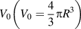 ${V_0}\left( {{V_0} = {4 \over 3}{\rm{\pi }}{R^3}} \right)$ are the wave vector, angular frequency, and particle volume; λ and R are the wave length and the metal NP's radii; ε0 and
 ${{\rm{\varepsilon }}_{\rm{m}}}\left( {{{\rm{\varepsilon }}_{\rm{m}}} = {{\rm{\varepsilon }}_{{{\rm{m}}_1}}} + i{{\rm{\varepsilon }}_{{{\rm{m}}_2}}}} \right)$ are the dielectric functions of the matrix and metal particle, respectively.
 ${{\rm{\varepsilon }}_{{{\rm{m}}_{\rm{1}}}}} = {n_{\rm{m}}} - {k_{\rm{m}}}$,
 ${{\rm{\varepsilon }}_{{{\rm{m}}_{\rm{2}}}}} = 2{n_{\rm{m}}}{k_{\rm{m}}}$, where n m and k m are the refraction index and the extinction coefficient of the metal, respectively. For inorganic materials
 ${{\rm{\varepsilon }}_0} = n_{\rm{s}}^2$, n s is the refractive index of SiO2. Therefore, α can be expressed asReference Belotelov, Carotenuto, Nicolais, Longo, Pepe, Perlo and Zvezdin9:
 $${\rm{\alpha }}\left( {\rm{\lambda }} \right) = 24{{\rm{\pi }}^2}n_{\rm{s}}^3{{{{\rm{\varepsilon }}_{{{\rm{m}}_{\rm{2}}}}}} \over {{{\left[ {{{\rm{\varepsilon }}_{{{\rm{m}}_{\rm{1}}}}}\left( {\rm{\omega }} \right) + 2n_{\rm{s}}^2} \right]}^2} + {\rm{\varepsilon }}_{{{\rm{m}}_{\rm{2}}}}^{\rm{2}}}}{R^3}\quad .$$
For the bimetallic system, considering, the system is obtained by layer-by-layer coating and step-by-step annealing, Au and Ag NPs distribute in different layers; hence, the absorption coefficient of AxBy/(SiO2)z composite film can be expressed as:
 $${\rm{\alpha }}\left( {\rm{\lambda }} \right) = {{\rm{\alpha }}_{\rm{A}}}\left( {\rm{\lambda }} \right) + {{\rm{\alpha }}_{\rm{B}}}\left( {\rm{\lambda }} \right)\quad ,$$
 $${{\rm{\alpha }}_{\rm{A}}}\left( {\rm{\lambda }} \right) = {{24{{\rm{\pi }}^2}xn_{\rm{s}}^3} \over {{\rm{\lambda }}\left( {x + y + z} \right)}}{{{\rm{\varepsilon }}_{{{\rm{m}}_2}}^{\rm{A}}} \over {{{\left[ {{\rm{\varepsilon }}_{{{\rm{m}}_{\rm{1}}}}^{\rm{A}} + 2n_{\rm{s}}^2} \right]}^2} + {\rm{\varepsilon }}_{{{\rm{m}}_2}}^{{{\rm{A}}^2}}}}R_{\rm{A}}^3\quad ,$$
 $${{\rm{\alpha }}_{\rm{B}}}\left( {\rm{\lambda }} \right) = {{24{{\rm{\pi }}^2}yn_{\rm{s}}^3} \over {{\rm{\lambda }}\left( {x + y + z} \right)}}{{{\rm{\varepsilon }}_{{{\rm{m}}_{\rm{2}}}}^{\rm{B}}} \over {{{\left[ {{\rm{\varepsilon }}_{{{\rm{m}}_1}}^{\rm{B}} + 2n_{\rm{s}}^2} \right]}^2} + {\rm{\varepsilon }}_{{{\rm{m}}_2}}^{{{\rm{B}}^2}}}}R_{\rm{B}}^3\quad ,$$
where αA, αB, and α are the optical absorption coefficients of A metal, B metal, and their total, respectively. The x, y, and z denote the molar fractions of the constituent materials, respectively, and x + y + z = 1. The R A and R B represent metal particle's radii. Here, the n m and k m of Au and Ag are cited from Ref. Reference Palik46. The particle radii used here is 20 nm, according to the TEM results.
Figure 6 shows the theoretical and experimental optical spectra of Ag0.1/(SiO2)0.4, Au0.1/(SiO2)0.4, and Au0.1Ag0.1/(SiO2)0.8 composite films. In theory, as shown in Fig. 6(a), the peaks related to the SPR of Ag and Au particles are revealed at about 410 and 515 nm, respectively. If bimetal NPs dispersed system is formed in Au0.1Ag0.1/(SiO2)0.8 composite film, two SPR peaks at 410 and 515 nm resulting from Ag and Au NPs are expected to be observed.Reference Sanchez-ramirez, Pal, Nolasco-Hernandez, Mendoza-Alvarez and Pescador-Rojas47 While, if Ag–Au alloy is formed in the composite film, there will be only one SPR peak and its position is revealed in-between the SPR peak positions of monometal Ag (410 nm) and monometal Au (515 nm) dispersed systems.Reference Sangpour, Akhavan and Moshfegh48Figure 6(b) shows the optical absorption spectra in experiment of Ag0.1/(SiO2)0.4, Au0.1/(SiO2)0.4, and Au0.1Ag0.1/(SiO2)0.8 composite films. For Ag0.1/(SiO2)0.4 and Au0.1/(SiO2)0.4 films, the SPR peaks appeared around 410 and 540 nm, respectively. For Au0.1Ag0.1/(SiO2)0.8 composite film, both peaks clearly appeared and confirmed the formation of bimetal NPs rather than alloy NPs. Notably, the two peaks have nearly equal intensity, which is rarely obtained by a chemical approach. In literature reports based on Au–Ag bimetal dispersed system, one SPR peak (usually Ag SPR peak or the peak location between Au and Ag) or two peaks with one in the form of a shoulder peak are shown. In our present work, two peaks with almost the same intensity are obtained. This result indicates that layer-by-layer coating and step-by-step annealing method affords a new opportunity for the preparation of bimetal NPs dispersed composite films.
Optical absorption spectra of Au0.1Ag0.1/(SiO2)0.8 composite film: (a) in theoretical and (b) in experimental. The sample is prepared by spin-coating one Au0.1/(SiO2)0.4 film and one Ag0.1/(SiO2)0.4 film.
Furthermore, the SPR peaks originating from Au NPs in Au0.1/(SiO2)0.4 and Au0.1Ag0.1/(SiO2)0.8 films show a red shift compared with the calculated one accordingly, maybe caused by large size distributions of Au NPs. It is known that the SPR frequency depends on the size, content, shape of the metal particles, and the dielectric properties of metal particles along with the surrounding matrix material.Reference Wang, Zhang, Yan and Deng37 Since Mie theory considers a case of perfect sphere metal particles with the same size and uniform distribution, it does not take into account the effects of the particle sizes, their distribution and the state of surrounding dielectrics.Reference Wang, Zhang, Yan and Deng37 However, it is reported that the size of Ag particles would induce the shift of the SPR peak when the size of particles becomes larger than 15 nm.Reference Kreibig and Frragstein49 The position shift and even the appearance of a shoulder at higher wave length were also observed when the silver nanocluster had two very different sizes.Reference Martinez-Castanon, Martinez, Zarzosa and Ruiz50 Likewise, as the size of Au NP increases, a red shift of SPR peak can be observed in the Au/SiO2 films, which is attributed to the enhancement of the screening of the d-electrons resulting from the decreasing surface-to-volume ratio of particles.Reference Lamarre, Yu, Harkati, Roorda and Martinu51 In fact, as shown in Fig. 3(a), the metal NPs have analogous special structure with large size distributions, but it is difficult to distinguish Au and Ag particles between them. But considering the Au/SiO2 film was annealed twice, at 190 and 500 °C, this annealing process may be caused by the larger particle size, which may induce a red shift of the experimental optical absorption spectra.
In addition, the relative intensity of Ag SPR peak for Au0.1Ag0.1/(SiO2)0.8 composite film is weaker than the calculated one, which may be caused by the experimental factors. In our present work, Au0.1Ag0.1/(SiO2)0.8 composite film is obtained by spin-coating Au0.1/(SiO2)0.4 and Ag0.1/(SiO2)0.4 layers one after another, so Au0.1/(SiO2)0.4 film is on the surface of glass substrate and primarily absorbs the incident light. This is because the incident light is illuminated from the glass substrate side. The intensity of incident light can be reduced due to the scattering and absorption phenomenon of Au0.1/(SiO2)0.4 film, which in turn gives rise to relative weaker intensity of incident light used for Ag0.1/(SiO2)0.4 film. Indeed, the calculated optical absorption does not consider these experimental factors; thus, the intensity of Ag SPR peak for Au0.1Ag0.1/(SiO2)0.8 composite film weakens to the calculated one. Just because of these experimental factors, it is possible to control the experiment condition and obtain two SPR peaks with almost the same intensity. However, it is reported that the particle size not only affects the position of SPR peak, but also the intensity and width.Reference Zhao, Zhang and Shang52,Reference Haruta53 When the particle size is larger to some extent, the SPR peak will be weak and broad.Reference Zhao, Zhang and Shang52 Taking into the account the large size distributions of metal NPs in the film, therefore, in addition to the experimental factors, the large particle size may be another considering factor contributing to the weaker intensity. Moreover, accompanied with the relative intensity change is also the broadening of the SPR peaks of Au0.1/(SiO2)0.4, Ag0.1/(SiO2)0.4, and Au0.1Ag0.1/(SiO2)0.8 composite films, which can be attributed to the broader distribution of their NPs.Reference Martinez-Castanon, Martinez, Zarzosa and Ruiz50
Figure 7(a) shows the optical absorption spectra of the AuxAg0.2−x/(SiO2)0.8 composite films, with x values from 0.02 to 0.1. All samples exhibit two absorption bands around 410 and 540 nm due to the SPR effect of Ag and Au NPs. The intensities of both peaks depend on their initial content. The intensity of the dominant peak located at 410 nm intensifies with increasing Ag content. A second peak at 540 nm develops with increasing Au content. The second peak appears as a shoulder in the relative lower content Au sample but is clearly discernible in the highest Au content sample. The variational trend of absorption spectra as the contents of two metal NPs is in accordance with the calculated one by the modified Mie theory [as shown in Fig. 7(a)]. Besides, the SPR peaks manifest a red shift trend with increasing Au content, given by the quantum size effect.Reference Wang, Zhang, Yan and Deng37,Reference Wang, Lau and Li54
Optical spectra of AuxAg0.2−x/(SiO2)0.8 composite films with different x values: (a) in theoretical and (b) in experimental. The sample is prepared by spin-coating one Aux/(SiO2)0.5−x film and one Agy/(SiO2)0.5−y film.
IV. CONCLUSION
In summary, we have prepared Ag–Au bimetal dispersed separate SiO2 composite films by a chemical solution approach combining a sol–gel with a spin-coating process and studied their SPR properties. The metal NPs have a spherical structure with large size distributions in the composite film. The presence of Ag and Au NPs in the composite film was confirmed in each metal state by XPS and UV-vis absorption spectra. It is noticeable that the two SPR peaks obtained in our work have almost the same intensity, which is rarely obtained by a chemical approach. Optical absorption spectra obtained from experimental ones accord well with the theoretical calculations except for a little difference in the location and intensity of SPR peaks. The intensity of both SPR peaks was also content-related. Increasing the metal's content intensifies the corresponding SPR peak.
ACKNOWLEDGMENTS
This work was supported by Beijing Natural Science Foundation of China (Grant No. 2112028), National Natural Science Foundation of China (Grant No. 51202011) and the Fundamental Research Funds for the Central Universities (FRT-TP-13-009A).