Introduction
Assessment of tumour extension by morphological criteria is known to be inadequate. Diagnosis by clinical examination is dependent on the experience of the physician, and requires a minimum tumour size. Conventional imaging such as computed tomography (CT) and magnetic resonance imaging (MRI) provide structural information; however, the contrast between muscular, connective and cancerous tissue is sometimes difficult to assess.
Tumours of the larynx are particularly difficult to interpret, and there are known inter-observer differences when estimating the tumour volume. Metastatic lymph nodes are usually assessed depending on their size (e.g. 8–15 mm) and shape (e.g. round), and there is the risk of missing small tumours.
The use of positron emission tomography (PET) scanning has a series of theoretical advantages. It enables imaging of metabolically active tissues by using 18-fluorine-fluoro-2-deoxyglucose (18F-FDG), a glucose analogue absorbed in a manner proportionate to the glycolytic activity of tissues (such activity is high in malignant neoplasms).Reference Wong, Lin, Schoder, Patel, Gonen and Wolden1 The increased uptake of FDG by cancer cells permits their differentiation from normal tissues, even in cases of post-treatment fibrosis or oedema. The association of 18F-FDG PET with CT scanning provides the necessary anatomical information to plan appropriate treatment.Reference Fukui, Blodgett and Meltzer2 The reported sensitivity and specificity of 18F-FDG PET for the detection of malignant head and neck tumours vary between 81 and 89 per cent and 38 and 95 per cent, respectively.Reference Hanasono, Kunda, Segall, Ku and Terris3, Reference Gordin, Golz, Keidar, Daitzchman, Bar-Shalom and Israel4
The aims of the present study were to determine: (1) the accuracy of PET-CT in the diagnosis of head and neck cancer; (2) the learning curve involved; and (3) whether the PET-CT results altered patient management.
Materials and methods
We designed a retrospective study of 80 patients with head and neck cancer who underwent PET-CT image fusion at Blackpool Victoria Hospital, Manchester. Patients were imaged between April 2006 and December 2009, and were identified from the PET-CT database.
The indications for PET-CT were divided into two categories: (1) tumour staging, including the identification of an unknown primary; and (2) detection of loco-regional recurrence.
Inclusion criteria
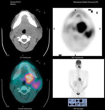
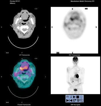
All patients had histological confirmation of carcinoma of the upper aero-digestive tract and/or metastatic cervical lymph nodes. All patients underwent high-resolution contrast-enhanced MRI or CT imaging of the neck and chest, followed by whole body PET-CT (Figures 1 and 2).
Screengrab showing head imaging: axial computed tomography (CT) scan (top left); axial positron emission tomography (PET) – CT scan showing increased fluoro-2-deoxyglucose uptake in the left oropharynx (top right); axial PET scan (bottom left); and coronal whole-body PET-CT scan (bottom right). Biopsy of the tonsil confirmed squamous cell carcinoma.

Screengrab showing head imaging: axial computed tomography (CT) scan (top left); axial positron emission tomography (PET) – CT scan showing increased fluoro-2-deoxyglucose uptake in the base of the tongue (top right); axial PET-CT scan (bottom left); and coronal whole-body PET-CT scan (bottom right). Biopsy of the tongue confirmed squamous cell carcinoma.

Exclusion criteria
We excluded patients with benign tumours, those in whom the primary tumour was found to be outside the head and neck, and those who had not undergone conventional imaging.
Technique
Patients were injected with FDG after a 6-hour fasting period. This was to ensure a low serum insulin level and therefore to minimise uptake into muscle, fat and myocardium. A whole body PET and non-contrast enhanced CT (GE discovery VCT system) with 64-slice CT capability) were performed in all patients 60 minutes after the injection of FDG.
After registration of the PET and CT scans, images were reconstructed in 3.2 mm slices for interpretation in axial, sagittal and coronal planes. The MRI, CT and PET-CT images were reported by two radiologists with an interest in head and neck pathology.
Data analysis
The biopsy, MRI, CT and PET-CT imaging results were correlated. Sensitivity, specificity, positive predictive value and negative predictive value were evaluated.
Results
Eighty PET-CT studies were reviewed. Thirty-two studies were performed for patients presenting with a neck mass, who underwent imaging for primary tumour diagnosis and accurate tumour staging, 21 were for staging in the presence of metastasis, and 27 were for detection of loco-regional recurrence.
In the 32 patients presenting with a neck mass, a total of 14 (44 per cent) unknown primaries were diagnosed, 10 (31 per cent) by PET-CT scan (Figures 1 and 2) and six (19 per cent) by conventional imaging (i.e. MRI or CT). Three (9 per cent) had a positive biopsy (no supporting positive imaging) and 18 (56 per cent) were not identified by imaging.
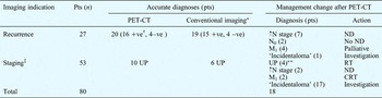
Of the 27 patients assessed for recurrence, 20 (74 per cent) were accurately diagnosed by PET-CT (16 positive and four negative) and 19 (70 per cent) were accurately diagnosed by conventional imaging (15 positive and four negative).
Eighteen patients had their original diagnostic stage modified, with a change of management, due to information obtained by PET-CT (Table I): sixteen had their diagnosis upgraded and two had their diagnosis downgraded with a change of management. Of these, four unknown primaries and six recurrences were diagnosed by PET-CT exclusively. Of the total group of 80 patients, PET-CT scanning led to an increase in nodal stage, resulting in neck dissection, in nine patients, while six patients were diagnosed with distant metastases resulting in palliative treatment. When searching for an unknown primary, PET-CT helped direct the biopsy, reducing the number of blind biopsies by 42 per cent.
Influence of PET-CT on H&N cancer management

*Computed tomography (CT) or magnetic resonance imaging. †Six recurrences diagnosed by postitron emission tomography – CT (PET-CT) only. ‡For investigation of unknown primary tumours (UP) and metastases. **Diagnosed by PET-CT only. H&N = head and neck; pts = patients; +ve = positive; –ve = negative; ↑= increased; N = node; ND = neck dissection; N0=node-negative; M1 = metastasis stage 1; RT = radiotherapy; CRT = chemo radiotherapy
Learning curve
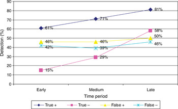
Patients were divided into three groups: 26 patients whose imaging was undertaken in the early period of using PET in the region (i.e. April 2006 to December 2007), 28 patients imaged in the middle period (January 2008 to December 2008) and 26 patients imaged in the last period (January 2009 to December 2009). There was a significant improvement in the true positive detection rate in the later group, compared with Conventional imaging (61 versus 81 per cent) (Figure 3).
The effect of the positron emission tomography – computed tomography learning curve on head and neck cancer detection. Note the improvement in detection of true positives (81 vs 61 per cent) and true negatives (58 vs 15 per cent), compared with early results; however, detection of false positive and false negative cases continued to be a problem.

Imaging accuracy and predictive value
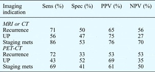
For PET-CT, the sensitivity and specificity were 70 and 42 per cent, respectively. The positive and negative predictive values were 60 and 53 per cent, respectively. For conventional imaging (i.e. either MRI or CT), the sensitivity and specificity were 73 and 51 per cent, respectively, and the positive and negative predictive values were 73 and 51 per cent, respectively. When divided by imaging indication (Table II), both imaging modes had poor sensitivity for detecting unknown primaries, but good sensitivity for detecting recurrence.
Accuracy and predictive value for MRI or CT, and PET-CT

MRI = magnetic resonance imaging; CT = computed tomography; PET = positron emission tomography; sens = sensitivity; spec = specificity; PPV = positive predictive value; NPV = negative predictive value; UP = unknown primary; mets = metastases
Positron emission tomography – computed tomography detected 66 per cent of the nodes found to be positive by histology, whereas conventional imaging detected 58 per cent.
‘Incidentalomas’
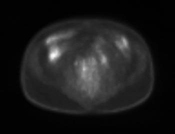
‘Incidentalomas’ (unexpected findings), independent of the head and neck tumour, were found in 18 of the 80 patients (23 per cent), nine of which needed investigation, resulting in delayed treatment (Table I and Figure 4).
Axial positron emission tomography – computed tomography abdominal scan, showing fluoro-2-deoxyglucose uptake in the ascending colon (right side). Colonoscopy showed villous adenoma.

Discussion
Treatment for head and neck cancer, in particular for advanced disease, still demands substantial functional, cosmetic and nutritional sacrifices in exchange for improved survival. Such morbidity may be limited by tailoring treatment as accurately as possible to the location and severity of the disease. Radiological examination is consequently of great importance in the assessment of patients with cancer in the head and neck, in whom the interpretation of findings is made specifically difficult by the increased uptake of contrast media in normal tissue, such as lymphoid tissue and the thyroid and salivary glands.
Findings in the literature
Positron emission tomography uses FDG. In the head and neck, PET can be used for a variety of indications, including whole body staging, mapping of tumour location and volume for radiotherapy or surgical treatment planning, detection of residual or recurrent disease, identification of an unknown primary, and post-treatment surveillance and re-staging.Reference Wong, Lin, Schoder, Patel, Gonen and Wolden1
The utility of conventional imaging techniques (e.g. CT and MRI) depends on criteria such as lymph node size and contrast enhancement. The interpretation of such features is limited by infection, tissue fibrosis, radiotherapy-related oedema, scarring and/or inflammation.Reference Sigg, Steinert, Gratz, Hugenin, Stoeckli and Eyrich5, Reference Curtin, Ishwararn, Mancuso, Dalley, Caudry and McNeill6 Positron emission tomography, however, is not limited by the formal size criteria (i.e. 8–15 mm) required to identify nodal metastasis; however, FDG can concentrate in areas of infection, inflammation and normal lymphoid tissue. Hence, focal asymmetric uptake is suggestive of a malignant process but non-specific.Reference Sigg, Steinert, Gratz, Hugenin, Stoeckli and Eyrich5–Reference Paulus, Sambon, Vivegnis, Hustinx, Moreau and Collignon7
Cervical node metastasis is an important prognostic factor. Management of the node-negative neck is still controversial. In a meta-analysis by Kyzas et al.,Reference Kyzas, Evangelou, Denaxa-Kyza and Ioannidis8 the sensitivity and specificity of detecting occult metastasis (in node-negative patients) using CT, MRI and PET scaning were as follows: for CT the sensitivity was 14–80 per cent and specificity 80–100 per cent, for MRI the sensitivity was 29–85 per cent and specificity 80–100 per cent, and for PET the sensitivity was 50 per cent and specificity 87 per cent. This confirms the notion that PET alone does not provide suitable guidance to avoid elective neck dissection in node-negative patients.
When we reviewed the above parameters for node-negative plus node-positive patients, several studies suggested improved accuracy for PET, with a sensitivity and specificity of 79–100 and 86–100 per cent, respectively, compared with a CT sensitivity of 82–89 per cent and specificity of 82–93 per cent, and an MRI sensitivity of 80 per cent and specificity of 79 per cent.Reference Kyzas, Evangelou, Denaxa-Kyza and Ioannidis8–Reference Martin, Fulham, Shannon, Hughes, Milross and Tin12
Primary tumour sites are normally detected by clinical and endoscopic examination, so PET is of less interest. After surgery or radiotherapy, diagnosis by examination and morphological imaging can be complicated due to post-therapy tissue reaction. If the patient has responded to treatment there will be a reduction in metabolic activity, but there might be minimal difference in uptake due to inflammation, and in some cases there can even be some increase in uptake early after treatment.Reference Weber13, Reference Wieder, Brucher, Zimmermann, Becker, Lordick and Beer14 Positron emission tomography assessment of response to therapy requires comparison with a pre-treatment scan.Reference Weber13 In the study by Wieder et al.,Reference Wieder, Brucher, Zimmermann, Becker, Lordick and Beer14 after effective chemo-radiotherapy, the uptake of FDG was reduced after one to three weeks. In a study by Martin et al.,Reference Martin, Fulham, Shannon, Hughes, Milross and Tin12 tumour recurrence was more accurately assessed if PET was performed at least 12 weeks after completing therapy.
Panendoscopy studies have shown the prevalence of synchronous primaries to be between 1.4 and 17 per cent. Due to limited special resolution, small and superficial growing tumours in the upper aero-digestive tract are missed by PET, so this imaging technique will not replace endoscopic examination and biopsy in these cases.Reference Lan, Zhang, Wu, Jia, Wei and Gao15 In a study by Strobel et al.,Reference Strobel, Haerle, Stoeckli, Schrank, Soyka and Veit-Haibach16 of 589 patients with head and neck cancer, 56 had confirmed synchronous primaries and PET diagnosed 93 per cent.
Strengths of the study and key findings
Our study evaluated PET-CT investigations of the head and neck undertaken in one centre only, and image reporting was undertaken by two radiologists with an interest in the head and neck. All patients had histological diagnoses.
We included primary tumours and recurrence, as well as metastasis. Patients with unknown primaries formed a significant proportion (32 of 80, 40 per cent) of those included in comparison with other studies.Reference Hanasono, Kunda, Segall, Ku and Terris3 In our study, 14 of 32 unknown primary lesions (44 per cent) were diagnosed by a variety of techniques; only four (13 per cent) were diagnosed exclusively by information provided by PET-CT. Imaging tools failed to identify 18 (56 per cent) primary tumours.
Although the literature suggests that PET is superior in detecting recurrence, in our study the difference between PET and conventional imaging was not significant. Twenty possible recurrences were evaluated accurately by PET-CT (16 positive and four negative), but only six (22 per cent) patients benefited from information exclusively from PET-CT. Combined PET-CT recognised 65 per cent of the nodes found to be positive by histology, whereas MRI plus CT recognised 58 per cent, but the difference was not significant.
Clinical relevance
The detection of an occult primary tumour usually results in a reduction in the volume of tissue irradiated. In this study, PET-CT failed to provide a diagnosis in 69 per cent of cases. Consequently, we found that the diagnosis of an occult primary tumour still required a combination of techniques (i.e. conventional imaging and endoscopic techniques).
Our findings showed that PET-CT has a sensitivity of 70 per cent and a specificity of 42 per cent in the detection of head and neck cancer. Therefore, if no cervical nodes are found on PET-CT, neck dissection is still indicated. This is particularly so if there is a non-FDG avid node larger than the accepted criteria used during CT scanning, or if the primary has a high incidence of occult metastasis. However, a positive study cannot be disregarded and can guide a biopsy more accurately, reducing the number of blind biopsies.
If PET-CT is carried out after treatment, to determine the response, we concur with the literature which states that imaging too early will result in frequent false positive results.Reference Martin, Fulham, Shannon, Hughes, Milross and Tin12, Reference Wieder, Brucher, Zimmermann, Becker, Lordick and Beer14 Consequently, we suggest a three-month wait.
An important point to consider is that the use of FDG PET-CT in the diagnosis of head and neck pathology has specific difficulties, due to large areas of physiological uptake, such as lymphoid tissue and the thyroid and salivary glands. Consequently, a combination of diagnostic techniques is commonly required to reach a diagnosis.
• This study found a high failure rate of positron emission tomography – computed tomography (PET-CT) in detecting occult primary tumour (69 per cent), necessitating combination with different techniques
• Alone, PET-CT did not provide suitable guidance to avoid neck dissection in node-negative patients
• It was more reliable in detecting recurrence (74 per cent diagnosed)
• Early PET-CT after treatment leads to frequent false positive results
• We recommend waiting for three months after treatment before PET-CT imaging
As with any new technique, there is a learning curve associated with the use of PET-CT. Experience will result in a higher detection rate of true positives, as seen in Figure 3 (61 vs 81 per cent), and a more reliable interpretation of physiological uptake and ‘incidentalomas’ (23 per cent in our study).
Conclusion
Although the learning curve has an important effect on its reliability, PET-CT is an effective diagnostic tool and plays an important role in the management of head and neck cancers. However, compared with MRI and CT its benefits may be limited to diagnosis of recurrence, as it is less hindered by tissue fibrosis, radiotherapy-related oedema, scarring and inflammation.