Introduction
‘Dead ear’ is a term used to describe profound sensorineural hearing loss with or without vertigo. When dead ear occurs after ear surgery, it is probably due to damage to delicate inner-ear structures as a result of mechanical manipulation, transmitted vibration or direct surgical disruption. Few complications of ear surgery are likely to cause more consternation. It is a problem said to occur following approximately 1 per cent of operations for otosclerosis, for cholesteatoma, and for chronic otitis media without cholesteatoma.
Dead ear is routinely mentioned in the consent process for middle-ear operations.
The dead ear may be predictable or come as a surprise. The despair felt by the patient and the surgeon after such an event is ironic, since one of the initial indications for the operation is to prevent the ear pathology from causing precisely this problem.
The low incidence of post-operative dead ear means that individual surgeons, especially general otolaryngologists treating only a small number of cholesteatoma cases, may be fortunate enough never to encounter this complication. However, few experienced surgeons will have encountered no cases of dead ear.
This report presents an audit of dead ear in a series of cholesteatoma cases from two hospitals in the county of Norfolk, UK.
Method
In order to identify patients who had developed dead ear post-operatively, the author scrutinised records compiled as part of the International Otology Audit (see http://www.ear-audit.net).Reference Van Rompaey, Yung and Van Heyning1 These records represented all otology patients operated upon under the care of the author at James Paget University Hospital and the Norfolk and Norwich University Hospital, Norfolk, between 2006 to 2012.
The International Otology Audit is a prospective, web-based audit of otology procedures which collects information about the nature and extent of disease and the operations performed. The audit also gathers prospective data about the post-operative course of the patient in the months and years after the procedure. It is free to use and is supported by ENT UK, the professional association of British ENT surgeons.
Analysis of the case records considered the following questions: (1) what was the incidence of dead ear in relation to the procedures performed?; (2) was the cause of the dead ear apparent, and if so was it predictable and avoidable?; (3) what was the impact of the dead ear on the patient, particularly with respect to hearing in the opposite ear?; (4) what rehabilitation was offered and what was the benefit?; and (5) was there any litigation as a result of the dead ear?
Results
The audit included 617 cases, covering the period 2006 to 2012. There were 83 cases of otosclerosis treated with stapedectomy, 254 cases of cholesteatoma (192 adults and 62 children under 16 years), and 280 cases treated for chronic otitis media without cholesteatoma by means of tympanoplasty or myringoplasty. Five of the 254 audit patients treated for cholesteatoma had a dead ear pre-operatively as a result of their cholesteatoma; these 5 cases were excluded from analysis, leaving 249 cases of cholesteatoma in the final calculations (187 adults and 62 children).
Six cases of post-operative dead ear were identified, giving an overall prevalence of 0.98 per cent. There were no cases of dead ear after the 83 stapedectomy operations. In 5 of the 6 post-operative dead ear cases, cholesteatoma was found, giving a prevalence of 5 post-operative dead ears amongst the 249 included cholesteatoma cases (2 per cent). One of the 280 chronic otitis media cases (0.36 per cent) suffered a post-operative dead ear but showed no peri-operative cholesteatoma, merely tympanic membrane collapse and granulations. There were no dead ears amongst the 62 children operated upon for cholesteatoma. Of the 187 included adults undergoing mastoidectomy for cholesteatoma, 5 had a post-operative dead ear, a prevalence of 2.7 per cent.
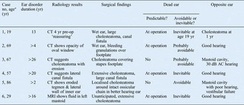
The individual records of the six patients with a post-operative dead ear were scrutinised. A case summary is presented below for each of these patients. The most salient features of all six cases are shown in Table I.
Case summary

*At ear surgery. Yr = years; CT = computed tomography; pre-op = pre-operatively; AC = air conduction; MRI = magnetic resonance imaging
Case one
The first patient was 19 years old and had cerebral palsy and learning difficulties. This patient had presented at the age of six years with deafness, treated with grommets; this presentation was repeated twice over the next few years.
At the age of 15 years, the patient had re-presented with a history of right ear discharge. Examination had revealed a dry attic crust on the right and a grommet in the left ear canal. Audiography had shown bilateral conductive hearing loss, worse on the right, to 50 dB average air conduction threshold. A computed tomography (CT) scan at this time had shown a normally pneumatised mastoid on the right side with a poor view of the tympanic membrane as a result of retraction. There had been sclerosis of the incudomalleolar joint but no definite radiological support for a diagnosis of cholesteatoma. The left side had shown opacity of the mastoid and attic but no evidence of bone destruction. In view of these radiological findings, and the fact that the discharge had settled, a conservative approach had been adopted. The patient had been lost to follow up.
The patient re-presented again aged 17 years, and examination under anaesthesia showed an attic cholesteatoma on the right. A CT scan was arranged but not performed, and the patient was not seen again until the following year.
At the age of 19 years, the patient was under the care of a neurologist for the investigation of ataxia. A magnetic resonance imaging (MRI) scan at this time showed a high T2 diffusion weighted signal in the right temporal bone, in keeping with cholesteatoma. Audiometry showed a 40 dB average air conduction threshold for the right ear.
The patient was admitted for mastoid surgery. At surgery, in addition to a wet ear, there was extensive cholesteatoma with a large defect in the posterior external auditory canal wall. The incus was absent and the stapes was buried in the cholesteatoma matrix. Elevation of the cholesteatoma matrix revealed a large defect in the lateral semicircular canal. The VIIth cranial nerve was widely exposed, and the disease was peeled off the perineural layer. An open cavity mastoidectomy was carried out, and a small cartilage and soft tissue plug was placed over the canal fistula. Fascia and cartilage were used for tympanoplasty.
The patient was dizzy for several days after the operation. Although the cavity healed well, the patient had a dead right ear. Subsequently, there was progression to cholesteatoma on the left side, with deterioration of hearing.
Observations on case one
This patient's dead ear was caused by removal of cholesteatoma from the eroded inner-ear structures. The occurrence of dead ear was predictable during the operation. It is tempting to think that the patient's cholesteatoma might have been avoided if it had been treated earlier. Four years before surgery, the diagnosis was ‘excluded’ by CT, and two years before surgery the patient did not have a requested CT performed, for unknown reasons, despite a clinical diagnosis of cholesteatoma. There was an over-reliance on CT scanning in this case. Surgery was delayed for two years, and was eventually prompted by results from a MRI head scan performed for other reasons. No further pre-operative CT scanning was performed; had it been, the surgeon might have been forewarned of the likely fistula to the labyrinth. The development of a dead ear was inevitable by this time, whether or not surgery was performed.
The patient was vertiginous for some weeks post-operatively. Assessment was problematic because of the patient's learning difficulties. One year post-operatively, the patient had hearing loss and cholesteatoma on the opposite side, and was wearing a hearing aid in the surviving ear.
The situation was fully explained to the patient, as far as it could be understood, and also to the family. There was no complaint or litigation.
Case two
This 65-year-old patient presented complaining of pressure in the left ear and intermittent discharge. Examination revealed a posterior tympanic membrane retraction pocket and granulations with a small anterior perforation. The patient was treated with topical antibiotic and steroid drops. The ear continued to discharge over a number of visits to the clinic. The patient was anticoagulated because of aortic stenosis, and a conservative approach to his ear disorder was adopted. The patient also had diabetes, controlled with oral hypoglycaemics.
At the age of 69 years, the patient underwent aortic valve replacement. The left ear continued to discharge. A CT scan of the temporal bone revealed a localised opacity filling the oval window, with no clear view of the stapes. The tympanic membrane was retracted but there was no radiological evidence of cholesteatoma, and the mastoid air cells were normal. Audiography showed left-sided hearing loss worse in the mid- and high tones.
An atticotomy and tympanoplasty were performed. The ear was wet, and the tympanic membrane was seen to have collapsed against the promontory and into the sinus tympani of the posterior middle ear. There was no cholesteatoma. The stapes footplate was identified together with a remnant of the posterior crus, but both were covered in bleeding granulations. As these were dissected from the oval window, the footplate was seen to lift slightly out of the oval window. This was replaced, with no obvious perilymph leak. The retracted tympanic membrane and granulations were removed from the ear. Limited drilling revealed a partially necrosed incus, which was not further disturbed. Cartilage and fascia were used to repair the tympanic membrane defect. No attempt at ossiculoplasty was made.
Post-operatively, the patient was dizzy for several weeks. Audiography revealed a dead left ear. Vestibular rehabilitation exercises were instituted and the patient's balance improved. At the time of writing, the patient's left ear continued to discharge intermittently, but less so than before the operation.
Observations on case two
This patient's dead ear was caused by elevation of the stapes footplate. After this happened, the development of a dead ear was predictable. The dead ear was probably avoidable, although it could not have been predicted that the strength of the granulation tissue attachment to the stapes remnant would be greater than the strength of the footplate attachment to the oval window. The CT scan showed opacity in the oval window, with stapes superstructure erosion, which alerted the surgeon.
The operation had been delayed for several years due to other medical problems, but the ear infection did not settle despite several years of out-patient treatment. The operation was performed to dry the ear and prevent complications. It achieved neither. Instead, there was complete loss of hearing in what had been the worst hearing ear. The patient's post-operative vertigo recovered with vestibular rehabilitation exercises.
The patient raised no complaint, and there was no litigation.
Case three
This patient presented at the age of 41 years with a history of right mastoid surgery many years earlier, and was found to have a large, right-sided cavity which was healthy, and a dry posterior retraction of the left tympanic membrane.
Eleven years later, aged 52 years, the patient re-presented with left ear discharge and pain. A posterior retraction was found, with a cholesteatoma which was cleaned by microsuction. It was noted that the patient ‘will require mastoid surgery’. The CT scan report at this time noted ‘intermediate attenuation material partially encasing the malleus and incus’. No surgery was performed. At a review some months later, the ear was seen to be dry and free of cholesteatoma. Hearing tests at this time showed normal right and left bone conduction thresholds. The average air conduction threshold was 30 dB on the right and 50 dB on the left. The patient was seen at 6- or 12-monthly intervals for the next several years for cleaning of the ears. Hearing was ‘adequate’ and no hearing aid was used.
At the age of 64 years, dry cholesteatoma matrix was noted in the left ear. This was removed under anaesthesia, to reveal a small cavity created by the disease (an ‘auto atticotomy’). Periodic out-patient follow up continued to the age of 67 years, by which time there was obvious cholesteatoma in the left ear. Audiography at this time showed an average air conduction threshold of 30 dB on the left and 30 dB on the right. A new CT was performed; the report noted soft tissue in the attic extending into the poorly pneumatised mastoid, with erosion of the scutum and the malleus and incus. There was thinning of the tegmen, and the horizontal facial nerve canal was intimately related to the soft tissue. The concluding diagnosis was a cholesteatoma with bone erosion as described.
In view of the clinical and CT findings, and after much thought by the patient and the surgeon, a mastoid operation was performed. The patient stated a clear preference at this time for any hearing improvement that might be surgically possible by means of ossicular reconstruction. Peri-operatively, an atticotomy and tympanoplasty were performed. The tympanic membrane was found to be collapsed on the promontory, with retraction into the sinus tympani. There was obvious cholesteatoma in the attic and aditus ad antrum. The ossicles were eroded, with absence of the incus long and short processes and the stapes arch. Cholesteatoma was covering the footplate and extending over the exposed horizontal facial nerve. The lateral semicircular canal was noted to be intact. The cholesteatoma was removed by gentle dissection, with no obvious mechanical trauma to the mobile stapes footplate. Atticotomy was performed and all disease removed. The drum was grafted with tragal cartilage and perichondrium, and a small strut of cartilage was placed between the graft and the footplate, supported by a ring of gelatin sponge. The ear was packed with sponge dressing and a bandage applied.
Eight weeks post-operatively, there was a small, well healed cavity. The patient suffered no post-operative vertigo. However, audiography revealed a dead left ear. The patient was fitted with a contralateral routing hearing aid.
Observations on case three
This was an unexpected and unanticipated dead ear. The cause was unknown, but perhaps the sponge packing of the ear caused the end of the cartilage to pass into the vestibule. It was unusual to use such sponge wicks to pack the ear post-operatively. The operation was carried out at a time when the normally used bismuth iodoform paraffin paste ribbon was unavailable as a result of a ‘national shortage’.
Surgery for cholesteatoma was long delayed in this patient; it had first been recommended some 15 years earlier. The operation revealed a destructive cholesteatoma which had caused erosion of the ossicular chain and the bone over the facial nerve. The operation resulted in one of the very same complications it was intended to prevent; however, one can only speculate on the likely course had no surgery been performed.
Case four
This patient presented at the age of 57 years with more than 20 years' history of an intermittently discharging left ear. Examination revealed an attic retraction with granulations. Over the following year, the patient was treated regularly with microsuction and topical antibiotic and steroid drops, but the discharge failed to settle.
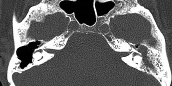
Audiography at this stage showed a 30 dB average air conduction threshold and 20 dB average bone conduction threshold in the left ear. Hearing in the right ear was normal. There was no vertigo. A CT scan revealed opacity throughout the left mastoid, with a suggestion of ossicular erosion and thinning or absence of the bone overlying the left lateral semicircular canal (Figure 1).
Axial computed tomography scan of case four, showing left incus erosion and a possible bony defect in the left lateral semicircular canal.

A tympanomastoidectomy was carried out. There was extensive cholesteatoma with ossicular erosion. As the matrix was slowly lifted away, a large (approximately 0.5 cm) defect was seen in the lateral semicircular canal. The defect was grafted with fascia and a small, open cavity created.
Post-operatively, the patient was vertiginous for several weeks. There was no hearing in the operated ear. The cavity healed well and there was no further discharge. A contralateral routing hearing aid proved very useful for this patient.
Observations on case four
A dead ear was perhaps inevitable at this stage of the patient's disease. The CT scan demonstrated the fistula well, and the dead ear was not a surprise post-operatively. If possible, an attempt to preserve cholesteatoma epithelium over the fistula is recommended,Reference Pulec2 but this was not practical in the present case. This patient, like several in the present series, had a very long history of aural discharge.
There was no complaint or litigation.
Case five
This 86-year-old patient presented with more than two years' history of intermittent vertigo and left purulent otorrhoea. A right mastoidectomy had been performed decades earlier. Examination revealed an attic retraction of the left tympanic membrane, which was infected and full of granulations and cholesteatoma. Audiography revealed a 55 dB average air conduction threshold on the left, with a minimal air–bone gap. On the right side, the average air conduction threshold was 85 dB. A CT scan of the temporal bones was reported as showing an erosive, soft tissue mass in the attic of the left ear with erosion of the ‘tegmen tympani and lateral wall of the inner ear at the level of the lateral semicircular canal and the facial nerve’ (Figure 2).
Axial computed tomography scan of case five, showing erosion of the left labyrinth and a large cavity on the right side. Note also probable erosion of the right lateral semicircular canal.

The dilemma was therefore how to manage an attic cholesteatoma with a CT suggestion of tegmen and inner ear erosion, in much the better hearing ear of a patient of 86 years.
The patient's discharge and episodic vertigo did not improve with topical treatments in the out-patient department.
It was decided to perform a mastoidectomy. This revealed cholesteatoma around the long process of the incus and the stapes superstructure. There was partial erosion of the incus body but the ossicular chain was intact. The attic and aditus were full of granulations but no further cholesteatoma. In view of this, a limited procedure to remove the cholesteatoma whilst preserving the ossicular chain was performed. The small cavity was grafted with cartilage and temporalis fascia.
The patient was vertiginous after the operation, with a dead ear and loud tinnitus. Subsequently, the cavity healed well, with no further discharge. However, the patient remained dizzy and did not respond to vestibular rehabilitation, and gained little benefit from a hearing aid in the right ear. Cochlear implantation was suggested but the patient refused to consider further surgery.
Observations on case five
This case presented a dilemma. There was symptomatic cholesteatoma and CT evidence of erosion of the inner ear and tegmen in the better hearing ear of an 86-year-old patient. At surgery, the extent of the cholesteatoma was less than suggested by the scan, but there were extensive granulations and inflammation of the temporal bone structures.
The patient's dead ear was caused by the surgery. It may have resulted from manipulation of the ossicular chain or from contact between the drill and the incus. Alternatively, dissection of the granulation tissue around the eroded labyrinth may have been the cause.
At the time of writing, the patient remained vertiginous, and further inspection of the CT scan suggested that the labyrinth in the opposite ear may also be non-functioning (Figure 2). Rehabilitation proved problematic regarding both balance and hearing.
There was no complaint or litigation.
Case six
This patient had moderate learning difficulties and presented at the age of 29 years with deafness and discharge from the left ear. There was a history of tympanostomy tube insertion at the age of 4 years and myringoplasty at 13 years. At 19 years, ear examination showed a large, dry perforation of the left tympanic membrane.
The patient was seeing a neurologist for epilepsy and for the investigation of left facial weakness. Examination at this time revealed lower motor neurone left facial paralysis, but also weakness of the right face and right sternomastoid muscle. There was reduced power of the left leg. The neurologist made a presumptive diagnosis of an underlying myopathy in conjunction with hearing difficulties and apparent focal neurological pathology, and investigations for a mitochondrial muscle disorder were instigated. An MRI head scan had been reported as revealing fluid in the left mastoid. There was extremely poor dental health and gross irregularity of the teeth, making assessment of facial movement awkward.
Otoscopy revealed a moist external auditory canal. Audiography showed a 70 dB average air conduction threshold and a 40 dB average bone conduction threshold in the left ear. The right ear had good hearing. There was a facial palsy of undocumented severity.
The patient was admitted for a myringoplasty. At operation, elevation of the tympanic membrane revealed the edge of an unanticipated, extensive cholesteatoma. The disease was dissected by means of mastoidectomy. The cholesteatoma completely surrounded the horizontal facial nerve and extended deep to the cochlea, inferior to the nerve as it passed toward the internal auditory meatus. There was wide exposure of the middle fossa dura by the disease. There was free flow of cerebrospinal fluid (CSF) below the cochlea as the dissection proceeded; this was sealed with a muscle flap. There was a small extension of inaccessible cholesteatoma within the petrous apex region. A mastoid cavity was created and packed.
Post-operatively, the patient had a dead ear and a facial palsy. There was no CSF leakage. Subsequently, the cavity healed well. Non-echo planar MRI scans done 6 and 18 months post-operatively revealed a small, white area anterior to the cochlea, which grew from 3 to 8 mm in diameter over the course of a year.
Observations on case six
This patient's large cholesteatoma was long-standing, and perhaps iatrogenic following tympanic membrane repair surgery at the age of 13 years. There was a dry perforation with no otoscopically obvious cholesteatoma. The significance of the patient's facial palsy was not appreciated by the surgeon in view of the neurological ‘diagnosis’. The extent of the disease meant that this patient's dead ear and facial palsy were probably inevitable.
There was no complaint or litigation.
Discussion
In 1995, results were published for a retrospective audit of ear surgery performed in 1991, for which 413 ENT surgeons working in England and Wales were asked to supply information. Fifty-five surgeons returned data on 611 mastoid operations. The mean number of mastoid operations performed by each surgeon in 1991 was 11 (range, 1–37). Seven dead ears were reported, giving an overall prevalence of 1.1 per cent. It is not known how many dead ears resulted from operations performed by the 354 surgeons who did not return data.Reference Harkness, Brown, Fowler, Grant, Ryan and Topham3
The present study identified 6 cases of dead ear, following an audit of slightly more than 600 ear operations performed over 6 years within a single consultant practice in 2 English hospitals. The prevalence was approximately 1 per cent, which corresponded to the information included in the hospital consent forms. However, this audit found that, if cholesteatoma was present, the risk of dead ear was almost 2 per cent overall and 2.7 per cent in adults. This information resulted in revision of the hospital consenting information.
The individual case analyses presented above are intended to provide some insight into the problem of surgical dead ear.
In half the cases (i.e. cases one, four and six), a dead ear was probably inevitable as a consequence of the pathology; however, in the remainder (cases two, three and five) the dead ear was the result of the surgery rather than the disease.
Dead ear may be predictable pre-operatively, or predictable as a result of a surgical finding (as in cases one, two, four and six). Cochlear jeopardy may be anticipated; alternatively, the occurrence of a dead ear may be a complete surprise (as in cases three and five).
A CT scan provides some guidance to the location and extent of cholesteatoma but cannot predict the development of a dead ear, since many other cases with CT scans suggestive of cochlear and labyrinthine erosion undergo surgery with no subsequent hearing loss. Also, CT scanning is not reliable in its ability to distinguish pathological processes, and there is the problem of volume averaging causing the illusion of fistula.
Banarjee and colleagues' statement that ‘the radiological findings of the CT scan determined the choice of surgical approach, but contributed less to the decision to operate and the prediction of potential hazards’ is in keeping with the findings of this audit.Reference Banarjee, Flood, Yates and Clifford4 Others have reported that CT scanning is very useful, with a 100 per cent sensitivity and specificity for the detection of fistula in 28 patients within a group of 317 undergoing mastoidectomy for cholesteatoma; in no case was there a surgical finding of a fistula not identified pre-operatively, and in no case was a CT fistula simply an artefact.Reference Stephenson and Saliba5
In most cases in the present series, there was a long history of ear disease with bone destruction, and in no case was surgery performed early in the course of the disease. In two cases, significant learning difficulties made assessment of symptoms less straightforward than usual, and this may have contributed to the delay in surgery.
The aim of removing the disease and drying the ear was usually achieved. None of the patients (or their carers) made a formal complaint or pursued litigation. The pre-operative counselling and the consenting process undergone by all patients may have helped in this respect, but candid explanation of the post-operative deafness and practical hearing rehabilitation were also important.
There are numerous reports presenting successful series of cholesteatoma surgery, often with an emphasis on comparison of different operative techniques.Reference Toner and Smyth6, Reference Sade7 However, a literature review failed to identify any other series of dead ears presented by a single surgical team able to report on a substantial number of operations. Since this complication might be considered to reflect poor surgical technique, the lack of previous, comparable studies is unsurprising. No surgeon would wish to gain a reputation for causing this problem. There are descriptions of safe cholesteatoma removal in hearing ears with known perilymphatic fistula, usually of the lateral semicircular canal, which is reported to occur in up to 5 per cent of cholesteatoma cases,Reference Dornhoffer and Milewski8 and this is technically feasible in a number of settings;Reference Pulec2, Reference Vartiainen9 however, there are circumstances, such as reported here, in which a dead ear is almost inevitable. The ironic circumstance in which surgery causes exactly the complication it is intended to prevent may be unavoidable, and not the result of poor surgical technique.
There is the question of whether to operate and also of when to operate.
In addition, the risk of complication must be set against the risk of not operating. The present series of 254 surgically treated cholesteatoma cases included 5 (2 per cent) cases of pre-operative dead ear due to the disease, which were excluded from analysis. An analogy may be drawn from the field of vascular surgery: carotid endarterectomy is intended to prevent stroke yet it carries a small risk of exactly this complication.
For some patients in the present series, the decision to operate was in retrospect regretted, for example, case 5, the 86-year-old who had apparently erosive disease in the better hearing ear. In this case, the decision to preserve the ossicular chain may have played a role in causation of the dead ear. The risk of sensorineural hearing loss is greater in the presence of an intact ossicular chain.Reference Vartiainen and Seppa10
The timing of surgery in the course of the disease is important. In some of the presented cases, surgery was performed many years after initial presentation, and the disease had progressed unchecked. It is noteworthy that two of our patients had learning difficulties, making assessment of symptoms difficult.
• In this series of adult mastoidectomy for cholesteatoma, post-operative dead ear occurred in 2.7 per cent
• Dead ear was inevitable in several cases due to disease progression
• Computed tomography was useful but not reliably predictive
• Even with meticulous surgical technique, rarely, iatrogenic dead ear occurs
• Careful consenting, and candid explanation and realistic rehabilitation if dead ear occurs, are crucial
The operations reported were performed by the author, an experienced otologist, and also by otology trainees under direct and continuous supervision. The audit recorded no cases of a dead ear occurring after an unsupervised procedure. Suggestion that the results of surgery performed by surgical registrars (i.e. fellows) are inferior has been previously refuted.Reference Mahendran, Bennett, Jones, Young and Prinsley11
Conclusion
In this series of adults undergoing mastoid surgery for cholesteatoma, the overall prevalence of post-operative dead ear was 2.7 per cent. This is a higher rate than stated on the hospital consent form (which was revised as a result).
In several cases, the disease had progressed to such a state that a dead ear was inevitable. Computed tomography scans, although useful, could not be relied upon to predict this complication.
Whilst meticulous surgical technique is always desirable, dead ear does occur as a result of the surgeon's actions on the patient in a small number of cases. Careful consent is crucial.
If a post-operative dead ear does occur, it is of great importance to offer the patient a candid explanation and realistic rehabilitation.