Implications
l-theanine, a unique nonproteinogenic amino acid found in tea, has recently received considerable attention for its various biological activities. This study provides evidence that dietary l-theanine supplementation can improve growth performance, immune function and intestinal health of duck, indicating that l-theanine can be used as a promising feed additive in duck production. Moreover, the current study also indicates that the optimum inclusion level of l-theanine in duck diet is 600 to 900 mg/kg.
Introduction
l-theanine (γ-glutamylethylamide), a natural analogue of glutamate, is a specific nonproteinogenic amino acid almost exclusively found in the tea plant (Camellia sinensis), and is synthesized in the tea root and then transferred to the leaves for accumulation (Liang et al., Reference Liang, Liu, Xiang and Zheng2015). It is the major amino acid component of green tea and constitutes approximately 1% to 2% of dry tea leaves weight (Borzelleca et al., Reference Borzelleca, Peters and Hall2006). It has been reported that l-theanine is absorbed quickly in intestinal tract through a sodium-coupled transporter (Terashima et al., Reference Terashima, Takido and Yokogoshi1999), and is degraded into glutamic acid and ethylamine in kidney (Unno et al., Reference Unno, Suzuki, Kakuda, Hayakawa and Tsuge1999).
Recently, increasing attention has been paid toward the exploration of the bioactive substance of plant source for the application to food industry and/or animal production, for example, resveratrol (Zhang et al., Reference Zhang, Wang, Zhao, Chen, Yang and Geng2017a), proanthocyanidins (Yang et al., Reference Yang, Zhang, Wang, Wu, Yue, Jiang and Qi2017) and grape seed extract (Farahat et al., Reference Farahat, Abdallah, Ali and Hernandez-Santana2017). Naturally, numerous studies have investigated the diverse biological activities of l-theanine. l-theanine can pass through the blood brain barrier quickly (Ben et al., Reference Ben, Zhang, Xuan, Sun, Shen, Gao, Cao, Zhou, Lan, Yin and Luo2015) and exhibit neuroprotective effect against cerebral ischemia-induced nerve cell apoptosis (Zukhurova et al., Reference Zukhurova, Prosvirnina, Daineko, Simanenkova, Petrishchev, Sonin, Galagudza, Shamtsyan, Juneja and Vlasov2013) and β-amyloid-induced mouse cognitive dysfunction (Kim et al., Reference Kim, Lee, Park, Choi, Ban, Park, Nam, Yun, Han, Oh and Hong2009). Deng et al. (Reference Deng, Xiao, Chen, Liu, Liu and Gong2016) reported that l-theanine can regulate the non-enzymatic antioxidative activity and increased catalase (CAT) messenger RNA (mRNA) level in liver of mouse. Previous studies in mouse also found that pre-treatment of l-theanine can prevent ethanol-induced (Li et al., Reference Li, Ye, Kang, Yao, Zhang, Jiang, Gao, Dai, Xin, Wang, Yin and Luo2012) and Escherichia coli-induced (Deng et al., Reference Deng, Xiao, Chen, Liu, Liu and Gong2016) oxidative stress injury of liver through restoring the antioxidant capability of hepatocytes including glutathione content, and superoxide dismutase (SOD), CAT and glutathione peroxidase (GSH-PX) activities and expression levels. In addition, several studies in rodents have shown that l-theanine has anti-inflammatory activity and can down-regulate the aroclor 1254-induced mRNA expressions of inflammatory cytokines (Sumathi et al., Reference Sumathi, Asha, Nagarajan, Sreenivas and Nivedha2016) and attenuate the ovalbumin-induced activation of nuclear factor κ B (NF- κ B) pathway (Hwang et al., Reference Hwang, Jin, Choi, Choi, Kim, Kim, Kim, Lee, Chung and Jeong2017). In a recent review, Liang et al. (Reference Liang, Liu, Xiang and Zheng2015) reviewed the other health benefits of l-theanine such as anti-tumor effects, anti-fatigue effects, anti-diabetic effects and hypotensive activity (Liang et al., Reference Liang, Liu, Xiang and Zheng2015). Besides, a 13-week dietary toxicity and toxicokinetic study in rats has been conducted to evaluate the safety of l-theanine and reported that the no-observed-adverse-effect-level for l-theanine can reach 4000 mg/kg BW/day (Borzelleca et al., Reference Borzelleca, Peters and Hall2006). Therefore, l-theanine has been used extensively as a supplement in beverages and foods nowadays.
Although studies in broilers reported that dietary l-theanine supplementation can enhance the immune function and antioxidant function of broilers (Wen et al., Reference Wen, Wei, Zhang, Hou, Xiao and He2012; Saeed et al., Reference Saeed, Xu, Hassan, Arain, Elhack, Noreldin and Sun2018), little other information is known about on the role of l-theanine as a feed additive in poultry and animal production, particularly for duck. Based on the previously reported favorable effects of l-theanine, we speculate that it may be an effective feed additive to improve some aspects of poultry and animal production. Therefore, the current study was designed to evaluate the effect of l-theanine on growth performance, immune function, blood biochemical parameters, and jejunum morphology and antioxidant status of ducks.
Material and methods
Experimental design and diets
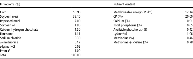
All experimental protocols were approved by the Animal Care and Use Committee of Anhui Agricultural University. A total of 600 1-day-old Chaohu ducks were used in this study. Chaohu duck is one of the most important local duck breeds in eastern China and has superior meat quality and a strong tolerance for coarse feed and poor feeding environment. Its average adult BW is around 3.1 kg for male and 2.8 kg for female, and its 72 weeks’ egg production can reach 160 to 200. Therefore, the breed is widely raised for both meat and eggs (Zhang et al., Reference Zhang, Ah Kan Razafindrabe, Chen, Zhao, Yang, Wang, Chen, Jin and Geng2018a). The selected ducks were banded and randomly allotted into five dietary treatments supplemented with 0 (control group), 300, 600, 900 or 1500 mg l-theanine per kg feed. Each treatment group included five replicates of 24 birds each (12 male and 12 female duck per replicate). The ducks were reared in a commercial production environment condition between August and September 2016. The pens (each 2×2.5 m2) were bedded with rice hulls. Room temperature was between 27 and 36°C, the relative humidity was about 70%, and the lighting period is 23 h/day. The test lasted for 28 days. Ducks had ad libitum access to powder feed and water. Basal diet was formulated according to the Feeding Standard of meat-type ducks of China (Criterion Code: NY/T 2122-2012), and the ingredient composition and nutrient content of diet are shown in Table 1. At the end of days 14 and 28, after a 12-h fasting period, BW of ducks and feed intake (FI) on a replicate basis were recorded. Then, four ducks (2 male and 2 female ducks) of average BW were selected from each experimental replicate pen for sample collection.
Composition and nutrient content of basal diet for ducks

1 Composition per kilogram of diet: vitamin D3, 3000 IU; vitamin A, 10 000 IU; biotin, 0.2 mg; vitamin E, 10 mg; vitamin K3, 2.7 mg; vitamin B1, 2.35 mg; vitamin B2, 12.5 mg; vitamin B6, 3 mg; vitamin B12, 0.03 mg; nicotinic acid, 50 mg; choline, 1400 mg; pantothenic acid, 11 mg; folic acid, 1 mg; Cu, 10 mg; Fe, 60 mg; Zn, 50 mg; Mn, 100 mg; I 0.4 mg; Se, 0.3 mg.
Sample collection and analytical determination
Blood was collected from wing vein by venipuncture and centrifuged at 2500×g for 12 min at 4°C. Then, serum samples were stored at −80°C until analysis. The activities of serum alanine aminotransferase (ALT) and aspartate transaminase (AST), and the contents of serum glucose (GLU), total protein (TP), albumin (ALB), uric acid (UA), triacylglycerol (TG), total cholesterol (TC), high-density lipoprotein cholesterol (HDL-C) and low-density lipoprotein cholesterol (LDL-C) were determined using colorimetric methods with a spectrophotometer (Biomate 5; Thermo Electron Corporation, Rochester, NY, USA) according to the instructions of the commercial kits (Jiancheng Bioengineering Institute, Nanjing, Jiangsu, China). Levels of immune globulin A (IgA), IgG, IgM, interferon-γ (IFN-γ), tumor necrosis factor-α (TNF-α), interleukin-2 (IL-2) and IL-6 in serum were measured using the corresponding commercial ELISA kits for duck (Nanjing Jiancheng Institute of Bioengineering, Nanjing, Jiangsu, China) according to the instructions of the manufacturer. Serum insulin concentration was measured using an insulin radioimmunoassay kit (intra-assay CV: 7%) for duck (Nanjing Jiancheng Institute of Bioengineering) according to the manufacturer’s instructions.
All selected ducks were individually weighed, euthanized by cervical dislocation and immediately exsanguinated by cutting the jugular artery. Liver, spleen, thymus and bursa of Fabricius were collected and weighed. The relative weight of organ was calculated as the ratio between fresh organ weight (mg) and duck BW (g) before slaughter. Segments of the mid-jejunum (about 3 cm) were removed and fixed in 10% formalin for morphology measurement. Adjacent jejunum sample (about 10 cm) was removed and then mucosa was collected by scraping using a sterile glass slide after washing in ice-cold physiological saline solution. The mucosa sample was rapidly frozen in liquid nitrogen and then stored at −80°C for the analysis of antioxidant capacity.
Formalin-fixed jejunal sample was dehydrated, embedded, and stained with hematoxylin and eosin for crypt depth and villus height measurements using an image processing and analysis system (Leica Imaging Systems Ltd, Cambridge, UK) according to a previous report (Al-Fataftah and Abdelqader, Reference Al-Fataftah and Abdelqader2014). Formalin-fixed jejunal sample was dehydrated, embedded and stained with periodic acid-Schiff regent, and then goblet cells were enumerated per 100 villus epithelial cells for each cross-section in the coincident visual field under an Olympus light microscope (Olympus Optical Co., Beijing, China) according to previous reports (Liu et al., Reference Liu, Fu, Yan, Xie, Li, Yu, He and He2016).
About 1 g of frozen intestinal mucosa sample was homogenized for 2 min in 9 ml of ice-cold saline solution and the homogenate was centrifuged at 2500×g for 10 min at 4°C. The clear supernatant was collected for antioxidant enzyme activity and malondialdehyde (MDA) content measurement. The activities of total superoxide dismutase (T-SOD), CAT and GSH-Px, and the content of MDA were determined using the colorimetric method with a spectrophotometer (Biomate 5) and the corresponding commercial kits (Nanjing Jiancheng Institute of Bioengineering) as previously described (Zhang et al., Reference Zhang, Wang, Zhao, Chen, Yang and Geng2017a and Reference Zhang, Zhao, Wang, Yang, Chen and Geng2017b).
Statistical analysis
All data were analyzed by one-way ANOVA procedure of the SPSS software (SPSS 18.0 for Windows; SPSS, Chicago, IL, USA) with each replicate as the experimental unit for growth performance and each bird as the experimental unit for other parameters. The model included the treatment effect and differences between treatment groups were determined by Duncan’s multiple range tests. Orthogonal polynomial contrasts were employed to assess the linear and quadratic effects of l-theanine levels. A P-value of <0.05 was considered significant, whereas a P-value of <0.10 was considered a tendency. Data are presented as mean and SEM.
Results
Growth performance
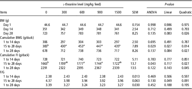
Growth performance data are shown in Table 2. l-theanine had no significant effect on BW at day 14 (P>0.05), while increased BW at day 28 (quadratic, P<0.05). From days 1 to 14, l-theanine had no significant effect on feed to gain ratio (F/G), cumulative BW gain (BWG) and cumulative FI (P>0.05). From days 15 to 28, l-theanine elevated cumulative BWG (linear and quadratic, P<0.05) and cumulative FI (quadratic, P<0.05) and decreased F/G (linear, P<0.05). From days 1 to 28, l-theanine elevated cumulative BWG (quadratic, P<0.05) and cumulative FI (quadratic, P<0.05).
Effect of dietary l-theanine supplementation on growth performance of ducks

BWG=BW gain; FI=feed intake; F/G=feed to gain ratio.
Data are expressed as mean and SEM (n=5).
a,bValues within a row with different superscripts differ significantly at P<0.05.
Relative organ weight
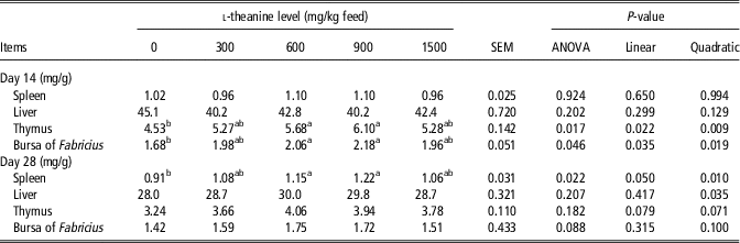
Relative organ weight data are shown in Table 3. l-theanine linearly and quadratically increased the relative weight of thymus and bursa of Fabricius at day 14 (P<0.05). l-theanine elevated the relative weight of spleen (linear and quadratic, P=0.05 and P<0.05, respectively) and liver (quadratic, P<0.05) at day 28. Moreover, l-theanine tended to linearly and quadratically increase the relative weight of thymus at day 28 (P=0.079 and P=0.071, respectively).
Effect of dietary l-theanine supplementation on the relative organ weight of ducks

Data are expressed as mean and SEM (n=20).
a,bValues within a row with different superscripts differ significantly at P<0.05.
Serum biochemical parameter
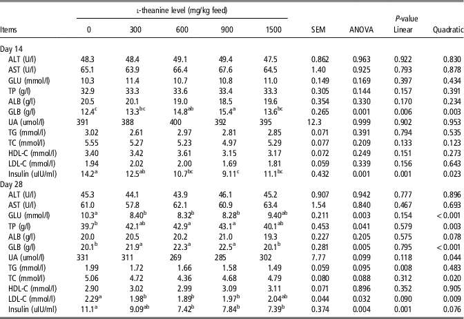
Data for serum parameter are shown in Table 4. l-theanine elevated serum globulin (GLB) content (linear and quadratic, P<0.05) and decreased serum insulin content (linear and quadratic, P<0.05) at day 14, while there were no distinct differences in serum ALT, AST, GLU, TP, ALB, UA, TG, TC, HDL-C and LDL-C content (P>0.05). l-theanine elevated serum TP and GLB contents (quadratic, P<0.05), and decreased serum GLU (quadratic, P<0.05), UA (quadratic, P<0.05), TG (linear, P<0.05), TC (quadratic, P<0.05), LDL-C (quadratic, P<0.05) and insulin contents (linear, P<0.05) at day 28, while there were no significant differences in serum ALT, AST, ALB and HDL-C contents (P>0.05).
Effect of dietary l-theanine supplementation on serum biochemical parameters of ducks

ALT=alanine transaminase; AST=aspartate transaminase; GLU=glucose; TP=total protein; ALB=albumin; GLB=globulin; UA=uric acid; TG=triacylglycerol; TC=total cholesterol; HDL-C=high-density lipoprotein cholesterol; LDL-C=low-density lipoprotein cholesterol.
Data are expressed as mean and SEM (n=20).
a,b,cValues within a row with different superscripts differ significantly at P<0.05.
Serum immune parameter
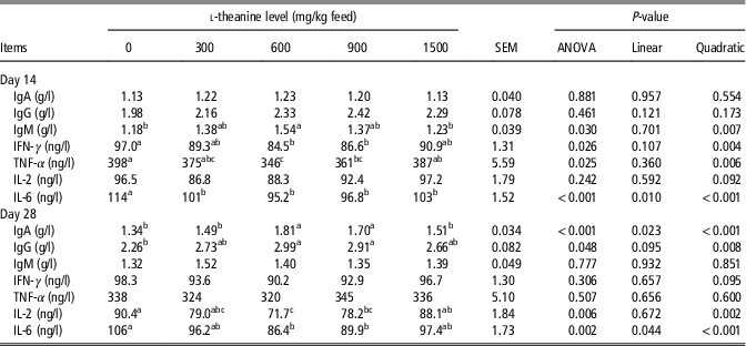
Data for serum immune parameters are displayed in Table 5. l-theanine elevated serum IgM content (quadratic, P<0.05) and decreased serum IFN-γ (quadratic, P<0.05), TNF-α (quadratic, P<0.05) and IL-6 (linear and quadratic, P<0.05) contents at day 14, while there were no distinct differences in serum IgA, IgG and IL-2 contents (P>0.05). l-theanine elevated serum IgG (quadratic, P<0.05) and IgA (linear and quadratic, P<0.05) contents, and decreased serum IL-2 (quadratic, P<0.05) and IL-6 (linear and quadratic, P<0.05) contents at day 28, while there were no distinct differences in serum IgM, INF-γ and TNF-α contents (P>0.05).
Effect of dietary l-theanine supplementation on serum immune measurements of ducks

IgA=immune globulin A; IgG=immune globulin G; IgM=immune globulin M; IFN-γ=interferon-γ; TNF-α=tumor necrosis factor-α; IL-2=interleukin-2; IL-6=interleukin-6.
Data are expressed as mean and SEM (n=20).
a,b,cValues within a row with different superscripts differ significantly at P<0.05.
Intestinal morphology
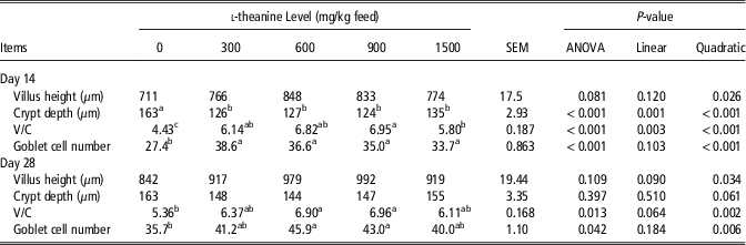
Data for jejunum morphology are displayed in Table 6. On day 14, l-theanine elevated villus height (quadratic, P<0.05), villus height to crypt depth ratio (V/C; linear and quadratic, P<0.05) and goblet cell number (quadratic, P<0.05), and decreased crypt depth (linear and quadratic, P<0.05). On day 28, l-theanine quadratically elevated villus height, V/C and goblet cell number (P<0.05). Moreover, l-theanine tended to quadratically decrease crypt depth at day 28 (P=0.061).
Effect of dietary l-theanine supplementation on jejunum morphology of ducks

V/C=villus height to crypt depth ratio.
Data are expressed as mean and SEM (n=20).
a,b,cValues within a row with different superscripts differ significantly at P<0.05.
Intestinal antioxidant index
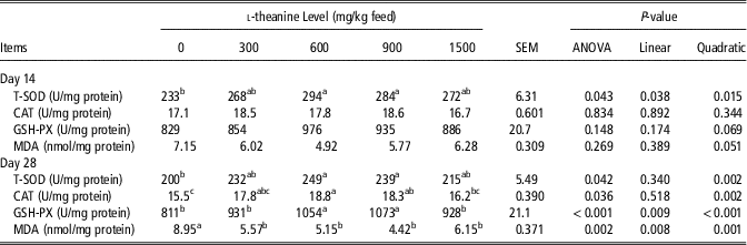
Data for intestinal antioxidant index are displayed in Table 7. On day 14, l-theanine increased T-SOD activity (linear and quadratic, P<0.05) and tended to quadratically increase GSH-PX activity (P=0.069) and decrease MDA content (P=0.051). On day 28, l-theanine increased T-SOD (quadratic, P<0.05), CAT (quadratic, P<0.05) and GSH-PX (linear and quadratic, P<0.05) activities and decreased MDA content (linear and quadratic, P<0.05).
Effect of dietary l-theanine supplementation on jejunum antioxidant status of ducks

T-SOD=total superoxide dismutase; CAT=catalase; GSH-PX=glutathione peroxidase; MDA=malondialdehyde.
Data are expressed as mean and SEM (n=20).
a,b,cValues within a row with different superscripts differ significantly at P<0.05.
Discussion
Previous study reported that l-theanine at the levels of 3000 and 4000 mg/kg BW per day markedly decreased the food consumption and BWG of rats (Borzelleca et al., Reference Borzelleca, Peters and Hall2006). In contrast, recent study reported that the average daily gain (ADG) of rats was significantly increased by l-theanine at levels from 50 to 400 mg/kg BW per day (Yan et al., Reference Yan, Tong, Tang, Tan, Han and Zhou2017). Moreover, Saeed et al. (Reference Saeed, Xu, Hassan, Arain, Elhack, Noreldin and Sun2018) reported that l-theanine at 300 mg/kg diet was able to improve the growth performance of broilers. However, Wen et al. (Reference Wen, Wei, Zhang, Hou, Xiao and He2012) did not observe significant differences in the ADG, average daily FI and F/G of yellow-feathered broilers when birds were fed diets containing 200 to 800 mg/kg l-theanine (Wen et al., Reference Wen, Wei, Zhang, Hou, Xiao and He2012). To the best of our knowledge, our present study is the first to evaluate the possible effect of l-theanine on ducks. We found that dietary l-theanine supplementation had no significant effect on growth performance of ducks from 1 to 14 days of age. However, the cumulative BWG and cumulative FI of ducks from 15 to 28 days of age and 1 to 28 days of age were quadratically improved by l-theanine, while F/G from 15 to 28 days of age was linearly decreased in the meantime. These results suggest that the dietary addition of l-theanine seems to have a time-dependent positive cumulative effect and the suitable l-theanine level in diet has positive impact on the growth performance of duck. The improvement of growth performance due to l-theanine supplementation may be attributed to its beneficial biological activity, such as antioxidative activities and immune modulating function (Saeed et al., Reference Saeed, Xu, Hassan, Arain, Elhack, Noreldin and Sun2018), which has caught our attention, making us want to explore it further.
The bursa of Fabricius, thymus and spleen are extensively involved in poultry cellular and humoral immunity and therefore these immune organs are absolutely vital for the development of poultry immune system (Liu et al., Reference Liu, He, Xie, Yang, Li and Zou2014). Thereinto, spleen is the vital peripheral immune organ, and thymus and bursa of Fabricius are the central immune organs. The immune organ to BW ratios are commonly measured and used as indicator in assessing the immune system of the poultry (Pope, Reference Pope1991). Previous studies reported that some environmental stress factors can cause immune organ dysplasia, while dietary supplementation with certain nutrients such as resveratrol, Forsythia suspensa extract and berberine are able to prevent immune organ dysfunction (Zhang et al., Reference Zhang, Piao, Zhang, Li, Yi, Liu, Li and Wang2013; Liu et al., Reference Liu, He, Xie, Yang, Li and Zou2014). A study in broilers found that l-theanine at levels of 200 and 400 mg/kg significantly improved the humoral immunity and tended to increase the relative weight of thymus and spleen (Wen et al., Reference Wen, Wei, Zhang, Hou, Xiao and He2012). Saeed et al. (Reference Saeed, Xu, Hassan, Arain, Elhack, Noreldin and Sun2018) also observed that briolers fed diets supplemented with l-theanine at 200 mg/kg has significantly higher spleen and bursa of Fabricius relative weight, and lower serum pro-inflammatory cytokine (IL-2 and IFN-γ) contents. Moreover, some researches in rodent and human reported that serum inflammatory cytokines contents and blood immunocompetent cells could be positively regulated by l-theanine administration (Murakami et al., Reference Murakami, Kurihara, Titchenal and Ohtani2010; Takagi et al., Reference Takagi, Kurihara, Higashi, Morikawa, Kase, Maeda, Arisaka, Shibahara and Akiyama2010; Hwang et al., Reference Hwang, Jin, Choi, Choi, Kim, Kim, Kim, Lee, Chung and Jeong2017). However, no experimental study has been published on the effect of l-theanine on the immune function of ducks. The current study, first, indicated that dietary addition of l-theanine quadratically increased the relative weight of thymus and bursa of Fabricius of ducks at 14 days of age and also quadratically increased the relative weight of spleen of ducks at 28 days of age. Moreover, GLB content in serum of ducks was quadratically increased by dietary l-theanine supplementation, which may suggest that the humoral immunity of ducks is improved because serum GLB content may be served as an indicator of body immune function. In order to confirm our assumption, we further measured some immune factors in serum. Excitingly, we found that serum pro-inflammatory cytokines (IFN-γ, TNF-α, IL-2 and IL-6) levels could be decreased by l-theanine, while serum IgA, IgG and IgM levels were increased. Therefore, l-theanine may be used as an effective feed additive for improving the immune function of duck. Nuclear factor κ B is a transcription factor which is closely related to the regulation of various inflammatory genes expression, its activation may lead to inflammatory reaction and immune dysfunction (Kim et al., Reference Kim, Lee, Park, Choi, Ban, Park, Nam, Yun, Han, Oh and Hong2009; Hwang et al., Reference Hwang, Jin, Choi, Choi, Kim, Kim, Kim, Lee, Chung and Jeong2017). Previous studies reported that the mRNA expression levels of cytokines in immune tissues of broilers (Saeed et al., Reference Saeed, Xu, Hassan, Arain, Elhack, Noreldin and Sun2018) and NF- κ B protein level in lung of mice (Hwang et al., Reference Hwang, Jin, Choi, Choi, Kim, Kim, Kim, Lee, Chung and Jeong2017) could be down-regulated by l-theanine. Therefore, the improvement of immune function in duck due to l-theanine supplementation may be partly attributed to its regulation activity on NF- κ B pathway, and further investigations are worthwhile.
The physiological and metabolic status of animals can be infered from serum biochemical parameters which can be changed by diet and environmental factors (Zhang et al., Reference Zhang, Ah Kan Razafindrabe, Chen, Zhao, Yang, Wang, Chen, Jin and Geng2018a). Previous study found that the insulin content in serum of rats was significantly decreased by l-theanine consumption (Yamada et al., Reference Yamada, Nishimura, Sakurai, Terashima, Okubo, Juneja and Yokogoshi2008), which was recently confirmed by Yan et al. (Reference Yan, Tong, Tang, Tan, Han and Zhou2017). Yan et al. (Reference Yan, Tong, Tang, Tan, Han and Zhou2017) also found that l-theanine administration could significantly decrease serum GLU content of rats. Consistently, we found that serum GLU content at day 28 and serum insulin content at days 14 and 28 were significantly decreased by dietary l-theanine supplementation. A study in mouse discovered that serum TG and non-esterified fatty acid levels were decreased by l-theanine administration (Zheng et al., Reference Zheng, Sayama, Okubo, Juneja and Oguni2004). Our present study found that TG, TC and LDL-C contents in serum of ducks at 28 days of age were decreased, which may indicate that the addition of l-theanine can regulate the lipid metabolism of ducks. Serum TP and UA contents are often used as the indicators of amino acid utilization and degree of protein breakdown (Donsbough et al., Reference Donsbough, Powell, Waguespack, Bidner and Southern2010). In the present study, we found that l-theanine quadratically increased TP content and decreased UA content in serum of ducks at 28 days of age, indicating that l-theanine at appropriate addition level has the potential to increase protein synthesis and retention which was in accordance with the increase of growth performance.
The villus–crypt axis in small intestine are of greatest importance in nutrient digestion and absorption (Yang et al., Reference Yang, Zhang, Wang, Wu, Yue, Jiang and Qi2017). It is well-known that villus height, crypt depth and V/C are usually used to assess the function status of small intestine. The crypt are regarded as the villus cells producer, and the lower crypt depth indicates that the intestinal villus is in good condition and has lower need for regeneration (Yason et al., Reference Yason, Summers and Schat1987). Therefore, it is generally considered that lower crypt depth, and higher villus height and V/C reflect a better state of intestinal structure and function which can lead to better nutrient digestion and absorption, improved disease resistance, and greater growth performance (Viveros et al., Reference Viveros, Chamorro, Pizarro, Arija, Centeno and Brenes2011; Zhang et al., Reference Zhang, Zhao, Yang, Chen, Jiang, Jin and Geng2017c). It is well documented that many nutrients, including some amino acids and plant extracts, have the function of regulating gut morphology (Min et al., Reference Min, Liu, Qu, Meng and Gao2017; Yang et al., Reference Yang, Zhang, Wang, Wu, Yue, Jiang and Qi2017; Zhang et al., Reference Zhang, Zhao, Yang, Chen, Jiang, Jin and Geng2017c). However, to the best of our knowledge, there is no relevant study conducted to evaluate the effect of l-theanine on gut morphology. Excitingly, our present study provided the first evidence that l-theanine could effectively improve jejunum morphology, reflecting by decreased crypt depth and increased villus height and V/C. Goblet cell is important for intestinal barrier function because it can secrete a lot of mucus on surface of intestinal mucosa, which play an important role in preventing pathogen attacks and environmental toxins (Zhang et al., Reference Zhang, Zhao, Yang, Chen, Jiang, Jin and Geng2017c). The present study indicated that goblet cell numbers in jejunum were linearly increased by dietary l-theanine supplementation. Deng et al. (Reference Deng, Xiao, Chen, Liu, Liu and Gong2016) reported that l-theanine could attenuate enterotoxigenic E. coli (ETEC) strain O78:K80-induced oxidative stress in mice, suggesting that l-theanine may possess antibacterial properties. Therefore, the beneficial effect of l-theanine on gut morphology may be partly attributed to the increased globet cell number and its antibacterial properties. However, further investigation needs to be conducted.
Reactive oxygen species (ROS) such as hydrogen peroxide, superoxide anions and hydroxyl radicals are inevitable by-product of normal animal metabolism and are essential for cell differentiation and animal growth (Samuel et al., Reference Samuel, Wang, Yue, Wu, Zhang, Duan and Qi2017). However, ROS production may be excessively induced by many stress factors which will cause protein and lipid oxidation and cellular damage, and ultimately affects animal health (Zhang et al., Reference Zhang, Yang, Zhao, Chen, Wang and Geng2018b). It is well documented that dietary supplementation with nutrients that have antioxidant function can eliminate ROS and guarantee animal health and growth, including intestinal health (Samuel et al., Reference Samuel, Wang, Yue, Wu, Zhang, Duan and Qi2017; Yang et al., Reference Yang, Zhang, Wang, Wu, Yue, Jiang and Qi2017; Zhang et al., Reference Zhang, Zhao, Yang, Chen, Jiang, Jin and Geng2017c). Li et al. (Reference Li, Ye, Kang, Yao, Zhang, Jiang, Gao, Dai, Xin, Wang, Yin and Luo2012) found that l-theanine prevented ethanol-triggered ROS and MDA generation in L02 cells and significantly inhibited ethanol-induced reduction of SOD and CAT activities in liver of mouse. Deng et al. (Reference Deng, Xiao, Chen, Liu, Liu and Gong2016) reported that l-theanine increased liver SOD activity and the mRNA expression levels of CuZn-SOD and GSH-PX in ETEC-infected mouse and also increased liver total antioxidant capacity and the mRNA expression of CAT in non-ETEC-infected mice. These studies indicated that l-theanine has antioxidant activity. In the present study, antioxidant markers, MDA, SOD, CAT and GSH-PX, were investigated to evaluate the antioxidant activity of l-theanine in jejunum of ducks. Results showed that MDA content in jejunum of ducks at 28 days of age was significantly decreased by l-theanine, which indicates a lower lipid oxidation. Moreover, the present study demonstrated that jejunal CAT and GSH-PX activities at 28 days of age and T-SOD activity at 14 and 28 days of age were significantly improved by dietary l-theanine supplementation, which suggest an improved antioxidative capacity and may partly explain why l-theanine can improve jejunum morphology.
Conclusion
To our best knowledge, this study provides the first evidence that l-theanine can serve as an effective and beneficial feed additive for improving growth performance of ducks, which may be partly attributed to the improvement of immune function and jejunum morphology and antioxidant capacity induced by l-theanine. Quadratic responses occur on growth performance, immune-related parameters, jejunum morphology and antioxidant capacity, and the optimum inclusion levels of l-theanine is 600 to 900 mg/kg based on the current experimental condition.
Acknowledgements
The present study was financially supported by the National Science and Technology Support Plan Program of China (2015BAD03B06) and the Science and Technology project of Anhui province (1604a0702009).
Declaration of interest
The authors declare that there is no conflicts of interest.
Ethics statement
All experimental protocols were approved by the Animal Care and Use Committee of Anhui Agricultural University.
Software and data repository resources
None of the data were deposited in an official repository.