


Introduction
Methicillin-resistant Staphylococcus aureus (MRSA) remains a major cause of community and healthcare-associated infections, with bacteremia mortality rates ranging from 18% to 32%. 1,Reference Bai, Lo and Komorowski2 Risk factors for severe MRSA infections include recent use of intravenous antibiotics, prolonged hospitalization, recent surgery, and immunocompromising conditions. Reference Lakhundi and Zhang3 Solid organ transplant (SOT) recipients frequently exhibit these risk factors. Historical studies report MRSA infection rates as high as 23% in liver transplant patients, with mortality as high as 60%. Reference Liu, Zhang and Wan4 Compared with nontransplant populations, SOT recipients have a higher prevalence of MRSA colonization and infection. Reference Malinis, Mawhorter and Jain5
In this high-risk population, empiric anti-MRSA therapy is often initiated despite concerns regarding the overuse of broad-spectrum antibiotics and the development of antimicrobial resistance. Determining which patients require empiric MRSA coverage and appropriate timing for de-escalation remains challenging among immunocompromised populations.
The MRSA nasal polymerase chain reaction (PCR) test offers a rapid, minimally invasive tool to guide antibiotic therapy. Its high negative predictive value (NPV) is well described in hospital-acquired pneumonia, often exceeding 95%, and, in other infectious sites, typically ranges from 92% to over 99%, though it may fall with higher MRSA-prevalent infections (ie, purulent cellulitis) or higher local MRSA prevalence. Reference Mergenhagen, Starr and Wattengel6–Reference Shaw, Zander and Ronnie10 The objective of this study was to evaluate the diagnostic performance of the MRSA nasal PCR in a diverse SOT population underrepresented in the current literature.
Methods
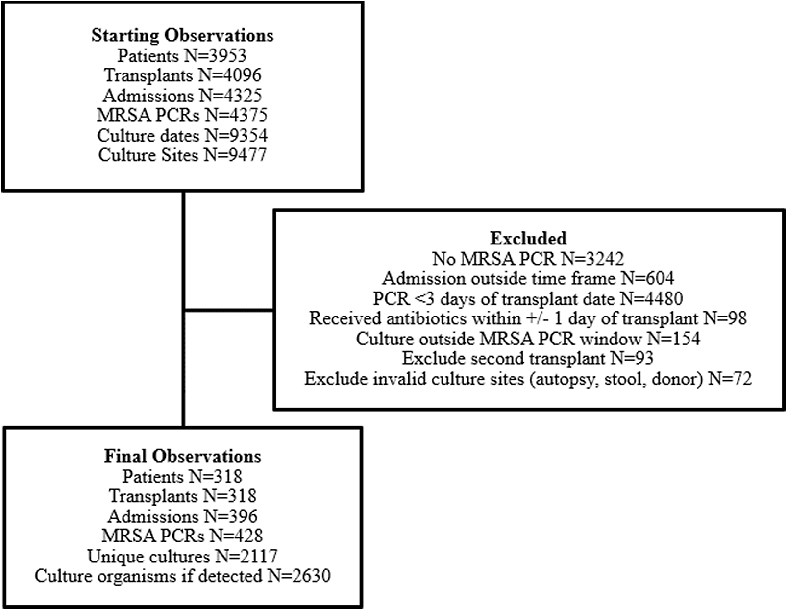
This single-center, retrospective cohort study was conducted at a quaternary care academic medical center performing over 500 SOTs annually, including heart, small intestine, kidney, liver, lung, and pancreas. During the study period, the institution utilized MRSA nasal PCR surveillance in select units with additional testing performed at providers’ discretion. Patients ≥18 years of age who received a MRSA nasal PCR between July 1, 2013, and October 1, 2022, at least 72 hours after SOT, with at least one clinical culture collected within 48 hours before or 14 days after MRSA nasal PCR collection were included (Figure 1). Patients were excluded if they received nasal mupirocin within 30 days; received anti-MRSA prophylaxis initiated within 24 hours of transplant during transplant index hospitalization only; or were pregnant, nursing, or incarcerated. Patients with repeat MRSA nasal PCRs during the same admission were included if criteria were met.
Consort Diagram.

All eligible cultures were collected. Cultures with mixed flora were routinely evaluated for S. aureus and reported, alongside susceptibilities, if present. MRSA nasal PCR testing was performed using the Cepheid Xpert® MRSA Assay. Primary endpoints were the sensitivity, specificity, positive predictive value (PPV), NPV, positive likelihood ratio (PLR), and negative likelihood ratio (NLR) of the MRSA nasal PCR for predicting MRSA in clinical cultures. Secondary endpoints evaluated performance by anatomical site, PCR-to-culture time, admission-to-PCR time, and transplant-to-PCR time, as well as the effect of prior anti-MRSA therapy within 7 days of the PCR and cystic fibrosis status. Data were extracted from the electronic health record, validated via manual chart review, and collected using Research Electronic Data Capture tools hosted by our institution. Descriptive statistics summarized demographics and clinical characteristics. We summarized continuous variables as median and interquartile range (IQR), and categorical variables as frequencies and percentages. Where applicable, Wilcoxon rank-sum or Kruskal-Wallis tests were used to compare continuous variables across groups. Fisher’s exact test or chi-squared test was used to compare categorical variables, as appropriate. For each aim, estimates of sensitivity, specificity, PPV, NPV, PLR, and NLR, along with their corresponding 95% CIs, were reported. All statistical tests used a fixed type I error rate of less than 0.05. Analyses were conducted using R version 4.1.1 or SAS version 9.4.
Results
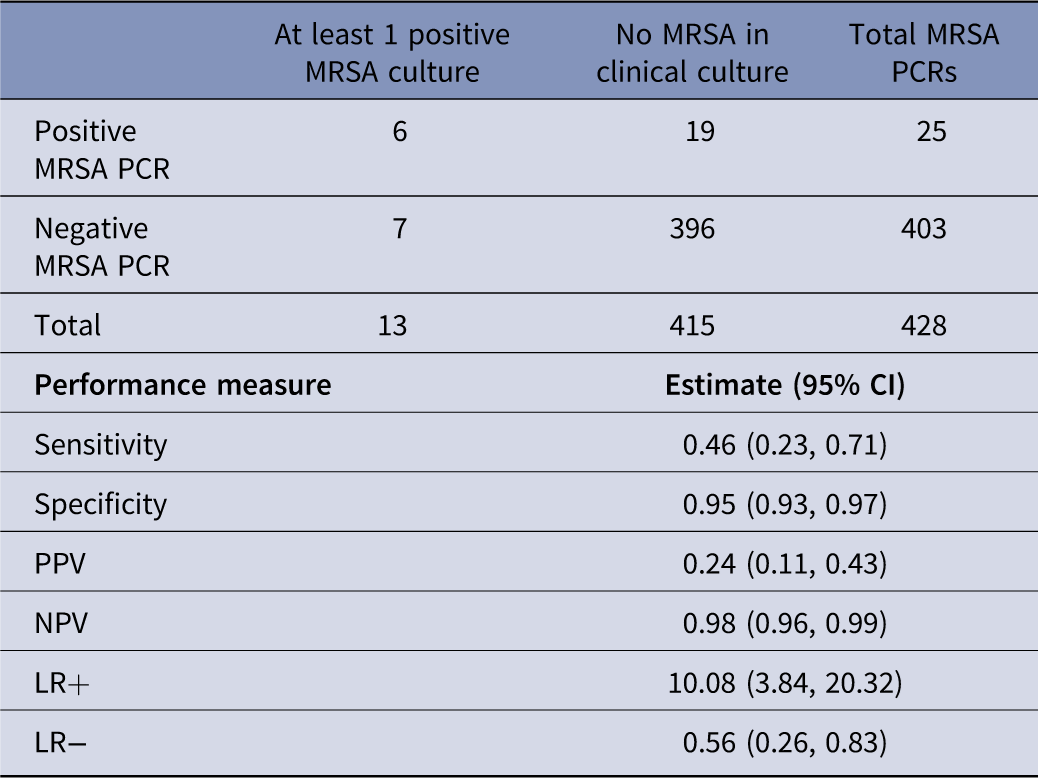
A total of 428 MRSA PCRs and 2,117 concomitant clinical cultures from 318 patients met the inclusion criteria (Supplementary Figure 1). Most patients were male (63.5%) and Caucasian (69.2%) (Table 1). The most common transplant types were lung (45.3%), heart (20.4%), kidney (18.6%), and liver (11.6%). Most patients underwent one PCR test during hospitalization (94.7%), and the median hospital length of stay was 10 days (IQR 5–29 days). MRSA was detected in 25 (5.8%) nasal swab PCRs. Of 2,117 cultures, most were blood (50%), respiratory (21.9%), and urine (15.4%). The specificity of the nasal PCR was 0.95 (95% CI: 0.93–0.97) and the NPV was 0.98 (95% CI: 0.96–0.99) (Table 2). Sensitivity was low at 0.46 (95% CI: 0.23–0.71), and the NLR was 0.56 (95% CI: 0.26–0.83). Performance was lower among patients with cystic fibrosis (NPV 0.85, 95% CI 0.69–0.93). Predictive measures by subgroups of interest are reported in Supplementary Table 3. Culture characteristics by culture and MRSA PCR result are reported in Supplementary Table 4.
Characteristics

Table 1. Long description
A table with 18 rows and 2 columns. The first column lists various patient characteristics and transplant details, and the second column provides corresponding values. Row 1: Total Patients, N 318. Row 2: Age, median years (IQR), 59 (46-66). Row 3: Female sex, n (%), 116 (36.5%). Row 4: Race, American Indian or Alaskan Native, 3 (0.9%). Row 5: Race, Asian, 3 (0.9%). Row 6: Race, Black or African American, 81 (25.5%). Row 7: Race, Caucasian/white, 220 (69.2%). Row 8: Race, Other/not reported, 11 (3.5%). Row 9: Transplant organ, Lung, 144 (45.3%). Row 10: Transplant organ, Heart, 65 (20.4%). Row 11: Transplant organ, Kidney, 59 (18.6%). Row 12: Transplant organ, Liver, 37 (11.6%). Row 13: Transplant organ, Kidney/pancreas, 7 (2.2%). Row 14: Transplant organ, Intestine, 4 (1.3%). Row 15: Transplant organ, Liver/intestine/pancreas, 1 (0.3%). Row 16: Transplant organ, Pancreas, 1 (0.3%). Row 17: Transplant year, 2013-2014, 109 (34.3%). Row 18: Transplant year, 2015-2016, 88 (27.7%). Row 19: Transplant year, 2017-2018, 42 (13.2%). Row 20: Transplant year, 2019-2020, 44 (13.8%). Row 21: Transplant year, 2021-2022, 35 (11%). Row 22: Underlying cystic fibrosis, 25 (7.9%). Row 23: Number of admissions during study period, Total Admissions, N 396. Row 24: Number of admissions during study period, 1, 259 (81.5%). Row 25: Number of admissions during study period, 2, 47 (14.8%). Row 26: Number of admissions during study period, 3, 7 (2.2%). Row 27: Number of admissions during study period, 4, 3 (0.9%). Row 28: Number of admissions during study period, 5, 2 (0.63%). Row 29: Hospital length of stay, median days (IQR), 10 (5-29). Row 30: Survival to hospital discharge, 332 (83.8%). Row 31: Number of MRSA PCRs during admission, Total MRSA PCRs, N 428. Row 32: Number of MRSA PCRs during admission, 1, 375 (94.7%). Row 33: Number of MRSA PCRs during admission, 2, 15 (3.8%). Row 34: Number of MRSA PCRs during admission, 3 or more, 6 (1.5%). Row 35: Received prior anti-MRSA antibiotic, initiated 0 to 7 days prior to MRSA PCR, 196 (46%). Row 36: Time from transplant to MRSA PCR, median days (IQR), 331.5 (113.5-1,083.5). Row 37: Location of MRSA PCR collection, Intensive care unit, 221 (51.6%). Row 38: Location of MRSA PCR collection, Floor, 157 (36.7%). Row 39: Location of MRSA PCR collection, Operating room, 6 (1.4%). Row 40: Location of MRSA PCR collection, Emergency department, 44 (10.3%). Row 41: Number of concomitant cultures, median (IQR), 4 (2-6).
Note: SD, standard deviation; IQR, interquartile range; MRSA, methicillin-resistant Staphylococcus aureus; Data are presented as n (%) unless otherwise noted.
PCR-given-culture predictive values, with 95% confidence intervals

Table 2. Long description
A table with four rows and three columns comparing PCR-given-culture predictive values for MRSA. The columns are labeled ‘At least 1 positive MRSA culture’, ‘No MRSA in clinical culture’, and ‘Total MRSA PCRs’. The rows are labeled ‘Positive MRSA PCR’, ‘Negative MRSA PCR’, ‘Total’, and ‘Performance measure’. Row 1: Positive MRSA PCR, 6, 19, 25. Row 2: Negative MRSA PCR, 7, 396, 403. Row 3: Total, 13, 415, 428. Row 4: Performance measure, Estimate (95% CI), Sensitivity 0.46 (0.23, 0.71), Specificity 0.95 (0.93, 0.97), PPV 0.24 (0.11, 0.43), NPV 0.98 (0.96, 0.99), LR+ 10.08 (3.84, 20.32), LR- 0.56 (0.26, 0.83).
Note: LR+, positive likelihood ratio; LR−, negative likelihood ratio; PPV, positive predictive value; NPV, negative predictive value; MRSA, methicillin-resistant Staphylococcus aureus.
Discussion
In this large cohort of SOT recipients with a low prevalence of MRSA in clinical cultures, MRSA nasal PCR demonstrated high NPV and specificity, consistent with prior studies conducted primarily in immunocompetent populations. These findings align with the rule-out utility of MRSA nasal PCR in a high-risk, immunocompromised population and support consideration of early de-escalation of anti-MRSA therapy in SOT recipients at low risk, particularly those without a history of MRSA and with a stable clinical course. However, the NLR of 0.56 indicates the MRSA swab provides at best modest rule-out discrimination and importantly, a negative PCR result does not fully exclude the possibility of MRSA in clinical cultures. Further, caution is warranted in high prevalence populations as well as critically ill patients, including those with septic shock, or high risk for MRSA infection.
This study has several limitations. As a retrospective review, we were unable to control timing of MRSA PCR collection relative to culture collection, or the number of cultures obtained per patient, and our analysis was based on growth in clinical cultures rather than adjudicated infections. MRSA nasal PCR testing was performed during hospitalization based on clinician discretion or unit-specific screening practices, introducing selection bias and limiting the generalizability of our findings. The small number of positive cultures and positive nasal PCR results preclude reliable estimation of PPV and PLR, and non-independence from repeat swabs and multiple culture-swab pairings per patient may introduce further bias. In lung transplant recipients, donor-derived MRSA may limit the utility of screening in the early posttransplant period. Additionally, patients with cystic fibrosis, although a small subset, had a lower NPV in our cohort; warranting caution in this population. Sample size limited our ability to detect differences by transplant type or timing of PCR collection. Future studies should also assess the durability of a negative nasal PCR over time in the SOT population.
Conclusion
Given the high NPV of MRSA nasal PCR testing observed in this study and in the literature, it may serve as a useful tool for antimicrobial stewardship and de-escalation of anti-MRSA therapy in low-risk SOT recipients. These exploratory findings require validation in cohorts with a higher MRSA prevalence. Results should be interpreted in the context of clinical judgment, culture history, source of infection, and illness severity. Further prospective studies are needed to define the role of MRSA nasal PCR-guided de-escalation in SOT recipients and other immunocompromised populations.
Supplementary material
The supplementary material for this article can be found at https://doi.org/10.1017/ice.2026.10550.
Financial support
We acknowledge support from the Biostatistics, Epidemiology, and Research Design Methods Core funded through Grant Award Number UL1TR002553 from the National Center for Advancing Translational Sciences, a component of the NIH. The content is solely the responsibility of the authors and does not necessarily represent the official views of the NIH.
Competing interests
Authors of this manuscript have nothing to disclose concerning possible financial or personal relationships with commercial entities that may have a direct or indirect interest in the subject matter.