A patient with giant ventricular septal defect associated with pulmonary hypertension is reported in this case study. The patient was tested with device closure of ventricular septal defect temporally with a decrease in pulmonary vascular resistance index from 10.3 to 4.1 Wood units. The patient then underwent successful corrective repair of the ventricular septal defect surgically.
Case report
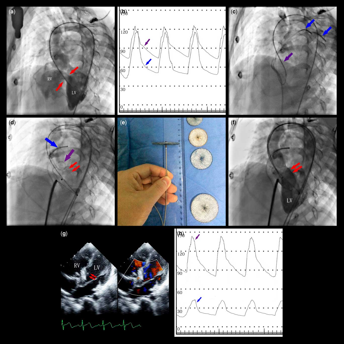
A 33-year-old woman with cyanosis and decreased activity tolerance has been diagnosed with perimembranous ventricular septal defect and pulmonary hypertension. The SPO2 is 88%, and echocardiography shows bidirectional shunt. Right ventricular catheterisation revealed a giant ventricular septal defect (32 mm) (Fig 1a) with a pulmonary artery pressure of 118/44 (68) mmHg (Fig 1b). The Qp/Qs ratio and Pp/Ps ratio were 1.18 and 0.97, respectively. The total pulmonary vascular resistance was 10.3 Woods units. After inhalation of lloprost for 10 min, the pulmonary artery pressure was 116/45 mmHg (68 mmHg), the Qp/Qs and Pp/Ps ratios were 1.18 and 1, and the pulmonary vascular resistance was 10.1 Woods units. The haemodynamics had no significant difference after acute pulmonary vasodilation test. After comprehensive assessment, physicians considered that the patient may have irreversible pulmonary artery changes complicated with Eisenmenger syndrome. With Ethics Committee approval and patient’s informed consent, the patient underwent a transcatheter device closure of ventricular septal defect to assess haemodynamic changes. Under local anaesthesia, access was established from the femoral artery, crossing the ventricular septal defect (Fig 1c). From the venous side, we delivered a newly developed occluder (Shape Memory Technology Co., Ltd, Shanghai, China) and achieved the temporary closure of the defect (Fig 1d). The occluder is a single disk made of nitinol wire with a diameter of 45 mm, containing two layers of blocking film (Fig 1e). At 15 minutes after closure, there was no residual shunt and regurgitation (Fig 1f and g) on either side confirmed by left ventricular angiography and echocardiography. The pulmonary arterial pressure was reduced to 43/21 (28) mmHg. The aortic pressure (Fig 1h) was increased. The total pulmonary resistance was reduced to 4.1 Woods units. The patient had no symptoms. The temporary occluder was then withdrawn and thoracotomy repair of ventricular septal defect was performed successfully. At 6 months after operation, the patient underwent cardiac ultrasound assessment. The pulmonary artery systolic pressure was estimated as 34 mmHg. The patient had no symptoms and other adverse cardiac events during the follow-up.
( a ) Left ventriculography (LV) with a pigtail catheter (left anterior oblique 50/20°). The contrast agent enters the right ventricle (RV) through the giant ventricular septal defect (red arrow) with a diameter of 32 mm. ( b ) The pressure of the systemic and pulmonary circulation was recorded and the total pulmonary resistance was calculated according to Fick’s formula. ( c ) The Infiniti angiographic catheter guided the 260-cm super slide wire into the right ventricle and positioned it to the pulmonary artery (purple arrow) through the ventricular septal defect. From the femoral vein, two angiographic catheters were delivered into the pulmonary artery through the right ventricle (blue arrow), of which one was used to monitor the pulmonary artery pressure and the other was used to deliver the snare. ( d ) Temporary occluder (purple arrow) was delivered with 9F Amplatzer arterial delivery system and expanded at the left side of the ventricular septal defect by gently pulling the connecting cable to close the ventricular septal defect (left anterior oblique 50/20°). The systemic (red arrow) and pulmonary (blue arrow) pressure were monitored in real time by the catheter. ( e ) NiTi wire weaved temporary occluder with the disk diameter from 20 to 100 mm, each interval of 5 mm as a model, the disc height is 3 mm. According to the size of the ventricular septal defect, we selected an occluder of 45-mm disk size in this case. ( f ) Left ventricular angiography indicates no residual ventricular septal defect shunt after full closure. ( g ) Echocardiographic image in 5-chamber view. There is no residual shunt and regurgitation. ( h ) After 15 minutes of ventricular septal defect closure, pulmonary artery pressure was reduced, with the mean pressure approaching the normal range. The aortic pressure was increased. This suggests that there is no reversible pulmonary artery elasticity change. The patent is suitable for assess whether the ventricular septal defect can be closed by surgery.

Discussion
In patients with patent ductus arteriosus and pulmonary hypertension, transcatheter closure of patent ductus arteriosus showed advantages compared with traditional treatment, as haemodynamics can be monitored in real time during the procedure.Reference Saurav, Kaushik and Mahesh Alla 1 However, in patients with the ventricular septal defect and pulmonary hypertension, the feasibility of transcatheter closure of ventricular septal defect is still under debate. Inappropriate closure of the ventricular septal defect surgically may increase the risk of mortality. Thus, the assessment before treatment – e.g. surgical repair of ventricular septal defect – is critical. Although the international consensus statement of pulmonary hypertension suggested that acute vasodilatation test is an important part for the assessment of pulmonary hypertension patients, there is no evidence to support the efficacy of this trial in predicting a proper closure. In this report, we presented a newly developed occluder for temporal closure ventricular septal defect. This large single-disk occluder was loaded into the ventricular septal defect delivery system and released successfully without valve clipping or III° atrio-ventricular block and other malignant arrhythmia. This case demonstrates that temporal device closure of ventricular septal defect to obtain quantitative measures of haemodynamics and PVR can be a safe and feasible approach for patients with ventricular septal defect and pulmonary hypertension, even in defects with larger diameter. This approach can assist surgeons to determine whether the patient needs surgical repair of ventricular septal defect. However, owing to the possibility of recurrence of pulmonary hypertension, the patient should maintain life-long follow-up.
Conclusion
This is the first attempt to use a temporary occluder to close a giant perivascular ventricular septal defect (32 mm), which is a clinical evidence of good haemodynamics in patients with severe pulmonary hypertension. This may provide an alternative assessment to guide cardiac surgeons in determining a definitive treatment.
Acknowledgements
None.
Financial Support
This research received no specific grant from any funding agency or commercial or not-for-profit sectors.
Conflicts of Interest
None.
Ethical Standards
None.